. 2010 May;120(5):1774-85.
doi: 10.1172/JCI41356. Epub 2010 Apr 19.

Small molecule BDNF mimetics activate TrkB signaling and prevent neuronal degeneration in rodents

Affiliations

Small molecule BDNF mimetics activate TrkB signaling and prevent neuronal degeneration in rodents

Stephen M Massa et al. J Clin Invest. 2010 May.

Abstract

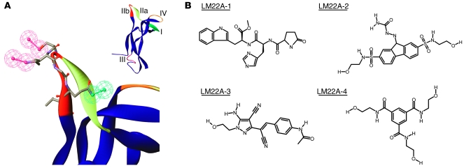
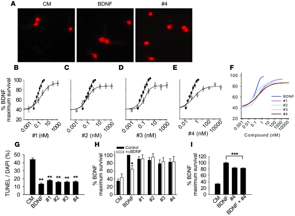
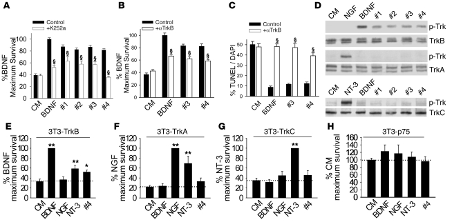
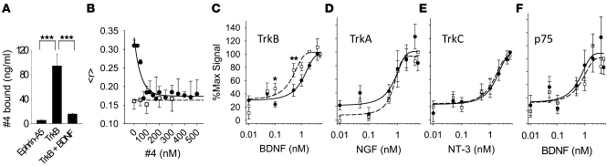
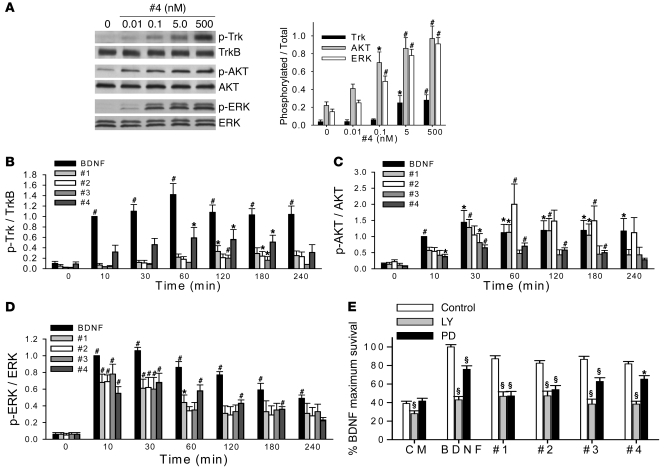
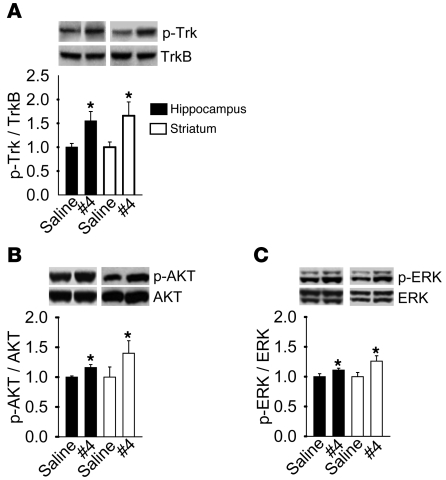
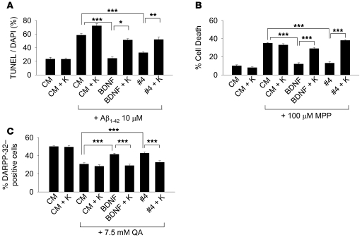
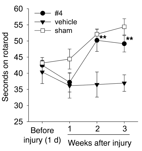
Brain-derived neurotrophic factor (BDNF) activates the receptor tropomyosin-related kinase B (TrkB) with high potency and specificity, promoting neuronal survival, differentiation, and synaptic function. Correlations between altered BDNF expression and/or function and mechanism(s) underlying numerous neurodegenerative conditions, including Alzheimer disease and traumatic brain injury, suggest that TrkB agonists might have therapeutic potential. Using in silico screening with a BDNF loop-domain pharmacophore, followed by low-throughput in vitro screening in mouse fetal hippocampal neurons, we have efficiently identified small molecules with nanomolar neurotrophic activity specific to TrkB versus other Trk family members. Neurotrophic activity was dependent on TrkB and its downstream targets, although compound-induced signaling activation kinetics differed from those triggered by BDNF. A selected prototype compound demonstrated binding specificity to the extracellular domain of TrkB. In in vitro models of neurodegenerative disease, it prevented neuronal degeneration with efficacy equal to that of BDNF, and when administered in vivo, it caused hippocampal and striatal TrkB activation in mice and improved motor learning after traumatic brain injury in rats. These studies demonstrate the utility of loop modeling in drug discovery and reveal what we believe to be the first reported small molecules derived from a targeted BDNF domain that specifically activate TrkB.We propose that these compounds constitute a novel group of tools for the study of TrkB signaling and may provide leads for developing new therapeutic agents for neurodegenerative diseases.


Figures

Figure 1
Figure 1. Pharmacophore and LM22A compound structures.
(A) Inset: Variable loop regions of a human BDNF monomer extracted from the crystallographically determined structure of a BDNF-NT3 heterodimer (56). The larger image shows the loop IIb (sequence SKGQL) pharmacophore hypothesis, as described in the text. Red, hydrogen bond donor; green, hydrogen bond acceptor. (B) Structures of the 4 LM22A compounds chosen for these studies. Compounds are abbreviated as #1–#4 in subsequent figures.
Figure 2
Figure 2. Neurotrophic activities of LM22A compounds.
(A) Fluorescence photomicrographs (original magnification, ×40) of GAP43-immunostained E16 mouse hippocampal neuronal cultures treated with culture medium (CM), BDNF, or LM22A-4 (#4) for 48 hours. (BE) Neuron survival dose-response curves for BDNF and LM22A compounds. Counts were normalized to survival achieved with 20 ng/ml (~0.7 nM) BDNF. For BDNF, survival values for each concentration were derived from n = 29–32 wells obtained from 12 separate experiments; for LM22A-1, -2, and -3, values for each concentration were derived from n = 11–19 wells obtained from 6 separate experiments; for LM22A-4, values for each concentration were derived from n = 13–21 wells obtained from 8 separate experiments. White circles, compound responses; black circles, BDNF responses (F) Comparison of survival curves from all compounds and BDNF. (G) Quantitation of the fraction of TUNEL-positive/DAPI-staining cells demonstrates that LM22A compounds decrease the number of TUNEL-positive cells to a degree similar to that of BDNF. A total of n = 93–95 fields per condition derived from 3 separate assays were counted. (H) Survival analysis of E16 hippocampal neurons treated with nonimmune serum (Control) or antibody to BDNF (Ab; 1:400) and BDNF (0.7 nM) or LM22A compounds (500 nM). Counts made in a total of n = 10–12 wells were derived from 5–6 experiments. (I) Survival of E16 hippocampal neurons treated with BDNF alone (0.7 nM), LM22A-4 alone (500 nM), or BDNF plus LM22A-4. n = 24–72 wells for each condition derived from 3 experiments were analyzed. *P < 0.05, **P < 0.01, ***P < 0.001.
Figure 3
Figure 3. LM22A compounds function through TrkB.
(A) Survival analysis of hippocampal neurons treated with BDNF (0.7 nM) or LM22A compounds (500 nM) with or without the Trk inhibitor K252a (200 nM). For BDNF, n = 37 wells derived from 7 experiments; for LM22A-1, -2, and -3, n = 17–21 wells derived from 6 experiments; and for LM22A-4, n = 28–57 wells derived from 4 experiments were assessed. (B) Survival analysis of hippocampal neurons treated with CM, BDNF (0.7 nM), or LM22A compounds (500 nM) with or without TrkBECD antibody. For each condition, n = 37–42 wells derived from 5 experiments were assessed. (C) TUNEL/DAPI analysis of E16 hippocampal neurons treated with CM, BDNF (0.7 nM), or LM22A compounds (500 nM) with or without TrkBECD antibody. For each condition, n = 17–31 fields derived from 3 experiments were assessed. (D) LM22A compounds or neurotrophic factors were incubated for 60 minutes with NIH-3T3 cells stably expressing specific Trk receptors. Western blot analysis (anti–pan-phospho-TrkY490 [p-Trk]) demonstrated that LM22A compounds activated TrkB (upper panels) but not TrkA (middle panels) or TrkC (lower panels), while all 3 Trks were activated by their cognate ligands. Activation patterns of 3 additional independent assays were identical. (EH) Trk- and p75NTR-specific NIH-3T3 cells were incubated in serum-free medium in the presence of the designated ligands for 72–96 hours, and survival was measured using the EnzyLight assay. n = 18–28 wells derived from 11–14 experiments. *P < 0.05, **P < 0.01, §P < 0.001.
Figure 4
Figure 4. LM22A-4 binds selectively to TrkB.
(A) LM22A-4 was incubated with ephrin A5-Fc receptor as a negative control or with TrkBECD-Fc in the absence or presence of BDNF (600 pmol). After rinsing of receptor complexes and subsequent ligand elution, LC/MS-MS demonstrated readily detectable LM22A-4 binding to TrkBECD-Fc that was blocked by BDNF. Values were derived from n = 3 independent binding studies. (B) BDNF (100 nM, black circles) or NGF (100 nM, white squares) was incubated with TrkBECD-Fc-Cy3B (100 nM) and increasing concentrations of LM22A-4; fluorescence anisotropy (<r>) was measured n = 6–9 times in single samples at each concentration. (CF) NIH-3T3 cells expressing TrkB, TrkA, TrkC, or p75NTR as indicated were incubated with increasing concentrations of the indicated neurotrophin in the absence (white squares, dashed line) or presence (black circles, solid line) of 100 nM LM22A-4. Symbols represent average fluorescent signal after background (i.e., binding to cells lacking expressed receptors) subtraction, as described in Methods. n = 14–18 wells from 7–9 experiments at each concentration. Significant rightward shifting of the binding curve by the compound was observed only with TrkB expressing cells. *P < 0.05, **P < 0.01, ***P < 0.001.
Figure 5
Figure 5. Comparison of BDNF’s and LM22A compound’s signaling activation kinetics and dependence of survival on downstream signaling.
(A) Left: Representative Western blot showing activation of Trk, AKT, and ERK in cultured E16 hippocampal neurons incubated with LM22A-4 at the indicated concentrations for 60 minutes. Right: Western blot analyses were quantitated for Trk, AKT, and ERK activation (ratio of p-TrkY490, using a pan–phospho-Trk antibody, to total TrkB; p-AKT to total AKT; and p-ERK to total ERK signal). n = 15–18 Western analyses from 4–9 independent protein preparations. (BD) Cultured E16 hippocampal neurons were incubated with BDNF (0.7 nM) or LM22A compounds (500 nM) for the indicated times, and quantitative analysis for Trk, AKT, and ERK activation was performed as in A. n = 8–11 Western blot analyses from 4–5 independent protein preparations. For each Western blot, the activation ratio induced by BDNF at the 10-minute time point was normalized to 1.0 and the other values adjusted accordingly. (E) E16 hippocampal neurons were incubated with BDNF (0.7 nM) or LM22A compounds (500 nM) for 48 hours in the presence or absence of the PI3K inhibitor LY294002 (LY, 10 μM) or the MAPK kinase inhibitor PD98059 (PD, 50 μM). For BDNF, n = 33–37 wells derived from 7 experiments were assessed. For LM22A-1, -2, and -3, n = 17–21 wells derived from 6 experiments were analyzed. For LM22A-4, n = 28–57 wells derived from 4 experiments were assessed. *P < 0.05, #P < 0.01, §P < 0.001.
Figure 6
Figure 6. LM22A-4 activates Trk, AKT, and ERK signaling in vivo.
LM22A-4 or saline control was administered intranasally (0.22 mg/kg/d) to adult mice for 7 days. Protein extracts of hippocampus and striatum were assessed by Western blot analysis for (A) Trk, (B) AKT, or (C) ERK phosphorylation. n = 11 mice for each assay. *P < 0.05.
Figure 7
Figure 7. LM22A-4 inhibits neuronal death in in vitro neurodegenerative disease models.
(A) Six- to 7-DIV hippocampal neurons from E16 mice were treated in the absence of Aβ with CM alone or CM + K252a (K) or in the presence of oligomeric Aβ with CM alone, CM + K252a, BDNF (0.7 nM), BDNF + K252a, LM22A-4 (500 nM), LM22A-4 + K252a. K252a was used at 200 nM. After 72 hours, cultures were assessed by TUNEL/DAPI staining. n = 28–29 fields for each condition derived from a total of 3 experiments. (B) SH-SY5Y human neuroblastoma cells were pretreated for 3 days in the following conditions: CM alone, CM + K252a, BDNF (0.7 nM), BDNF + K252a, LM22A-4 (500 nM), LM22A-4 + K252a. MPP+ (100 μM) was added, and 48 hours later cell survival was assessed by MTT/cell count assay. (C) Six- to 7-DIV striatal neurons from E16 mice were pretreated for 2 hours in the following conditions: CM, CM + K252a, BDNF (0.7 nM), BDNF + K252a, LM22A-4 (500 nM), LM22A-4 + K252a. QA (7.5 mM) was added, and after 24 hours numbers of DARPP-32–positive/total surviving cells was determined. n = 50–100 fields for each condition derived from a total 5 experiments. *P < 0.05, **P < 0.01, ***P < 0.001.
Figure 8
Figure 8. LM22A-4 restores motor learning after TBI.
Adult male Sprague-Dawley rats were trained on an accelerating rotarod task and subjected to parietal controlled cortical impact injury (filled symbols) or sham surgery (open symbols). Injured animals were treated intranasally with vehicle alone or containing LM22A-4 beginning immediately after injury then daily for 2 weeks, as indicated. Symbols indicate the mean ± SEM of the dwell time on the rod from the average of 2 trials per animal/week; n = 5 animals for vehicle and sham, and n = 6 for LM22A-4; significance was determined by repeated-measures ANOVA and post-hoc Holm-Sidak analysis. **P < 0.01.

Comment in

Similar articles

Cited by

References

    1. Huang EJ, Reichardt LF. Trk receptors: roles in neuronal signal transduction. Annu Rev Biochem. 2003;72:609–642. doi: 10.1146/annurev.biochem.72.121801.161629. - DOI - PubMed
    1. Kaplan DR, Miller FD. Neurotrophin signal transduction in the nervous system. Curr Opin Neurobiol. 2000;10(3):381–391. doi: 10.1016/S0959-4388(00)00092-1. - DOI - PubMed
    1. Chao MV. Neurotrophins and their receptors: a convergence point for many signalling pathways. Nat Rev Neurosci. 2003;4(4):299–309. doi: 10.1038/nrn1078. - DOI - PubMed
    1. Mamidipudi V, Li X, Wooten MW. Identification of interleukin 1 receptor-associated kinase as a conserved component in the p75-neurotrophin receptor activation of nuclear factor-kappa B. J Biol Chem. 2002;277(31):28010–28018. doi: 10.1074/jbc.M109730200. - DOI - PubMed
    1. Roux PP, Bhakar AL, Kennedy TE, Barker PA. The p75 neurotrophin receptor activates Akt (protein kinase B) through a phosphatidylinositol 3-kinase-dependent pathway. J Biol Chem. 2001;276(25):23097–23104. doi: 10.1074/jbc.M011520200. - DOI - PubMed

Publication types

Substances