

Hypertrophy in mesenchymal stem cell chondrogenesis: effect of TGF-beta isoforms and chondrogenic conditioning
- PMID: 20407224
- PMCID: PMC2968769
- DOI: 10.1159/000313399
Hypertrophy in mesenchymal stem cell chondrogenesis: effect of TGF-beta isoforms and chondrogenic conditioning
Abstract
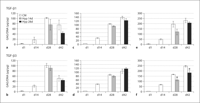
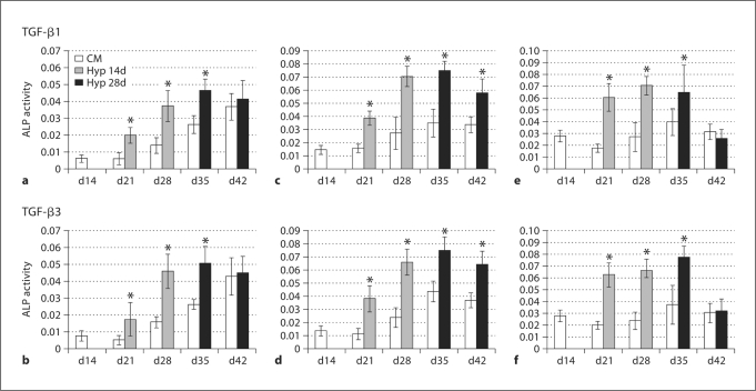
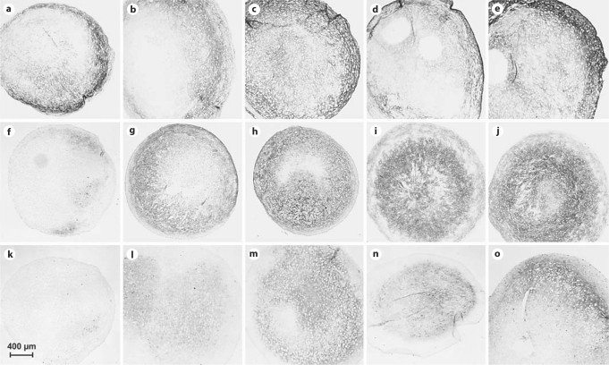
Induction of chondrogenesis in mesenchymal stem cells (MSCs) with TGF-beta leads to a hypertrophic phenotype. The hypertrophic maturation of the chondrocytes is dependent on the timed removal of TGF-beta and sensitive to hypertrophy-promoting agents in vitro. In this study, we have investigated whether TGF-beta3, which has been shown to be more prochondrogenic compared to TGF-beta1, similarly enhances terminal differentiation in an in vitro hypertrophy model of chondrogenically differentiating MSCs. In addition, we tested the impact of the time of chondrogenic conditioning on the enhancement of hypertrophy. MSCs were chondrogenically differentiated in pellet culture in medium containing TGF-beta1 or TGF-beta3. After 2 or 4 weeks, chondrogenic medium was switched to hypertrophy-inducing medium for 2 weeks. Aggregates were analyzed histologically and biochemically on days 14, 28 and 42. The switch to hypertrophy medium after 14 days induced hypertrophic cell morphology and significant increase in alkaline phosphatase activity compared to the chondrogenesis only control using both TGF-beta1 and TGF-beta3. After 28 days predifferentiation, differences between hypertrophic and control groups diminished compared to 14 days predifferentiation. In conclusion, chondrogenic conditioning with both TGF-beta isoforms similarly induced hypertrophy in our experiment and allowed the enhancement of the hypertrophic chondrocyte phenotype by hypertrophic medium. Enhancement of hypertrophy was seen more clearly after the shorter chondrogenic conditioning. Therefore, to utilize this experimental model as a tool to study hypertrophy in MSC chondrogenesis, a predifferentiation period of 14 days is recommended.
Copyright 2010 S. Karger AG, Basel.
Figures

References
-
- Bahrami S., Plate U., Dreier R., DuChesne A., Willital G.H., Bruckner P. Endochondral ossification of costal cartilage is arrested after chondrocytes have reached hypertrophic stage of late differentiation. Matrix Biol. 2001;19:707–715. - PubMed
-
- Ballock R.T., Heydemann A., Wakefield L.M., Flanders K.C., Roberts A.B., Sporn M.B. TGF-β1 prevents hypertrophy of epiphyseal chondrocytes: regulation of gene expression for cartilage matrix proteins and metalloproteases. Dev Biol. 1993;158:414–429. - PubMed
-
- Ballock R.T., O'Keefe R.J. Physiology and pathophysiology of the growth plate. Birth Defects Res C Embryo Today. 2003;69:123–143. - PubMed
-
- Barry F., Boynton R.E., Liu B., Murphy J.M. Chondrogenic differentiation of mesenchymal stem cells from bone marrow: differentiation-dependent gene expression of matrix components. Exp Cell Res. 2001;268:189–200. - PubMed
-
- Cheung J.O., Hillarby M.C., Ayad S., Hoyland J.A., Jones C.J., Denton J., Thomas J.T., Wallis G.A., Grant M.E. A novel cell culture model of chondrocyte differentiation during mammalian endochondral ossification. J Bone Miner Res. 2001;16:309–318. - PubMed
