. 2010 May 19;29(10):1659-73.
doi: 10.1038/emboj.2010.54. Epub 2010 Apr 20.

Protein Tpr is required for establishing nuclear pore-associated zones of heterochromatin exclusion

Affiliations

Protein Tpr is required for establishing nuclear pore-associated zones of heterochromatin exclusion

Sandra Krull et al. EMBO J. .

Abstract

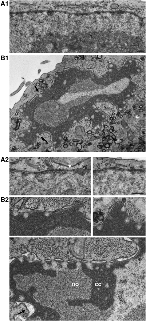
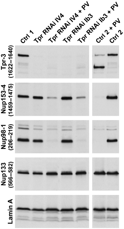
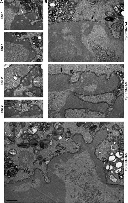
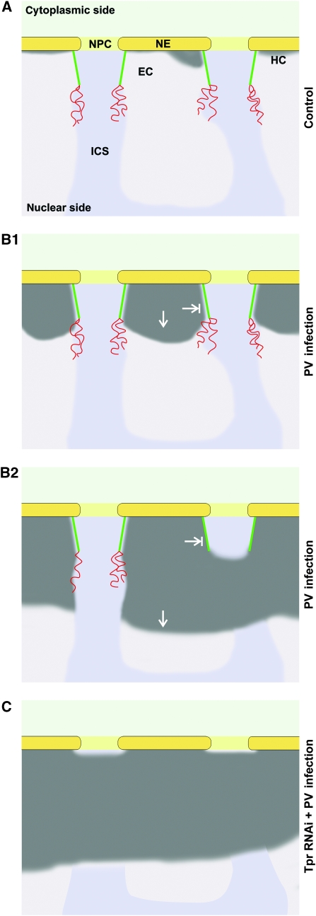
Amassments of heterochromatin in somatic cells occur in close contact with the nuclear envelope (NE) but are gapped by channel- and cone-like zones that appear largely free of heterochromatin and associated with the nuclear pore complexes (NPCs). To identify proteins involved in forming such heterochromatin exclusion zones (HEZs), we used a cell culture model in which chromatin condensation induced by poliovirus (PV) infection revealed HEZs resembling those in normal tissue cells. HEZ occurrence depended on the NPC-associated protein Tpr and its large coiled coil-forming domain. RNAi-mediated loss of Tpr allowed condensing chromatin to occur all along the NE's nuclear surface, resulting in HEZs no longer being established and NPCs covered by heterochromatin. These results assign a central function to Tpr as a determinant of perinuclear organization, with a direct role in forming a morphologically distinct nuclear sub-compartment and delimiting heterochromatin distribution.


Conflict of interest statement

The authors declare that they have no conflict of interest.

Figures

Figure 1
Figure 1
NPC-associated HEZs of distinct size and shape withstand the expansion of condensed chromatin after PV infection. (A1) TEM of a perpendicular NE cross-section of a non-infected HeLa cell in mid-interphase, with a thin layer of NE-associated heterochromatin between the NPCs. (B1) A PV-infected HeLa cell (assembled from two micrographs) at 12 h post-infection, illustrating the characteristic nuclear distortion and emergence of NPC-associated HEZs (arrows). (A2, B2) Higher magnification views of NE cross-sections of non-infected HeLa cells (A2) and of cells 12 h after PV infection (B2), illustrating the contouring of NPC-associated HEZs by condensed chromatin (cc). Nucleolar materials (no) were excluded from these zones too. Chromatin hyper-condensation was also accompanied by gradual loss of the electron-dense NPC midplanes seen in non-infected cells (white arrowhead), and often by dilation of the NE lumen (arrow) late in the infection process. Bars: 200 nm; same magnification for panels A1 and B1, and panels A2 and B2.
Figure 2
Figure 2
Stereometric size approximation of the NPC-associated minimal HEZ. (A) A perpendicularly but non-diametrically sectioned NPC-associated HEZ enclosed by condensed chromatin, from the late stage of PV infection. Bar: 50 nm. (B1) Schematic depiction of a non-diametrically sectioned HEZ and measuring tracks (double-headed arrows) of intra-membrane NPC channel diameter (2rn), of HEZ-section diameter at the base (2rb) and parallel to the base at 40 nm distance (2r40), and of HEZ-section height (hy). A total of 121 perpendicularly cross-sectioned HEZs from the late stages of PV infection were measured and normalized for non-diametric section planes as described in Supplementary Figure S2. (B2) Schematic depiction of the mean normalized diametric HEZ section, with a basis diameter 2Rb of 119 nm and heights hyfr and hapex of 74 and 96 nm, respectively. (C) The 3D shape of the corresponding mean HEZ, showing a cone with flattened top. Schemes are drawn in scale to an NPC with an intra-membrane channel diameter set to 84 nm; for possibly smaller channel diameters in PV-infected cells and the effect on calculated HEZ dimensions, see Supplementary Figure S2.
Figure 3
Figure 3
Nup153 and Nup98 appear largely degraded upon PV infection whereas the coiled-coil rod domain of protein Tpr remains intact. (A) SDS–PAGE and Coomassie staining of whole-protein extracts from HeLa cells before and at 2–12 h post-infection, showing the bulk of cellular proteins unaffected by PV-induced proteolysis. The extracts were from the same HeLa cell cultures analysed by TEM in Figure 1. (B) Immunoblotting of extracts used in panel A, showing that lamin A and lamin C, like lamin B (not shown), remain unaffected. This indicates that rearrangements within the nuclear lamina or its NE attachments, but not lamin proteolysis, are required for the NE upfolding observed. (C) Epitope sites of Nup153, Nup98, and Tpr antibodies indicated by arrowheads. Different antibodies targeting the same protein are numbered as in panels D and E. (D, E) Immunoblotting of Nup153, Nup98, and Tpr, using cell extracts shown in panel A; (see also Supplementary Figure S3). Target regions are given in parentheses; Δ indicates mAbs for which actual epitopes within defined protein segments are unknown. (D) At 8–12 h post-infection, when HEZs are visible at almost all NPCs, Nup153 (full-length proteins marked by arrows) appears largely degraded, except for a small segment (double-asterisk) comprising at least part of the Tpr-binding region. Additionally, only minor amounts of a 120-kDa degradation product (asterisk) and some unspecific cross-reactions (u) are seen with some Nup153 antibodies. Nup98 is degraded more rapidly, with only its C-terminal domain (asterisk) resisting proteolysis slightly longer. (E) Whereas Tpr's C-terminal domain is being degraded around 8 h post-infection, its entire rod domain (double-asterisk) withstands proteolysis. The membrane marked 1622–1640 was first incubated with rb-anti-Tpr-4 (2063–2084), then stripped and re-incubated with gp-anti-Tpr-3 (1622–1640).
Figure 4
Figure 4
The coiled-coil rod domain of Tpr remains anchored to the NPC even late after PV infection. IFM of HeLa cells at 10 h post-infection, showing that antibodies against Tpr's coiled-coil domain (anti-Tpr-2 (636–655), -3 (1622–1640), -5 (1370–1626Δ)) and the Tpr-binding domain of Nup153 (anti-Nup153-2 (391–404)) still label the NE. At this time point, and earlier ones (not shown), the other parts of Nup153, and the FG-repeat domain of Nup98, are no longer detectable whereas the NPC anchor of Nup98 and Tpr's C-terminal tail are still present at some NEs. Nucleoporins of the Nup107 subcomplex (also Supplementary Figure S3), representing direct and indirect anchor sites for Tpr, Nup153, and Nup98, remain bound to the NPC. DNA staining and differential interference contrast (DIC) micrographs show nuclear-peripheral chromatin accumulation and cell rounding, characteristic for later stages of PV infection. Bar: 10 μm; same magnification for all the micrographs.
Figure 5
Figure 5
RNAi-mediated Tpr knockdown in HeLa cells. (A) Confocal IFM of Tpr at day 4 after transfection with different Tpr siRNAs (Ib3, IV2, IV4) or target-less control siRNAs. Only traces of Tpr staining are seen in most cells after Tpr RNAi; bright nuclear rim staining shown as reference is visible only in cells that remained untransfected. For occasionally observed dots of residual Tpr staining at otherwise largely Tpr-deficient NEs, see Supplementary Figure S6. Bar: 20 μm. (B1) SDS–PAGE and Coomassie staining of serial dilutions of whole-protein extracts from non-transfected cells (Ctrl 2), and cells treated with Tpr siRNAs (Ib3, III4, III5, IV2, IV4) or transfection reagent alone (Ctrl 1), showing that the bulk of cellular proteins remains unaffected by siRNA treatment. (B2) Immunoblotting of identical loadings as in panel B1. Incubations with anti-Tpr and anti-Nup98 (asterisks: unrelated cross-reactions) were on different halves of the same membrane. Efficient Tpr knockdown was achieved with all Tpr siRNAs without eliciting distinct effects on other NPC proteins, including Nup93, Nup107, Nup133, and gp210 (not shown). Cells transfected with III4 and III5, however, were later found to not allow for a normal PV-infection process, so that these siRNAs were not used further. Of the infection-compatible siRNAs, Ib3 and IV4 were used for all subsequent PV-infection experiments in parallel. (C) TEM of non-transfected cells, and of cell populations at day 4 after treatment with transfection reagent alone, and transfection with Tpr siRNAs Ib3, or non-target control siRNAs (Ctrl 3). NPCs juxtaposed to heterochromatic or nucleolar material (arrows) in control cells remained characterized by HEZs but mostly lacked such exclusion zones after Tpr RNAi. Bars: 200 nm; same magnification for all the images.
Figure 6
Figure 6
Post-transcriptional tpr gene silencing by RNAi does not impair subsequent PV infection and degradation of nucleoporins. Four days after transfection with Tpr siRNAs or mock transfection with non-target control siRNAs (Ctrl 1), or after incubation with transfection reagent alone (Ctrl 2), cells were either infected with PV or not, and harvested 10 h later. Total cell proteins were analysed by immunoblotting. Regardless of whether Tpr had already been eliminated by RNAi before PV infection or not, Nup153 and Nup98 were again degraded in the infected cells whereas the NPC core protein Nup133 and lamin A remained unaffected. PV-induced degradation of nucleoporins such as Nup35, and stability of others such as Nup107 and gp210, was also similar in control and Tpr-RNAi cells (not shown).
Figure 7
Figure 7
NPC-associated HEZs are no longer maintained after in vivo depletion of Tpr, resulting in NPC coating by heterochromatin. (A) After mock transfection (Ctrl 1) or treatment with transfection reagent alone (Ctrl 2), HeLa controls were PV-infected, harvested 9.5–10 h post-infection, and then analysed by TEM, in parallel to the specimens shown in panel B. The hyper-condensed chromatin masses contoured but did not trespass the borderlines of the NPC-associated HEZs (white arrows) that persisted in these cells. (B) Four days after transfection with Tpr siRNAs, HeLa cells were infected with PV and harvested 9.5–10 h later. Amassments of condensed chromatin were found across the entrance of most NPCs (black arrows). Beside HEZ loss in the Tpr-knockdown cells, the staining of the hyper-condensed chromatin often appeared slightly lighter than in parallel controls. Whether such decreased affinity for heavy metal stains correlates with altered chromatin condensation, and how this might be caused by Tpr deficiency, remains unknown. Bar: 500 nm; same magnification for all the images.
Figure 8
Figure 8
A model for Tpr as an essential structural element of perinuclear compartmentalization. Schemes summarizing the spatial relationships between NPCs and nuclear-peripheral heterochromatin (HC) in HeLa control cells (A), and between NPCs and hyper-condensed chromatin in PV-infected cells with (B) and without Tpr (C). NEs, NPCs, and HEZs are shown in diametric cross-section and approximately to scale. The light and medium grey areas represent the euchromatic regions (EC) and the inter-chromosomal space (ICS). The coiled-coil rod domains of Tpr contribute to forming a fibrillar HEZ scaffold (green) that corresponds to the NB seen in other cell types. The unfolded Tpr tail domains (red), or other yet unidentified appendices, may project deeper into the nucleus. (A) In HeLa cells, nuclear-peripheral heterochromatin is limited to a thin layer only a few nanometres thick. Whether such heterochromatin is actually excluded from the NPC entrance zones is unclear at this point. (B1) Early upon PV infection, a process of chromatin compaction appears to spread towards the nuclear interior, thereby outlining the NPC-proximal parts of HEZs reminiscent of those observed in different types of tissue cells. (B2) Later after infection, the HEZs can be completely engirded by condensed chromatin excluded from this region. By then, the Tpr tails or any other appendices are either largely degraded or have collapsed onto the residual HEZ scaffold comprising the intact Tpr rods. Similarly shaped HEZs are observed in terminally differentiated cells with high heterochromatin content. (C) In Tpr-deficient cells, the nuclear entrances of the NPCs are covered by condensed chromatin after PV infection. It remains unknown though whether the HEZs are in fact trespassed by expanding masses of heterochromatin, or whether euchromatin possibly present in these regions after loss of Tpr is condensed upon PV infection.

Similar articles

Cited by

References

    1. Balasundaram D, Benedik MJ, Morphew M, Dang VD, Levin HL (1999) Nup124p is a nuclear pore factor of Schizosaccharomyces pombe that is important for nuclear import and activity of retrotransposon Tf1. Mol Cell Biol 19: 5768–5784 - PMC - PubMed
    1. Bancaud A, Huet S, Daigle N, Mozziconacci J, Beaudouin J, Ellenberg J (2009) Molecular crowding affects diffusion and binding of nuclear proteins in heterochromatin and reveals the fractal organization of chromatin. EMBO J 28: 3785–3798 - PMC - PubMed
    1. Bangs P, Burke B, Powers C, Craig R, Purohit A, Doxsey S (1998) Functional analysis of Tpr: identification of nuclear pore complex association and nuclear localization domains and a role in mRNA export. J Cell Biol 143: 1801–1812 - PMC - PubMed
    1. Batisse J, Batisse C, Budd A, Bottcher B, Hurt E (2009) Purification of nuclear poly(A)-binding protein Nab2 reveals association with the yeast transcriptome and a messenger ribonucleoprotein core structure. J Biol Chem 284: 34911–34917 - PMC - PubMed
    1. Beck M, Forster F, Ecke M, Plitzko JM, Melchior F, Gerisch G, Baumeister W, Medalia O (2004) Nuclear pore complex structure and dynamics revealed by cryoelectron tomography. Science 306: 1387–1390 - PubMed

Publication types