. 2010 Jun 2;29(11):1903-15.
doi: 10.1038/emboj.2010.76. Epub 2010 Apr 20.

PRR5 regulates phosphorylation, nuclear import and subnuclear localization of TOC1 in the Arabidopsis circadian clock

Affiliations

PRR5 regulates phosphorylation, nuclear import and subnuclear localization of TOC1 in the Arabidopsis circadian clock

Lei Wang et al. EMBO J. .

Erratum in

  • EMBO J. 2010 Sep 1;29(17):3033

Abstract

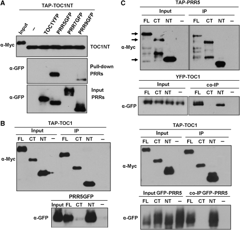
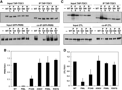
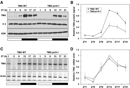
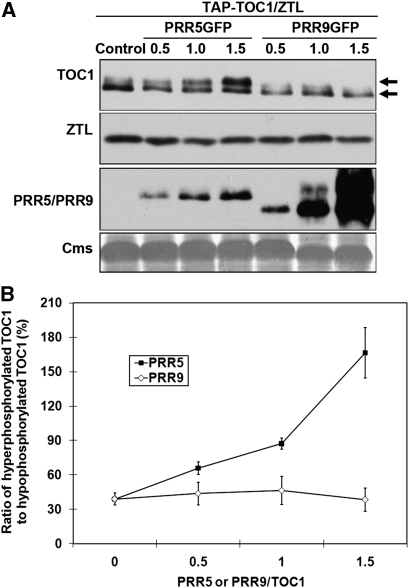
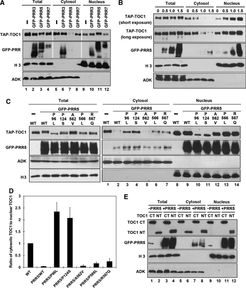
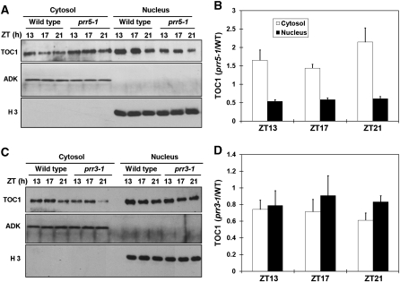
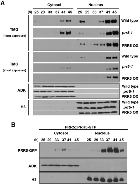
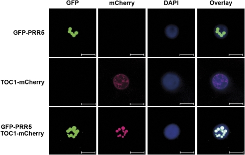
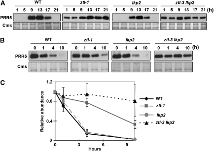
Many core oscillator components of the circadian clock are nuclear localized but how the phase and rate of their entry contribute to clock function is unknown. TOC1/PRR1, a pseudoresponse regulator (PRR) protein, is a central element in one of the feedback loops of the Arabidopsis clock, but how it functions is unknown. Both TOC1 and a closely related protein, PRR5, are nuclear localized, expressed in the same phase, and shorten period when deficient, but their molecular relationship is unclear. Here, we find that both proteins interact in vitro and in vivo through their conserved N-termini. TOC1-PRR5 oligomerization enhances TOC1 nuclear accumulation two-fold, most likely through enhanced nuclear import. In addition, PRR5 recruits TOC1 to large subnuclear foci and promotes phosphorylation of the TOC1 N-terminus. Our results show that nuclear TOC1 is essential for normal clock function and reveal a mechanism to enhance phase-specific TOC1 nuclear accumulation. Interestingly, this process of regulated nuclear import is reminiscent of similar oligomeric pairings in animal clock systems (e.g. timeless/period and clock/cycle), suggesting evolutionary convergence of a conserved mechanism across kingdoms.


Conflict of interest statement

The authors declare that they have no conflict of interest.

Figures

Figure 1
Figure 1
PRR5 interacts with TOC1 in vitro and in vivo. (A) Test of TOC1 interactions with Arabidopsis extracts expressing specific PRR clock-related proteins using TAP-tagged amino-terminal TOC1 (TAP-TOC1NT, aa 1–242). (B) The TOC1 N-terminus is necessary and sufficient to interact with PRR5 in vitro and in vivo. Only full length and amino-terminal TOC1 resin are able to pull down PRR5 in vitro (left panel). TOC1 interaction with PRR5 in vivo was detected by co-immunoprecipitation after transient co-expression of PRR5-GFP with full-length TOC1 (FL) or N-terminal (NT) or C-terminal (CT) TOC1 in N. benthamiana (right panel). (C) The PRR5 N-terminus (NT, aa 1–171) is necessary and sufficient to interact with TOC1 in vivo. Assay performed as in (B) except full length and deleted forms of TAP-PRR5 (NT, CT) were co-expressed with full-length YFP-TOC1 in N. benthamiana. Arrows indicate the migration position of the three forms of PRR5-TAP proteins (FL, CT and NT) with the vertical order of the arrows corresponding, from left to right, to the running position of TAP-tagged PRR5-FL, CT and NT, respectively. The lower bands in some lanes in the upper portion of the panel may be due to partial degradation of PRR5.
Figure 2
Figure 2
Interactions between TOC1 and PRR5 and ZTL are disrupted by specific PRR domain point mutations in TOC1. (A) Interactions between TOC1 and PRR5 in vivo were detected by co-immunoprecipitation after transient co-expression in N. benthamiana of PRR5-GFP with wild-type (WT) and various single amino-acid mutants of TOC1 in the PRR domain (P96L, P124S) and C-terminal region (A562V, P566L and R567Q). After resin binding and enzymatic release, TAP-TOC1 was detected by anti-myc and PRR5 detected by anti-GFP antibodies, respectively. (B) Quantification of the densitometric ratio of co-immunoprecipitated (co-IP) PRR5 to immunoprecipitated TOC1 in (A). Error bars indicate s.d. (n=3). (C) Co-expression and co-IP experiments conducted as in (A) but with ZTL as the test partner for interaction with TOC1. Western blots as in (A) using an anti-ZTL antibody. (D) Quantification of densitometric ratio of co-immunoprecipitated ZTL to immunoprecipitated TOC1 in (C). Error bars indicate s.d. (n=3).
Figure 3
Figure 3
PRR5 promotes post-transcriptional accumulation of TOC1. (A, B) Immunodetection of TOC1-YFP in TOC1TOC1-YFP (TMG) wild-type and prr5-1 mutant plants. TOC1 protein levels are significantly decreased in the prr5-1 mutant. Representative data are shown in (A). Quantitation of (A) shown in (B) with values normalized to the maximum expression level (ZT13). Histone H3 and ADK were used as loading controls for nuclear and cytosolic compartments, respectively. (C, D) TOC1 mRNA levels are not significantly affected by the absence of PRR5. Semi-quantitative RT–PCR experiments were repeated three times from the same tissue used in (A). Representative data are shown in (C). Quantitation of (C) are shown in (D) with values normalized to the maximum expression level (ZT13). All error bars are s.d. (n=3). Seedlings were entrained in 12-h light/12-h dark cycles and harvested at the indicated times (ZT; Zeitgeber time indicating the number of hours since lights-on.). White and black bars indicate light and dark periods, respectively.
Figure 4
Figure 4
PRR5 enhances accumulation of hyperphosphorylated TOC1. (A) Full-length PRR5 and PRR9 were co-expressed transiently in N. benthamiana with TOC1 and ZTL in a dosage series. Representative of three independent trials. Numbers indicate the ratio of initial Agrobacteria volume of PRR5 or PRR9 to that of TOC1 used for infiltration. (B) The densitometric determination of hyperphosphorylated TOC1 to hypophosphorylated TOC1 in (A). Error bars indicate s.d. (n=3). Cms: Coomassie-stained membrane indicating protein loading. Arrows indicate mobility shifts. ZTL was co-expressed to help enhance the detection of both phosphorylated and unphosphorylated TOC1.
Figure 5
Figure 5
PRR5 promotes TOC1 cytosolic depletion and nuclear accumulation. (A) PRR5 alone is able to alter the TOC1 nucleocytoplasmic distribution. GFP-PRR3, GFP-PRR5 and GFP-PRR7 were transiently co-expressed with TAP-TOC1 in N. benthamiana and tissues were processed for cytosolic and nuclear protein fractions at ZT13 2 days after infiltration. Representative of three trials. (B) PRR5 promotion of TOC1 cytosolic depletion and nuclear accumulation is dose dependent. Numbers indicate the ratio of initial Agrobacteria infiltration volume of PRR5 to that of TOC1. (C) Point mutations in the TOC1 PRR domain specifically reduce TOC1 nuclear accumulation. Left panel shows the total protein expression for each infiltration combination; right panel shows TOC1 and PRR5 accumulation in cytosolic and nuclear fractions. (D) Quantitation of relative cytosolic-to-nuclear TOC1 levels from (C). See Materials and methods for explanation. Error bars indicate s.d. (n=2). (E) TOC1 N-terminus is nuclear localized by PRR5. TOC1 NT and CT as in Figure 1. Histone H3 and ADK antibodies used are as in Figure 3. All immunoblots performed with anti-myc (TAP-TOC1) and anti-GFP (PRR5) antibodies.
Figure 6
Figure 6
Nucleocytoplasmic ratio of TOC1 distribution is altered in prr5-1. (A) Subcellular determination of cytosolic and nuclear TOC1-YFP (TOC1) in Arabidopsis wild-type and prr5-1 mutant. Anti-GFP antibody was used for immunodetection of TOC1-YFP. Histone H3 and ADK antibodies used are as in Figure 3. (B) prr5-1/WT ratio of quantitated cytosolic and nuclear TOC1 from (A). A value of 1.0 would indicate identical TOC1 levels in both backgrounds. (C) Subcellular determination of cytosolic and nuclear TOC1-YFP (TOC1) in Arabidopsis wild-type and prr3-1 mutant. (D) prr3-1/WT ratio of quantitated cytosolic and nuclear TOC1 from (C). Error bars indicate s.d. (n=3). Seedlings were entrained in 12-h light/12-h dark cycles and harvested at the indicated ZT times.
Figure 7
Figure 7
Phase-specific effect of PRR5 expression on TOC1 nucleocytoplasmic distribution. (A) Cytosolic and nuclear TOC1-YFP (TMG) levels in Arabidopsis wild-type, prr5-1 and PRR5 OX backgrounds under free-running constant light conditions. (B) Cytosolic and nuclear PRR5-GFP levels in Arabidopsis wild type under free-running constant light conditions. Seedlings were entrained in 12-h light/12-h dark cycles then maintained in constant white light for the number of hours indicated until harvested. Different immunoblot exposure levels are shown for TMG (anti-GFP) panels in (A) to allow comparisons between earlier and later time points within and between genotypes. SDS–PAGE fractionated protein extracts from all genotypes and time points within each TMG-probed panel were immunoblotted and probed together and are directly comparable, relative to the cytosolic and nuclear loading controls. Direct comparisons of abundance between TMG and PRR5-GFP panels are not appropriate. All data are representative of two independent trials. Histone H3 and ADK antibodies used are as in Figure 3.
Figure 8
Figure 8
PRR5 and TOC1 co-localize in the nucleus. 35S∷TOC1-mCherry and 35S∷GFP-PRR5 were individually or co-expressed in N. bentamiana leaves as indicated. Signals from GFP, mCherry, 4′,6-diamidino-2-phenylindole (DAPI), and the merged signals (overlay) are shown. Bars=10 μm.
Figure 9
Figure 9
LKP2 contributes to PRR5 turnover. (A) Diurnal oscillation of PRR5 is further diminished in the ztl lkp2 background. Time course of PRR5GFP protein levels in 10-day-old plants grown under 12/12L/D cycles probed with anti-GFP antibodies. Coomassie-stained regions (Cms) shown as loading controls. Blots are representative of more than three trials. (B) PRR5 stability regulated by both ZTL and LKP2. Plant growth and tissue processing as in (A) with plants transferred to continuous light at ZT0 and treated with 100 μM cyclohexamide (CHX) after 19 h. Plants were harvested at 0, 1, 4 and 10 h after adding CHX. PRR5GFP protein was detected by anti-GFP antibodies. (C) Quantitation relative to Coomassie-stained regions and normalized to time 0. Blots representative of three trials. Means of three trials±s.e.m. are shown.

Similar articles

Cited by

References

    1. Alabadi D, Oyama T, Yanovsky MJ, Harmon FG, Mas P, Kay SA (2001) Reciprocal regulation between TOC1 and LHY/CCA1 within the Arabidopsis circadian clock. Science 293: 880–883 - PubMed
    1. Baker CL, Kettenbach AN, Loros JJ, Gerber SA, Dunlap JC (2009) Quantitative proteomics reveals a dynamic interactome and phase-specific phosphorylation in the Neurospora circadian clock. Mol Cell 34: 354–363 - PMC - PubMed
    1. Bell-Pedersen D, Cassone VM, Earnest DJ, Golden SS, Hardin PE, Thomas TL, Zoran MJ (2005) Circadian rhythms from multiple oscillators: lessons from diverse organisms. Nat Rev Genet 6: 544–556 - PMC - PubMed
    1. Benito J, Zheng H, Ng FS, Hardin PE (2007) Transcriptional feedback loop regulation, function, and ontogeny in Drosophila. Cold Spring Harb Symp Quant Biol 72: 437–444 - PMC - PubMed
    1. Blau J (2008) PERspective on PER phosphorylation. Genes Dev 22: 1737–1740 - PMC - PubMed

Publication types

MeSH terms