. 2010 May 5;29(9):1573-84.
doi: 10.1038/emboj.2010.49. Epub 2010 Apr 20.

TRF2/RAP1 and DNA-PK mediate a double protection against joining at telomeric ends

Affiliations

TRF2/RAP1 and DNA-PK mediate a double protection against joining at telomeric ends

Oriane Bombarde et al. EMBO J. .

Abstract

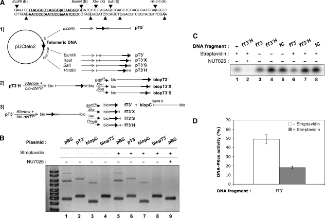
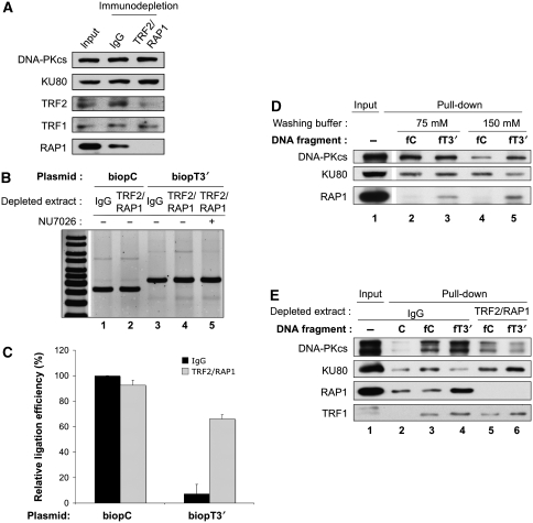
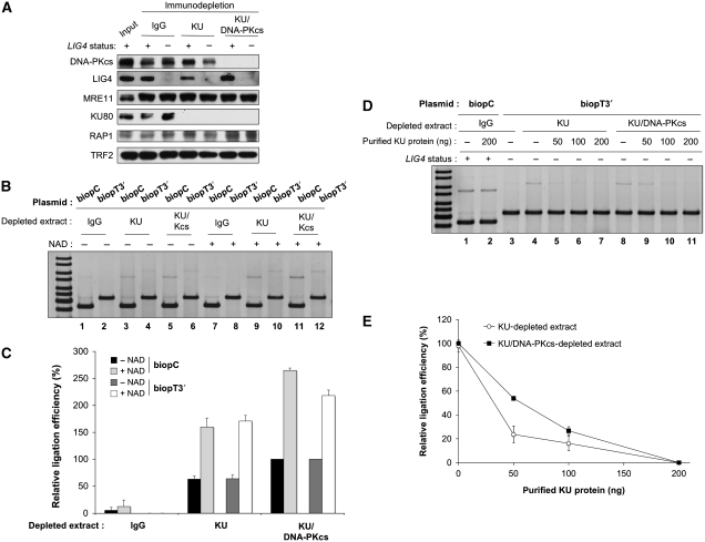
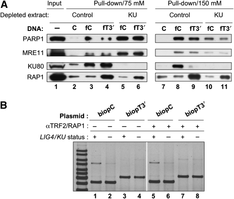
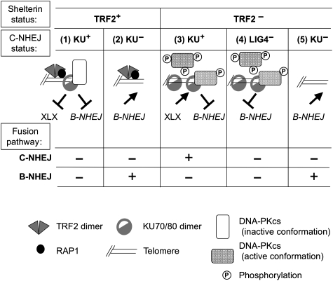
DNA-dependent protein kinase (DNA-PK) is a double-strand breaks repair complex, the subunits of which (KU and DNA-PKcs) are paradoxically present at mammalian telomeres. Telomere fusion has been reported in cells lacking these proteins, raising two questions: how is DNA-PK prevented from initiating classical ligase IV (LIG4)-dependent non-homologous end-joining (C-NHEJ) at telomeres and how is the backup end-joining (EJ) activity (B-NHEJ) that operates at telomeres under conditions of C-NHEJ deficiency controlled? To address these questions, we have investigated EJ using plasmid substrates bearing double-stranded telomeric tracks and human cell extracts with variable C-NHEJ or B-NHEJ activity. We found that (1) TRF2/RAP1 prevents C-NHEJ-mediated end fusion at the initial DNA-PK end binding and activation step and (2) DNA-PK counteracts a potent LIG4-independent EJ mechanism. Thus, telomeres are protected against EJ by a lock with two bolts. These results account for observations with mammalian models and underline the importance of alternative non-classical EJ pathways for telomere fusions in cells.


Conflict of interest statement

The authors declare that they have no conflict of interest.

Figures

Figure 1
Figure 1
DNA substrates construction and EJ and kinase assays with telomeric DNA. (A) Construction scheme of the plasmids and DNA fragments used. pUCtelo2 plasmid contains a 648 bp telomeric sequence inserted between EcoRI and BamHI sites as detailed in the upper part of the figure. Track 1: non-biotinylated plasmids. pUtelo2 was digested with the indicated enzymes to produce linearized plasmid bearing a telomeric sequence at the 5′ end (pT5′), the 3′ end (pT3′) or moved at various distance inward from the 3′ end (pT3′X, pT3′S, pT3′H). Track 2: biotinylated plasmids. Biotinylation followed by appropriate restriction produced the same 3′ended-telomeric plasmids but containing biotin at the opposite end (biopT3′, biopT3′X, biopT3′S). Track 3: biotinylated fragments. Biotinylation followed by appropriate restriction produced fragments with the telomeric sequence at various distance inward from the 3′ end (fT3′, fT3′X, fT3′S, fT3′H) and containing biotin at the opposite end. The control plasmid (biopC) corresponds to the pUCtelo2 plasmid without telomeric sequence and biotinylated at the 5′ end. A control non-telomeric 502 bp fragment biotinylated or not at the 5′-end was amplified by PCR from pBluescript-KS-II(−) (see Materials and methods). (B) EJ assay catalysed under standard reaction conditions with the indicated plasmids and HeLa extracts, in the presence or not of streptavidin or DNA–PK-specific inhibitor NU7026. DNA ligation products were separated by agarose gel electrophoresis followed by SYBR-Green staining. pBS stands for pBluescript-KS-II(−). Ligation efficiency (% of multimers versus monomer) was 30.2, 17.9, 13.7, 2.7 for lanes 1–4 (without streptavidin) and 31, 15.2, 12.7, 1.6 for lanes 5–8 (with streptavidin), respectively (C) DNA–PK assay catalysed under standard conditions with the indicated DNA fragments and HeLa extracts, in the presence or not of streptavidin or DNA–PK-specific inhibitor NU7026. DNA–PK peptide substrate was isolated by polyacrylamide denaturing gel electrophoresis followed by auto-radiography of the gel. (D) Quantification of independent experiments as shown in (C) (n=3). Relative DNA–PKcs activity was calcultated as the % of radiolabel incorporation in the peptide substrate obtained with fT3′ fragment as activating DNA compared with the incorporation obtained with fT3′H fragment, after subtraction in each case of the background incorporation obtained without DNA. Error bars correspond to s.e.m.
Figure 2
Figure 2
TRF2/RAP1complex mediates C-NHEJ inhibition at telomeric ends through hindrance at the KU loading and DNA–PK activation steps. (A) Western blotting analysis of HeLa protein extracts after immuno-depletion as indicated. Protein samples were denatured and separated on 8% SDS–PAGE gel followed by electrotransfer on membrane and blotting with the antibodies as indicated. (B) EJ assay catalysed under standard reaction conditions with the indicated plasmids and HeLa extracts depleted as specified, in the presence or not of NU7026. DNA ligation products were separated by agarose gel electrophoresis followed by SYBR-Green staining. (C) Quantification of independent experiments as shown in (B) (n=3). Relative ligation efficiency was calcultated as the % of ligation obtained in each case related to the ligation obtained on the control biopC plasmid with the control IgG-depleted extracts. Error bars correspond to s.e.m. Black bars correspond to control IgG-depleted extracts and grey bars to TRF2/RAP1-depleted extracts. Absolute ligation efficiency was 4.6±0.8% s.e.m. for IgG-depleted extracts on bioPC plasmid. (D) Pull-down experiment under standard conditions with HeLa extracts mixed with the indicated biotinylated DNA fragments. Salt molarity during washing of the beads is specified. The initial amount of protein used during the pull-down experiment was loaded as input. Protein samples were denatured and separated on 8% SDS–PAGE gel followed by electrotransfer on membrane and blotting with the antibodies as indicated. (E) Pull-down experiment as in (D) but with immuno-depleted HeLa extracts as indicated and washing under high salt conditions. C stands for the 502 bp non-biotinylated control DNA fragment.
Figure 3
Figure 3
KU and DNA–PKcs prevent B-NHEJ at telomeric ends. (A) Western blotting analysis of Nalm-6 and N114P2 protein extracts after immuno-depletion as indicated. Protein samples were denatured and separated on 8% SDS–PAGE gel followed by electrotransfer on membrane and blotting with the antibodies as indicated. (B) EJ assay catalysed under standard reaction conditions with the indicated plasmids and N114P2 LIG4- extracts depleted as specified, in the presence or not of β-NAD. DNA ligation products were separated by agarose gel electrophoresis followed by SYBR-Green staining. (C) Quantification of independent experiments as shown in (B) (n=3). For each plasmid series (biopC control or biopT3′ telomeric plasmid), relative ligation efficiency was calcultated as the % of ligation obtained in each case related to the ligation obtained with the KU/DNA–PKcs-depleted extracts without NAD. Error bars correspond to s.e.m. (D) EJ assay catalysed under standard reaction conditions with the indicated plasmids and Nalm-6 LIG4+ or N114P2 LIG4 extracts depleted as specified, in the presence or not of the indicated amount of purified KU protein. DNA ligation products were separated by agarose gel electrophoresis followed by SYBR-Green staining. (E) Quantification of independent experiments as shown in (D) (n=3). Relative ligation efficiency was calcultated as the % of ligation obtained in each case related to the ligation obtained without KU added to the extracts. Error bars correspond to s.e.m.
Figure 4
Figure 4
TRF2/RAP1complex does not impair loading of candidate B-NHEJ proteins or B-NHEJ catalysed EJ at telomeric ends. (A) Pull-down experiment under standard conditions with control or Ku-depleted N114P2 extracts mixed with the indicated DNA fragments. Salt molarity during washing of the beads is specified. The initial amount of protein used during the pull-down experiment was loaded as input. Protein samples were denatured and separated on 8% SDS–PAGE gel followed by electrotransfer on membrane and blotting with the antibodies as indicated. C stands for the 502 bp non-biotinylated control DNA fragment. (B) EJ assay catalysed under standard reaction conditions with the indicated plasmids and Nalm-6 LIG4+ or N114P2 LIG4 extracts depleted as specified, in the presence or not of a mixture of antibodies against TRF2 and RAP1. DNA ligation products were separated by agarose gel electrophoresis followed by SYBR-Green staining. The ratio of ligation efficiency with TRF2/RAP1 antibodies versus without antibodies was 0.9, 1, 8.4 and 1.2 for pair of lanes 5/1, 6/2, 7/3 and 9/4, respectively.
Figure 5
Figure 5
Model for the interplay between shelterin and EJ proteins at telomeres. See ‘Discussion' section for details. XLX refers to the ligation complex of the C-NHEJ pathway (XRCC4/LIG4/Cernunnos–XLF).

Similar articles

Cited by

References

    1. Adachi N, Ishino T, Ishii Y, Takeda S, Koyama H (2001) DNA ligase IV-deficient cells are more resistant to ionizing radiation in the absence of Ku70: implications for DNA double-strand break repair. Proc Natl Acad Sci USA 98: 12109–12113 - PMC - PubMed
    1. Amiard S, Doudeau M, Pinte S, Poulet A, Lenain C, Faivre-Moskalenko C, Angelov D, Hug N, Vindigni A, Bouvet P, Paoletti J, Gilson E, Giraud-Panis MJ (2007) A topological mechanism for TRF2-enhanced strand invasion. Nat Struct Mol Biol 14: 147–154 - PubMed
    1. Audebert M, Salles B, Calsou P (2004) Involvement of poly(ADP-ribose) polymerase-1 and XRCC1/DNA ligase III in an alternative route for DNA double-strand breaks rejoining. J Biol Chem 279: 55117–55126 - PubMed
    1. Bae NS, Baumann P (2007) A RAP1/TRF2 complex inhibits nonhomologous end-joining at human telomeric DNA ends. Mol Cell 26: 323–334 - PubMed
    1. Bailey SM, Brenneman MA, Halbrook J, Nickoloff JA, Ullrich RL, Goodwin EH (2004) The kinase activity of DNA-PK is required to protect mammalian telomeres. DNA Repair (Amst) 3: 225–233 - PubMed

Publication types