

The proportion of tumour cells is an independent predictor for survival in colorectal cancer patients
- PMID: 20407439
- PMCID: PMC2869173
- DOI: 10.1038/sj.bjc.6605674
The proportion of tumour cells is an independent predictor for survival in colorectal cancer patients
Abstract
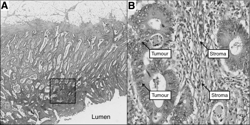
Background: The proportion of epithelial and stromal cells in tumours is thought to have an important role in the progression of epithelial malignancy. We aimed to determine whether the relative proportion of tumour (PoT) was related to survival in colorectal cancer.
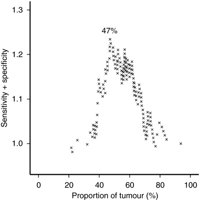
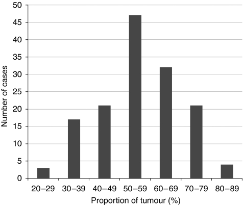
Methods: The PoT at the luminal surface was measured by point counting using virtual tissue sections in a series of 145 colorectal cancer cases. The relationship of PoT to clinicopathological parameters including cancer-specific survival was analysed. Modified receiver operating characteristic curves were used to determine the optimum cut off points to dichotomise the data for survival analyses.
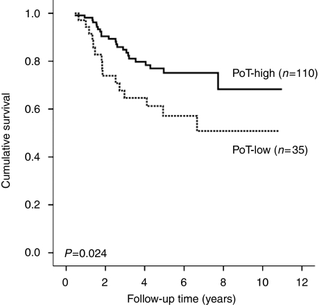
Results: Tumours with PoT-low (<or=47%) were associated with significantly lower cancer-specific survival when compared to PoT-high (hazard ratio (HR)=2.087, 95% CI=1.088-4.003, P=0.024). On sub-analysis, the prognostic effect remained significant in colonic tumours (HR=2.474, 95% CI=1.132-5.408, P=0.019) and tumour, node, metastasis stage III disease (HR=3.480, 95% CI=0.325-9.136, P=0.007). Multivariate Cox regression analysis demonstrated that PoT was an independent prognostic marker when adjusted for age, T stage, N stage and extramural vascular invasion (P=0.017).
Conclusion: This study suggests that a low proportion of tumour cells in colorectal cancer is related to poor cancer-specific survival. A relatively quick, inexpensive and well-established method such as point counting on diagnostic tissue sections could be used to identify a subset of patients who may benefit from adjuvant therapy.
Figures
References
-
- Baak JPA, Van Dop H, Kurver PHJ, Hermans J (1985) The value of morphometry to classic prognosticators in breast cancer. Cancer 56: 374–382 - PubMed
-
- Baak JPA, Langley FA, Hermans J (1991) Classification and prognosis for new cases: some aspects of univariate and multivariate analysis. In Manual of Quantitative Pathology in Cancer Diagnosis and Prognosis Baak JPA (ed), pp 189–209. Springer-Verlag: Berlin
-
- Breuninger H, Schaumburg-Lever G, Holzschuh J, Horny HP (1997) Desmoplastic squamous cell carcinoma of skin and vermilion surface. A highly malignant subtype of skin cancer. Cancer 79: 915–919 - PubMed
-
- Cancer Research UK (2009) (Accessed March 2009) Cancer Stats. http://info.cancerresearchuk.org/cancerstats/types/bowel/
-
- Chalkley HW (1943) Methods for quantitative morphological analysis of tissue. J Natl Cancer Inst 4: 47–53
Publication types
MeSH terms
LinkOut - more resources
Full Text Sources
Medical
Molecular Biology Databases
