. 2010 Feb 12;2(2):181-9.

Attenuation of diabetic retinopathy by enhanced inhibition of MMP-2 and MMP-9 using aspirin and minocycline in streptozotocin-diabetic rats

Affiliations

Attenuation of diabetic retinopathy by enhanced inhibition of MMP-2 and MMP-9 using aspirin and minocycline in streptozotocin-diabetic rats

Lokesh Kumar Bhatt et al. Am J Transl Res. .

Abstract

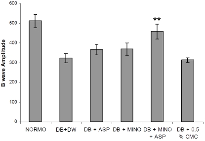
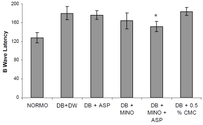
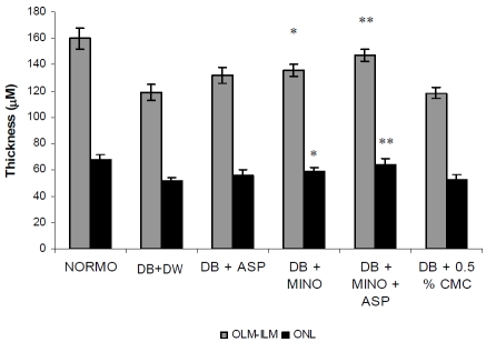
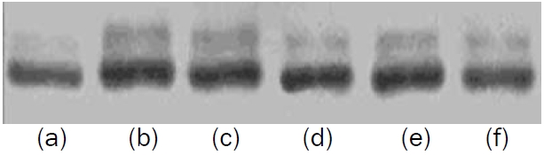
Interruptions of Matrix Metalloproteinase-2 (MMP-2) and Matrix Metalloproteinase-9 (MMP-9) have been shown to reduce the ensuing threatening risk factors of vascular complications of diabetes by alteration in Extracellular Matrix (ECM). We hypothesized that minocycline induced MMP-2 and MMP-9 inhibition can be enhanced by aspirin, a non-selective COX and tPA inhibitor and this combination can reduce progression of diabetic retinopathy. Diabetes was induced in male Wistar rats by streptozotocin (55 mg/kg i.p.). Four weeks after diabetes induction rats were treated with minocycline (50 mg/kg, p.o.) per se, aspirin (50 mg/kg, p.o.) per se, or minocycline in combination with aspirin for a period of four weeks. At the end of eighth week rats were anesthetized and electroretinograms were recorded. B-wave latency, B-wave amplitude and retinal permeability were measured. Histology was done and retinal thickness was measured. Zymography was carried out for MMP-2 and MMP-9 level determinations. B-wave amplitude was significantly decreased while B- wave latency was significantly increased in diabetic group when compared with normo-glycemic rats. Treatment with combination of minocycline and aspirin significantly reversed B-wave amplitude and latency compared with vehicle-treated diabetic controls. Blood retinal permeability and retinal thickness were also significantly attenuated by the treatment of minocycline in combination with aspirin. Results of the present study suggest that MMP-2 and MMP-9 inhibition in presence of COX inhibitor prevents the development of experimental diabetic retinopathy in rats and can be a potential approach for the treatment.

Keywords: Matrix metalloproteinase 2; diabetic retinopathy; extracellular matrix; matrix metalloproteinase 9.


Figures

Figure 1
Figure 1
Effect of four week treatment with MINO, ASP and MINO in combination with ASP on B wave amplitude. ** p<0.01 vs vehicle treated diabetic rats, (n = 6 for all groups). DB + DW (Diabetic group treated with distilled water), DB + MINO (Diabetic group treated with Minocycline per se), DB + ASP (Diabetic group treated with Aspirin per se), DB + ASP + MINO (Diabetic group treated with Minocycline and Aspirin), DB + 0.05 % CMC (Diabetic group treated with 0.5 % Carboxymethyl Cellulose solution)
Figure 2
Figure 2
Effect of four week treatment with MINO, ASP and MINO in combination with ASP on B wave latency. * p<0.05 vs vehicle treated diabetic rats, (n = 6 for all groups) DB + DW (Diabetic group treated with distilled water), DB + MINO (Diabetic group treated with Minocycline per se), DB + ASP (Diabetic group treated with Aspirin per se), DB + ASP + MINO (Diabetic group treated with Minocycline and Aspirin), DB + 0.05 % CMC (Diabetic group treated with 0.5 % Carboxymethyl Cellulose solution).
Figure 3
Figure 3
Effect of four week treatment with MINO, ASP and MINO in combination with ASP on retinal thickness. * p<0.05, ** p<0.01 vs vehicle treated diabetic rats. (n = 6 for all groups) OLM -ILM (Outer Limiting Membrane to Inner Limiting Membrane), ONL (Outer Nuclear Layer), DB + DW (Diabetic group treated with distilled water), DB + MINO (Diabetic group treated with Minocycline per se), DB + ASP (Diabetic group treated with Aspirin per se), DB + ASP + MINO (Diabetic group treated with Minocycline and Aspirin), DB + 0.05 % CMC (Diabetic group treated with 0.5 % Carboxymethyl Cellulose solution).
Figure 4
Figure 4
Effect of three week treatment with MINO, ASP and MINO in combination with ASP on retinal protein (72 KDa) level. (a) Normal (b) Diabetic + DW (Diabetic group treated with distilled water), (c) Diabetic + 0.5 % CMC (Diabetic group treated with 0.5 % Carboxymethyl Cellulose solution) (d) Diabetic + ASP (Diabetic group treated with Aspirin per se) (e) Diabetic + MINO (Diabetic group treated with Minocycline per se) (f) Diabetic + ASP + MINO (Diabetic group treated with Minocycline and Aspirin)
Figure 5
Figure 5
Effect of three week treatment with MINO, ASP and MINO in combination with ASP on retinal protein (92 KDa) level. (a) Normal (b) Diabetic + DW (Diabetic group treated with distilled water), (c) Diabetic + 0.5 % CMC (Diabetic group treated with 0.5 % Carboxymethyl Cellulose solution) (d) Diabetic + ASP (Diabetic group treated with Aspirin per se) (e) Diabetic + MINO (Diabetic group treated with Minocycline per se) (f) Diabetic + ASP + MINO (Diabetic group treated with Minocycline and Aspirin).
Figure 6
Figure 6
Effect of four week treatment with MINO, ASP and MINO in combination with ASP on retinal permeability. * p<0.05, ** p<0.01 vs vehicle treated diabetic rats. (n = 6 for all groups) DB + DW (Diabetic group treated with distilled water), DB + MINO (Diabetic group treated with Minocycline per se), DB + ASP (Diabetic group treated with Aspirin per se), DB + ASP + MINO (Diabetic group treated with Minocycline and Aspirin), DB + 0.05 % CMC (Diabetic group treated with 0.5 % Carboxymethyl Cellulose solution)

References

    1. Cunha-Vaz JG, Fonseca JR, de Abreu JF, Ruas MA. Detection of early retinal changes in diabetes by vitreous fluorophotometry. Diabetes. 1979;28:16–19. - PubMed
    1. Ishibashi T, Tanaka K, Taniguchi Y. Disruption of blood-retinal barrier in experimental diabetic rats: An E.M. study. Exp Eye Res. 1980;30:401–410. - PubMed
    1. Cogan DG, Toussaint D, Kuwabara T. Retinal vascular pattern. IV. Diabetic retinopathy. Arch Ophthalmol. 1961;66:366–378. - PubMed
    1. Papachristodoulou D, Heath H, Kang SS. The development of retinopathy in sucrose-fed and streptozotocindiabetic rats. Diabetologia. 1976;12:367–374. - PubMed
    1. Antonetti DABA, Bronson SK, Freeman WM, Gardner TW, Jefferson LS, Kester M, Kimball SR, Krady JK, LaNoue KF, Norbury CC, Quinn PG, Sandirasegarane L, Simpson IA. JDRF Diabetic Retinopathy Center Group (2006) Diabetic retinopathy: seeing beyond glucose-induced mi-crovascular disease. Diabetes. 2006;55:2401–2411. - PubMed

LinkOut - more resources