. 2010 Mar 3:4:5.
doi: 10.3389/neuro.04.005.2010. eCollection 2010.

The Neuronal Splicing Factor Nova Co-Localizes with Target RNAs in the Dendrite

Affiliations

The Neuronal Splicing Factor Nova Co-Localizes with Target RNAs in the Dendrite

Claudia Racca et al. Front Neural Circuits. .

Abstract

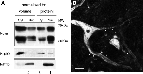
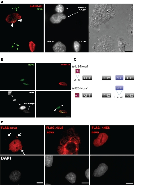
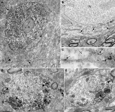
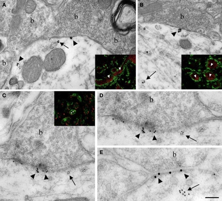
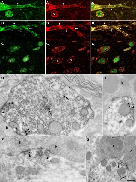
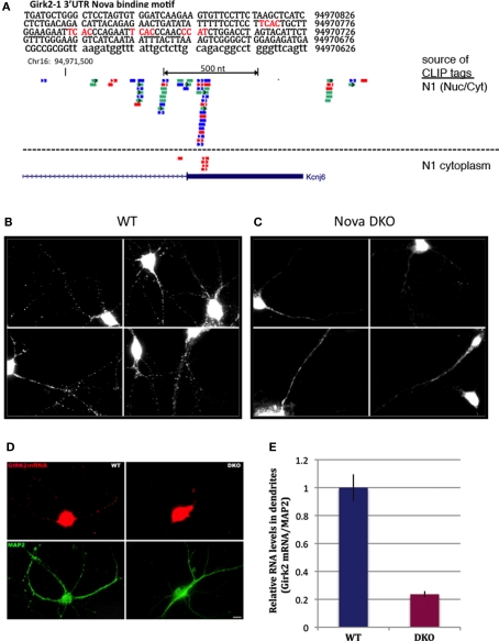
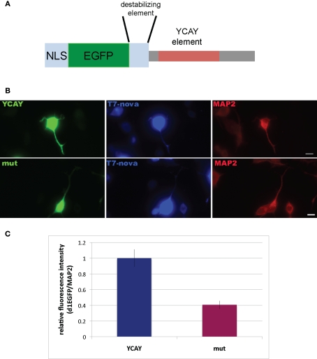
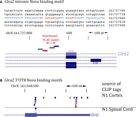
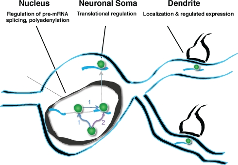
Nova proteins are neuron-specific RNA binding proteins targeted by autoantibodies in a disorder manifest by failure of motor inhibition, and they regulate splicing and alternative 3' processing. Nova regulates splicing of RNAs encoding synaptic proteins, including the inhibitory glycine receptor alpha2 subunit (GlyRalpha2), and binds to others, including the GIRK2 channel. We found that Nova harbors functional NES and NLS elements, shuttles between the nucleus and cytoplasm, and that 50% of the protein localizes to the soma-dendritic compartment. Immunofluoresence and EM analysis of spinal cord motor neurons demonstrated that Nova co-localizes beneath synaptic contacts in dendrites with the same RNA, GlyRalpha2, whose splicing it regulates in the nucleus. HITS-CLIP identified intronic and 3' UTR sites where Nova binds to GlyRalpha2 and GIRK2 transcripts in the brain. This led directly to the identification of a 3' UTR localization element that mediates Nova-dependent localization of GIRK2 in primary neurons. These data demonstrate that HITS-CLIP can identify functional RNA localization elements, and they suggest new links between the regulation of nuclear RNA processing and mRNA localization.

Keywords: GIRK2; HITS-CLIP; RNA localization; RNA splicing; glycine receptor; nova; shuttling protein; spinal motor neuron.


Figures

Figure 1
Figure 1
Subcellular distribution of Nova proteins. (A) Immunoblot analysis of Nova distribution in cytoplasmic and nuclear fractions from mouse brain (equal volumes (20 μl) of each fraction were loaded in lanes 1 and 2; equal protein amounts (50 μg) were loaded in lanes 3 and 4). Hsp90 is used as a cytoplasmic marker, and brPTB as a nuclear marker. The antibody used detects both Nova 1 (∼55 kD) and Nova-2 (∼75 kD) isoforms. (B) Nova signal is detected within the nucleus, somatic cytoplasm, and neurites of ventral horn spinal cord neurons. Within neurites, the signal is observed along the plasma membrane (arrowheads). Scale bar: 10 μm (B).
Figure 2
Figure 2
Nova proteins shuttle between the nucleus and cytoplasm. (A) IMR32 and COS7 cells were fused with PEG 3350, and anti-hnRNPC1 and anti-Nova antibodies were used to detect endogenous proteins. In this field one cell has been fused with COS7 (top; see phase contrast, right panel), and two unfused cells are evident (bottom); cell types can be distinguished with DAPI staining (middle panel). Nova proteins were detected in IMR32 and fused COS7 cells (arrowheads), but no signal in isolated COS7 cells. DAPI staining showed IMR32 cells and COS7 cells, respectively. (B) Shuttling of endogenous Nova from SK-N-BE(2) neuroblastoma cells into mouse NIH 3T3 cells; Nova, hnRNP-C12 and DAPI stains are shown as in (A). (C) Schematic of Flag-tagged Nova NLS and NES domains and deletion constructs generated. (D) COS7 cells were transfected with the indicated Flag-Nova1 plasmid constructs and stained with anti-flag antibody to visualize flag-Nova1 (top panels), and DAPI to visualize nuclei (bottom panels). Nova1 can be seen in the cytoplasm in cells transfected with the WT (left panel; arrows) but not in cells transfected with the ΔNES construct (right panel); in contrast the ΔNLS construct is largely excluded from nuclei (middle panel).
Figure 3
Figure 3
Ultrastructural localization of Nova in the somata and dendrites of ventral horn spinal cord neurons. (A,B) Micrographs of pre-embedding immunocytochemistry experiments. (C) Micrograph of a post-embedding immunocytochemistry experiment. (A) Distribution of Nova visualized with electron dense HRP reaction product (arrows). The nucleus is heavily stained but not the nucleolus (n). Presence of electron dense HRP reaction product (arrowhead) in front of a synaptic contact (b). (B) Gold particles associated with Nova are found within the nucleus (N), as well as within the somatic cytoplasm (arrows). (C) High magnification micrograph of the nuclear periphery. In spinal neuron nuclei (N), where the chromatin is condensed at the nuclear periphery in proximity of the inner side of the nuclear membrane, Nova (15 nm gold particles) accumulates over chromatin (crossed arrows) and can be found within nuclear pores (arrow). (D,E) Electron microscopic immunolabeling of Nova in dendrites (HRP reaction product) showing Nova tendency to accumulate peripherally to the dendritic center, and close to synapses (arrowheads). Axons are devoid of staining. Boutons (b). Scale bar: 4 μm (A); 2 μm (B); 0.3 μm (C); 1 μm (D,E).
Figure 4
Figure 4
Localization of Nova at inhibitory synapses. (A,B) Nova signal detected by gold labeling (arrows) is observed in dendrite and opposed to boutons (b) at the dendritic periphery (arrowheads). Insets in (A) and (B), Double-immunofluorescence detection of Nova (in red) and synapsin (in green) show that Nova immunoreactivity is present opposite to synaptic contacts (arrowheads). (C,D,E) Simultaneous detection of Nova and gephyrin immunoreactivity within the postsynaptic cytoplasm. Nova immunoreactivity (10 nm gold particles; arrows) is found beneath postsynaptic regions where gephyrin immunoreactivity (15 nm gold particles; arrowheads) decorates and identifies inhibitory synapses. (D) High magnification of the postsynaptic cytoplasm of (C). Inset in (C), Double-immunofluorescence of Nova (in green) and gephyrin (in red) showed that Nova immunoreactivity is observed in proximity of gephyrin immunoreactivity synaptic contacts (arrowheads). Scale bar: 0.2 μm (A); 0.6 μm (B); 0.2 μm (C,E); 0.4 μm (D); 9 μm (insets in A,B); 6 μm (inset in C).
Figure 5
Figure 5
Nova and GlyRα2 mRNAs, its nuclear alternative splicing RNA target, colocalize outside the nucleus within dendrites. Fluorescence labeling for Nova immunoreactivity (in green; A,B,C) and ISH signal (in red) for GlyRα1 (A1) or GlyRα2 mRNAs (B1,C1). (A) Within the somatic and dendritic cytoplasm of ventral horn neurons, GlyRα1/2 (A1,B1) mRNAs and Nova (A,B) labeling co-localizes (in yellow; A1,B1). The labeling pattern of Nova mirrors that of GlyRα1/2 mRNAs. Both are also unevenly distributed within the dendritic cytoplasm and sometimes accumulate at the dendritic periphery (A,B; arrowheads) and branch points (B; arrows). Note that the signal corresponding to Nova protein or GlyRα1/2 mRNAs are separated in some areas. (C) in neurons of the dorsal horn, where GlyRα2 mRNA (C1) is restricted to the somatic cytoplasm, Nova immunoreactivity is detected in nuclei (N) and to a lesser extent in somatic cytoplasm (C) resulting in a less accentuated co-localization (C2). (D–G) Ultrastructural simultaneous detection of Nova immunoreactivity and GlyRα2 mRNAs. Nova (HRP immunolabeling) and GlyRα2 mRNA ISH signal (gold particles) colocalize within the dendritic cytoplasm (arrows) and in front of synaptic boutons (arrowheads, b). Note in (E,G) the association of Nova and mRNA signals with small cisternae. Scale bar: 15 μm (A–C); 0.2 μm (D–F); 0.35 μm (G).
Figure 6
Figure 6
Nova regulates localization of the CLIP target GIRK2-1 mRNA isoform in dendrites. (A) Location of Nova1 CLIP tags in the 3′ UTR of the GIRK2-1 transcript. The RNA sequence of the GIRK2-1 3′ UTR in the region of the Nova binding site is shown, with YCAY elements highlighted in red. CLIP tags from mouse cortex (Nuc/Cyt) or cytoplasmic extracts of mouse brain (N1 cytoplasm) are shown separately. Nova tags are colored, with each color representing tags from a different mouse brain. Several other robust Nova CLIP target sites rich in YCAY elements were found in upstream sites, most notably in intron 2 ∼187,000nt upstream (see text). (B,C) Fluorescence ISH (FISH) using GIRK2-1 specific probes to detect mRNA in WT (B) or Nova DKO (C) primary cultured neurons obtained from E18.5 mouse cortex. No signal was seen with control ISH probes. (D) Comparison of GIRK2-1 FISH and MAP2 immunoreactivity in WT and Nova DKO primary neurons, as indicated. (E) Quantitation of fluorescence intensity from (D); MAP2 fluorescence signal was counted beginning 5–10 μm from the cell body and extended to the distal dendrites, and then the signal from GIRK2-1 was obtained from the same dendrite; at least 10 neurons were counted in each of three experiments. The results from a three independent experiments are plotted as the sum of the dendritic signal from GIRK2-1 (Cy3) divided by MAP2 (Cy5); error bars represent standard deviation (p < 0.05; Student's t-test). The MAP2 signal alone was not significantly changed in WT vs. KO neurons (data not shown). Sense strand oligonucleotides were used as a negative control and showed no signal under identical conditions. Scale bar: 10 μm (D).
Figure 7
Figure 7
Binding of Nova to the GIRK2-1 YCAY element is necessary for proper localization. (A) Schematic of a reporter encoding destabilized d1EGFP with an M9 nuclear localization sequence (NLS) and the 3′ UTR GIRK2-1 YCAY element. An identical construct in which the four YCAY sequences were mutated to YGUY was also made. (B) Comparison of EGFP and MAP2 immunoreactivity in cells co-transfected with plasmid constructs encoding the YCAY or the mutant (“mut”) YGUY element, as indicated, and a construct expressing T7-tagged Nova. The WT Nova target mRNA (green channel, left panel) was localized in distal neurites in transfecting cells expressing the YCAY but not mutant GIRK2-1 element; T7-tagged Nova itself, detected with T7 antibody (middle panel) also appeared more localized in distal dendrites, while MAP2 (right panel) was unchanged in either condition. (C) Quantitation of EGFP fluorescence intensity from (B); Region of Interest (ROI) was chosen based on neurite in which MAP2 fluorescence signal was detected, beginning 5 μm from the cell body and extending outwards. The signal from d1EGFP was obtained from the same ROI; more than 10 cells were counted. The results are plotted as relative ratio of the sum of the neurite signal from d1EGFP (Cy2) divided by MAP2 (Cy3) in YCAY element or mutated constructs; error bars represent standard deviation (p < 0.05); results are from three independent experiments. Scale bar: 10 μm (B).
Figure 8
Figure 8
Location of Nova CLIP tags in the GlyRα2 transcript. (A) The sequence of the previously characterized Nova binding YCAY motif upstream of GlyRα2 exon 3A is shown (blue); mutation of these YCAY elements (red) abrogates E3A splicing (Buckanovich and Darnell, 1997). Lower panel shows Nova HITS-CLIP tags in this intronic region. (B) Nova HITS-CLIP tags in the GlyRα2 3′ UTR. In (A,B) Nova tags are colored as in Figure 6.
Figure 9
Figure 9
Model of Nova action. Nova binds to pre-mRNA in the nucleus. This binding can, but need not be intronic (pathway labeled “1”). If binding sites are in the region of alternatively spliced exons, Nova can mediate alternative splicing, according to the rules of a functional binding map, such that the position of Nova binding determines the outcome of alternative exon inclusion. Subsequently, Nova may multimerize in cis upon the same transcript (pathway 1), or may be independently deposited upon additional mRNA binding sites, such as those in the 3′ UTR (pathway labeled “2”). 3′ UTR binding sites may determine additional processing steps (such as mediating alternative polyadenylation), and mediate soma and dendritic mRNA localization (as in the case of GIRK2-1 and perhaps GlyRα2 mRNA). It is presumed, based for example on the studies of Singer and colleagues (Lawrence and Singer, ; Kislauskis et al., ; Rodriguez et al., 2008), that such RNA localization would be coupled to the regulation of translation. In neurons, coupling pre-mRNA binding to mRNA localization and translation offers the possibility of linking information content generated in the nucleus (alternative splicing or polyadenylation) with differential gene expression in dendrites.

Similar articles

Cited by

References

    1. Altschuler R. A., Betz H., Parakkal M. H., Reeks K. A., Wenthold R. J. (1986). Identification of glycinergic synapses in the cochlear nucleus through immunocytochemical localization of the postsynaptic receptor. Brain Res. 369, 316–32010.1016/0006-8993(86)90542-1 - DOI - PubMed
    1. Arn E. A., Cha B. J., Theurkauf W. E., Macdonald P. M. (2003). Recognition of a bicoid mRNA localization signal by a protein complex containing Swallow, Nod, and RNA binding proteins. Dev. Cell 4, 41–5110.1016/S1534-5807(02)00397-0 - DOI - PubMed
    1. Ashraf S. I., McLoon A. L., Sclarsic S. M., Kunes S. (2006). Synaptic protein synthesis associated with memory is regulated by the RISC pathway in Drosophila. Cell 124, 191–20510.1016/j.cell.2005.12.017 - DOI - PubMed
    1. Bassell G. J., Singer R. H. (2001). Neuronal RNA localization and the cytoskeleton. Results Probl. Cell Differ. 34, 41–56 - PubMed
    1. Bassell G. J., Warren S. T. (2008). Fragile X syndrome: loss of local mRNA regulation alters synaptic development and function. Neuron 60, 201–21410.1016/j.neuron.2008.10.004 - DOI - PMC - PubMed