. 2010 Aug;130(4):484-93.
doi: 10.1111/j.1365-2567.2010.03269.x. Epub 2010 Apr 12.

Simvastatin induces Foxp3+ T regulatory cells by modulation of transforming growth factor-beta signal transduction

Affiliations

Simvastatin induces Foxp3+ T regulatory cells by modulation of transforming growth factor-beta signal transduction

Yong Chan Kim et al. Immunology. 2010 Aug.

Abstract

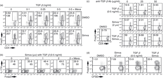
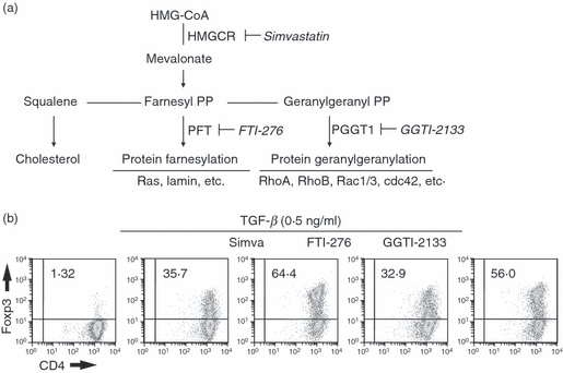
Statins are widely used drugs for the treatment of hypercholesterolaemia. A number of recent studies have suggested that statins also have pleiotropic effects on immune responses and statins have proven to be effective in the treatment of autoimmune diseases in animal models. Foxp3(+) T regulatory cells are a unique subset of CD4(+) T cells that mediate immunosuppression. Foxp3(+) T cells develop in the thymus, but can also be induced in peripheral sites in the presence of transforming growth factor-beta (TGF-beta). We demonstrate here that simvastatin blockade of the mevalonate pathway can mediate induction of mouse Foxp3(+) T cells and that simvastatin can synergize with low levels of TGF-beta to induce Foxp3(+) T cells. The effects of simvastatin are secondary to a blockade of protein geranylgeranylation, are mediated at late time-points after T-cell activation, and are associated with demethylation of the Foxp3 promoter. One major effect of simvastatin was inhibition of the induction of Smad6 and Smad7, inhibitory Smads that inhibit TGF-beta signalling. Our results suggest that one mechanism responsible for the immunosuppressive effects of statins is the ability to promote the generation of Foxp3(+) T regulatory cells.


Figures

Figure 1
Figure 1
Simvastatin induces Foxp3 expression in the absence or presence of transforming growth factor-β (TGF-β). (a) CD4+Foxp3 T cells isolated from T-cell receptor (TCR) transgenic RAG−/− mice were stimulated for 72 hr with plate-bound anti-CD3/CD28 (4 and 2 μg/ml, respectively), 100 U/ml interleukin-2 (IL-2), in the presence of vehicle alone, TGF-β, simvastatin (2 μm), separately or together. Mevalonate (200 μm) was added to some cultures as indicated. Expression of Foxp3 was measured by intracellular staining. (b) Same protocol as in panel (a) except that a neutralizing anti-TGF-β monoclonal antibody (mAb) was added at the concentrations indicated. (c) Same protocol as in panel (a), except that the concentration of simvastatin was varied. (d) The CD4+ Foxp3 T cells were labelled with carboxyfluorescein succinimidyl ester (CFSE), stimulated for 72 hr under the in vitro activating condition used in (a). CFSE dilution and Foxp3 expression were measured by fluorescence-activated cell sorting analysis. The numbers in black indicate the percentage of Foxp3+ cells and numbers in italics represent the mean fluorescence intensity (MFI) of the Foxp3+ cells. These results are representative of four different experiments.
Figure 2
Figure 2
Simvastatin-induced Foxp3-expressing CD4+ T cells exhibit normal suppressive function. Fluorescence-activated cell-sorted CD4+ GFP T cells were isolated from Foxp3gfp mice, stimulated with plate-bound CD3/CD28 antibodies and interleukin-2 (IL-2) in the presence of transforming growth factor-β (TGF-β) alone or with TGF-β and simvastatin. After the initial 72 hr of culture, converted GFP+ cells were re-sorted and mixed with CD4+ Foxp3 responder cells, T-cell-depleted splenocytes, and soluble anti-CD3 antibody at the indicated cell : cell ratios (x-axis). [3H]Thymidine incorporation was assayed 96 hr after stimulation. Results are presented as counts/min (CPM) ± SEM of triplicate cultures. Similar results were observed in two other experiments.
Figure 3
Figure 3
The synergistic effects of simvastatin on transforming growth factor-β (TGF-β) -induced Foxp3 induction are mediated by blocking protein geranylgeranylation. (a) Schematic diagram of the mevalonate pathway is shown. Names in bold indicate metabolic enzymes, names in italics are specific inhibitors of those enzymes. (b) CD4+ Foxp3 T cells were stimulated as in Fig. 1 in the presence of simvastatin or inhibitors of the downstream branches of the mevalonate pathway (20 μm of FTI-276 or 20 μm of GGTI-2133) for 72 hr. Foxp3 expression was measured by intracellular staining. Data are representative of three other experiments.
Figure 4
Figure 4
The synergistic effects of simvastatin on transforming growth factor-β (TGF-β) -induced Foxp3 induction are mediated at a late stage of T-cell activation. (a) CD4+ Foxp3 T cells were stimulated with plate-bound anti-CD3/28, interleukin-2 (IL-2), in the presence of TGF-β, simvastatin, or both together. Fluorescence-activated cell sorting analysis of Foxp3 expression was performed at the indicated hours after stimulation. Similar results were observed in one other experiment. (b) CD4+ Foxp3 T cells were stimulated with plate-bound anti-CD3/CD28, IL-2, in the presence of TGF-β alone (0.2 ng/ml, closed squares) or together with simvastatin (2 μm, open squares). After initiation of T-cell stimulation, simvastatin (2 μm, closed symbols) or mevalonate (200 μm, open symbols) were added at the indicated time-points (x-axis). After 72 hr, all cultures were simultaneously harvested and the percentage of Foxp3-expressing cells was determined by intracellular staining. Similar results were observed in one other experiment. (c) CD4+ Foxp3 T cells were stimulated as in Fig. 1(d) in the presence of vehicle alone or TGF-β. Simvastatin (2 μm) was added for the entire 72 hr of culture (continuous) or after 24 hr and then neutralized at 48 hr with mevalonate (200 μm, pulsed). Foxp3 expression was measured by intracellular staining at 72 hr. Similar results were observed in two other experiments.
Figure 5
Figure 5
Simvastatin increases Foxp3 messenger RNA and inhibits DNA methylation of the Foxp3 promoter in the presence of transforming growth factor-β (TGF-β). (a) CD4+ Foxp3 T cells were stimulated as in Fig. 1(d) in the presence of dimethylsulphoxide, TGF-β (0.5 ng/ml) or simvastatin (2 μm) together. After the indicated times, the cultures were harvested and the total RNA extracted. Foxp3 mRNA levels were determined by quantitative RT-PCR and expression levels normalized to 18S ribosomal RNA. Similar results were observed in one other experiment. (b) Foxp3+ and Foxp3- cells were FACS-sorted from lymph node cells of male Foxp3gfp male mice (FACS profile). The methylation status of each CpG site is displayed as closed circles (methylated) and opened circles (demethylated). Each group of circles is arranged in the order of the position of the CpG sites (number on top) and represents an individual cloned sample. (c) Cultures were stimulated as in (a), and genomic DNA was extracted at 72 hr. Methylation status of the Foxp3 promoter region was analysed by disulphite modified CpG sequencing. Blue bold numbers indicate the percentage of methylated sites in each sample after 72 hr. Foxp3 expression was measured at 72 hr of activation by intracellular staining. Similar results were observed in three other experiments.
Figure 6
Figure 6
Simvastatin has no effect on transforming growth factor-β (TGF-β) -induced phosphorylation of Smad3/4, but blocks TGF-β-induced expression of Smad6 and Smad7. (a) CD4+ Foxp3 T cells were stimulated as in Fig. 1(d) in the presence of dimethylsulphoxide, TGF-β (5 ng/ml), simvastatin (2 μm, Simva) or TGF-β and simvastatin. Proteins were extracted with lysis buffer and analysed by Western blot for phospho-Smad3 (pSmad3) as well as total Smad3 and Smad4. (b) Total proteins from cells stimulated as in (a) were extracted at the indicated times and relative changes of Smad6 and Smad7 expression were determined by Western blotting. (c) CD4+ Foxp3 and CD4+ Foxp3+ cells were isolated from lymph nodes of female Foxp3gfp mice by fluorescence-activated cell sorting. Western blot analysis of the expression of Smad6 and Smad7 was performed on resting cells or following activation for 72 hr with plate-bound anti-CD3 and interleukin-2 in the presence or absence of TGF-β. Similar results were observed in three other experiments.

References

    1. Shevach EM. From vanilla to 28 flavors: multiple varieties of T regulatory cells. Immunity. 2006;25:195–201. - PubMed
    1. Davidson TS, DiPaolo RJ, Andersson J, Shevach EM. Cutting edge: IL-2 is essential for TGF-beta-mediated induction of Foxp3+ T regulatory cells. J Immunol. 2007;178:4022–6. - PubMed
    1. Fontenot JD, Gavin MA, Rudensky AY. Foxp3 programs the development and function of CD4+CD25+ regulatory T cells. Nat Immunol. 2003;4:330–6. - PubMed
    1. Hori S, Nomura T, Sakaguchi S. Control of regulatory T cell development by the transcription factor Foxp3. Science. 2003;299:1057–61. - PubMed
    1. Tone Y, Furuuchi K, Kojima Y, Tykocinski ML, Greene MI, Tone M. Smad3 and NFAT cooperate to induce Foxp3 expression through its enhancer. Nat Immunol. 2008;9:194–202. - PubMed

Publication types

MeSH terms