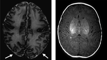
Imaging selective vulnerability in the developing nervous system
- PMID: 20408904
- PMCID: PMC2992418
- DOI: 10.1111/j.1469-7580.2010.01226.x
Imaging selective vulnerability in the developing nervous system
Abstract
Why do cells in the central nervous system respond differently to different stressors and why is this response so age-dependent? In the immature brain, there are regions of selective vulnerability that are predictable and depend on the age when the insult occurs and the severity of the insult. This damage is both region and cell population specific. Vulnerable cell populations include the subplate neurons and oligodendrocyte precursors early in development and the neurons closer to the end of human gestation. Mechanisms of injury include excitotoxicity, oxidative stress and inflammation as well as accelerated apoptosis. Advanced imaging techniques have shown us particular patterns of injury according to age at insult. These changes seen in the newborn at the time of injury on magnetic resonance imaging correlate well with the neurodevelopmental outcome. New questions about how the injury evolves and how the newborn brain adapts and repairs itself have emerged as we now know that injury in the newborn brain can evolve over days and weeks, rather than hours. The ability to follow these processes has allowed us to investigate the role of repair in attenuating the injury. Neurogenesis and angiogenesis exist in response to ischemic injury and can be enhanced by processes that are known to protect the brain. The injury response in the developing brain is a complex process that evolves over time and is amenable to repair.
© 2010 The Authors. Journal of Anatomy © 2010 Anatomical Society of Great Britain and Ireland.
Figures


Similar articles
-
Unbiased Quantification of Subplate Neuron Loss following Neonatal Hypoxia-Ischemia in a Rat Model.Dev Neurosci. 2017;39(1-4):171-181. doi: 10.1159/000460815. Epub 2017 Apr 22. Dev Neurosci. 2017. PMID: 28434006 Free PMC article.
-
Magnetic resonance imaging of brain injury in the high-risk term infant.Semin Perinatol. 2010 Feb;34(1):67-78. doi: 10.1053/j.semperi.2009.10.007. Semin Perinatol. 2010. PMID: 20109974 Review.
-
Brain development in rodents and humans: Identifying benchmarks of maturation and vulnerability to injury across species.Prog Neurobiol. 2013 Jul-Aug;106-107:1-16. doi: 10.1016/j.pneurobio.2013.04.001. Epub 2013 Apr 11. Prog Neurobiol. 2013. PMID: 23583307 Free PMC article. Review.
-
Telomerase reverse transcriptase: a novel neuroprotective mechanism involved in neonatal hypoxic-ischemic brain injury.Int J Dev Neurosci. 2011 Dec;29(8):867-72. doi: 10.1016/j.ijdevneu.2011.07.010. Epub 2011 Aug 2. Int J Dev Neurosci. 2011. PMID: 21827847 Review.
-
MR imaging of the developing brain. Introduction.Semin Perinatol. 2015 Mar;39(2):71-2. doi: 10.1053/j.semperi.2015.01.001. Epub 2015 Mar 10. Semin Perinatol. 2015. PMID: 25765904 No abstract available.
Cited by
-
Role of PPAR-β/δ/miR-17/TXNIP pathway in neuronal apoptosis after neonatal hypoxic-ischemic injury in rats.Neuropharmacology. 2018 Sep 15;140:150-161. doi: 10.1016/j.neuropharm.2018.08.003. Epub 2018 Aug 4. Neuropharmacology. 2018. PMID: 30086290 Free PMC article.
-
cGAS/STING Pathway Activation Contributes to Delayed Neurodegeneration in Neonatal Hypoxia-Ischemia Rat Model: Possible Involvement of LINE-1.Mol Neurobiol. 2020 Jun;57(6):2600-2619. doi: 10.1007/s12035-020-01904-7. Epub 2020 Apr 6. Mol Neurobiol. 2020. PMID: 32253733 Free PMC article.
-
Hypoxic-Ischemic Encephalopathy: Impact on Retinal Neurovascular Integrity and Function.J Ophthalmic Vis Res. 2021 Jul 29;16(3):317-319. doi: 10.18502/jovr.v16i3.9427. eCollection 2021 Jul-Sep. J Ophthalmic Vis Res. 2021. PMID: 34394859 Free PMC article. No abstract available.
-
Role of Vitamin E in Neonatal Neuroprotection: A Comprehensive Narrative Review.Life (Basel). 2022 Jul 20;12(7):1083. doi: 10.3390/life12071083. Life (Basel). 2022. PMID: 35888171 Free PMC article. Review.
-
Comparison of fractional anisotropy and apparent diffusion coefficient among hypoxic ischemic encephalopathy stages 1, 2, and 3 and with nonasphyxiated newborns in 18 areas of brain.Indian J Radiol Imaging. 2017 Oct-Dec;27(4):447-456. doi: 10.4103/ijri.IJRI_384_16. Indian J Radiol Imaging. 2017. PMID: 29379241 Free PMC article.
References
-
- Azzopardi DV, Strohm B, Edwards AD, et al. Moderate hypothermia to treat perinatal asphyxial encephalopathy. N Engl J Med. 2009;361:1349–1358. - PubMed
-
- Back SA, Tuohy TM, Chen H, et al. Hyaluronan accumulates in demyelinated lesions and inhibits oligodendrocyte progenitor maturation. Nat Med. 2005;11:966–972. - PubMed
Publication types
MeSH terms
Grants and funding
LinkOut - more resources
Full Text Sources
Other Literature Sources
Medical

