

When cortical development goes wrong: schizophrenia as a neurodevelopmental disease of microcircuits
- PMID: 20408906
- PMCID: PMC2992411
- DOI: 10.1111/j.1469-7580.2010.01231.x
When cortical development goes wrong: schizophrenia as a neurodevelopmental disease of microcircuits
Abstract
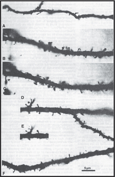
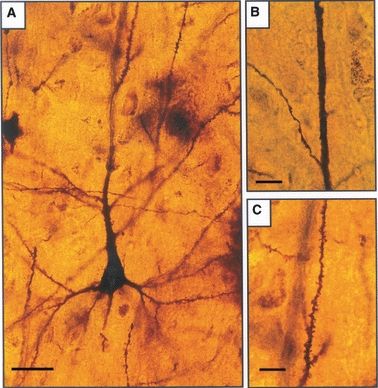
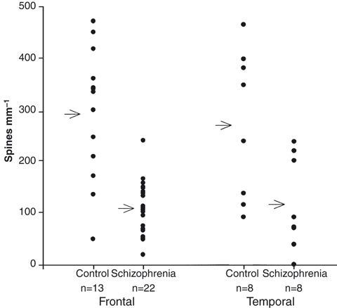
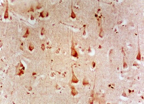
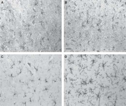
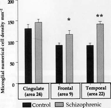
Schizophrenia probably has a developmental origin. This review refers to three of our published series of studies related to this hypothesis: loss of dendritic spines on cerebral neocortical pyramidal neurons, decreased numerical density of glutamatergic neurons, and microgliosis. First, brains of schizophrenic patients and non-schizophrenic controls were obtained post mortem and blocks of multiple cortical areas impregnated with a Rapid Golgi method. Spines were counted on the dendrites of pyramidal neurons of which the soma was in layer III (which takes part in corticocortical connectivity) and which met strict criteria for impregnation quality. Data were obtained blind: diagnoses were only revealed by a third party after measurements were completed. The mean spine count in all cortical areas studied in the control series was 243 mm(-1) of dendrite and in the schizophrenics 108. Measurements in frontal and temporal association cortex showed the greatest reduction in spine number in schizophrenia (299 in control frontal cortex and 101 in schizophrenics, and 276 mm(-1) in control temporal cortex and 125 in schizophrenics). There was no correlation of spine loss with age at death. Our results support the concept of a neurodevelopmental defect in the neuropil affecting glutamatergic neurons in schizophrenia and may help to explain loss of cortical volume without loss of neurons. In a second part of our study we used an antibody to the kainate receptor subunit GluR 5/6/7 and showed a decrease in numerical density of presumed glutamatergic neurons in schizophrenic orbitofrontal cortex. Finally, as glia play a major role in the developing nervous system, we investigated whether schizophrenia was associated with glial changes in frontal and temporal cortex. Astroglia and microglia were identified in schizophrenic and control brains, using antibodies to glial fibrillary acidic protein (GFAP) and class II human leucocyte antigen (HLA-DR), respectively. Significant increases were found in microglial numerical density in schizophrenics compared with controls: 28% in frontal area 9 (115 cells mm(-2) compared with 89), and a 57% increase in temporal area 22 (139 cells mm(-2) compared with 88). For both areas, astroglia showed no significant differences between schizophrenics and controls. No significant differences were found in cortical thickness or total neuronal numerical density between the two groups. This specific increase in numerical density of microglia in temporal and frontal cortex of chronic schizophrenics, not related to aging, could be related to possible changes in cortical neuropil architecture as revealed by loss of dendritic spines.
© 2010 The Author. Journal of Anatomy © 2010 Anatomical Society of Great Britain and Ireland.
Figures
Similar articles
-
Reduced dendritic spine density on cerebral cortical pyramidal neurons in schizophrenia.J Neurol Neurosurg Psychiatry. 1998 Oct;65(4):446-53. doi: 10.1136/jnnp.65.4.446. J Neurol Neurosurg Psychiatry. 1998. PMID: 9771764 Free PMC article.
-
Increase in HLA-DR immunoreactive microglia in frontal and temporal cortex of chronic schizophrenics.J Neuropathol Exp Neurol. 2000 Feb;59(2):137-50. doi: 10.1093/jnen/59.2.137. J Neuropathol Exp Neurol. 2000. PMID: 10749103
-
Decreased numerical density of kainate receptor-positive neurons in the orbitofrontal cortex of chronic schizophrenics.Exp Brain Res. 2006 Aug;173(2):234-42. doi: 10.1007/s00221-006-0396-8. Epub 2006 Feb 28. Exp Brain Res. 2006. PMID: 16505999
-
(Micro)Glia as Effectors of Cortical Volume Loss in Schizophrenia.Schizophr Bull. 2018 Aug 20;44(5):948-957. doi: 10.1093/schbul/sby088. Schizophr Bull. 2018. PMID: 30124942 Free PMC article. Review.
-
A GABAergic cortical deficit dominates schizophrenia pathophysiology.Crit Rev Neurobiol. 2004;16(1-2):1-23. doi: 10.1615/critrevneurobiol.v16.i12.10. Crit Rev Neurobiol. 2004. PMID: 15581395 Review.
Cited by
-
Three-dimensional alteration of neurites in schizophrenia.Transl Psychiatry. 2019 Feb 12;9(1):85. doi: 10.1038/s41398-019-0427-4. Transl Psychiatry. 2019. PMID: 30755587 Free PMC article.
-
Mice lacking collapsin response mediator protein 1 manifest hyperactivity, impaired learning and memory, and impaired prepulse inhibition.Front Behav Neurosci. 2013 Dec 27;7:216. doi: 10.3389/fnbeh.2013.00216. eCollection 2013. Front Behav Neurosci. 2013. PMID: 24409129 Free PMC article.
-
Neuroimaging schizophrenia: a picture is worth a thousand words, but is it saying anything important?Curr Psychiatry Rep. 2013 Mar;15(3):345. doi: 10.1007/s11920-012-0345-0. Curr Psychiatry Rep. 2013. PMID: 23397252 Review.
-
PCB-95 promotes dendritic growth via ryanodine receptor-dependent mechanisms.Environ Health Perspect. 2012 Jul;120(7):997-1002. doi: 10.1289/ehp.1104832. Epub 2012 Apr 25. Environ Health Perspect. 2012. PMID: 22534141 Free PMC article.
-
Modelling Cortical Laminar Connectivity in the Macaque Brain.Neuroinformatics. 2022 Jul;20(3):559-573. doi: 10.1007/s12021-021-09539-2. Epub 2021 Aug 14. Neuroinformatics. 2022. PMID: 34392433
References
-
- Akbarian S, Bunney WE, Potkin SG, et al. Altered distribution of nicotinamide-adenine dinucleotide phosphate-diaphorase cells in frontal lobe of schizophrenics implies disturbances of cortical development. Arch Gen Psychiatry. 1993;50:169–177. - PubMed
-
- Andreasen NC, Smith MR, Jacoby CG, et al. Ventricular enlargement in schizophrenia: definition and prevalence. Am J Psychiatry. 1982;139:292–296. - PubMed
-
- Benes FM, Paskevich PA, Davidson J, et al. Synaptic rearrangements in medial prefrontal cortex of haloperidol-treated rats. Brain Res. 1985;348:15–20. - PubMed
-
- Benes FM, Todtenkopf MS, Taylor JB. A shift in tyrosine hydroxylase-immunoreactive varicosities (TH-IRv) from pyramidal (PN) to nonpyramidal (NP) neurons occurs in layer II of the anterior cingulate cortex of schizophrenics. Abstr Soc Neurosci. 1995;21:259.
Publication types
MeSH terms
LinkOut - more resources
Full Text Sources
Medical
Research Materials
Miscellaneous
