

LCP-Tm: an assay to measure and understand stability of membrane proteins in a membrane environment
- PMID: 20409473
- PMCID: PMC2856142
- DOI: 10.1016/j.bpj.2009.12.4296
LCP-Tm: an assay to measure and understand stability of membrane proteins in a membrane environment
Abstract
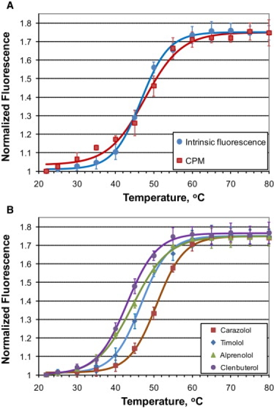
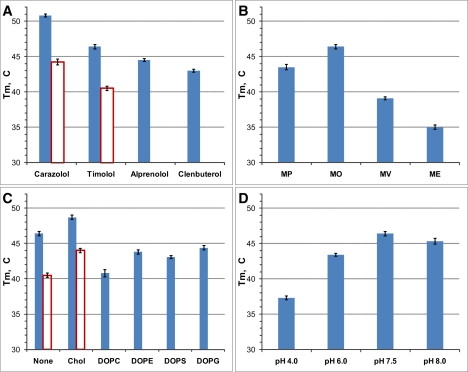
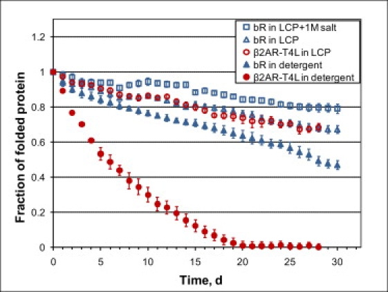
Structural and functional studies of membrane proteins are limited by their poor stability outside the native membrane environment. The development of novel methods to efficiently stabilize membrane proteins immediately after purification is important for biophysical studies, and is likely to be critical for studying the more challenging human targets. Lipidic cubic phase (LCP) provides a suitable stabilizing matrix for studying membrane proteins by spectroscopic and other biophysical techniques, including obtaining highly ordered membrane protein crystals for structural studies. We have developed a robust and accurate assay, LCP-Tm, for measuring the thermal stability of membrane proteins embedded in an LCP matrix. In its two implementations, protein denaturation is followed either by a change in the intrinsic protein fluorescence on ligand release, or by an increase in the fluorescence of a thiol-binding reporter dye that measures exposure of cysteines buried in the native structure. Application of the LCP-Tm assay to an engineered human beta2-adrenergic receptor and bacteriorhodopsin revealed a number of factors that increased protein stability in LCP. This assay has the potential to guide protein engineering efforts and identify stabilizing conditions that may improve the chances of obtaining high-resolution structures of intrinsically unstable membrane proteins.
Copyright 2010 Biophysical Society. Published by Elsevier Inc. All rights reserved.
Figures
Similar articles
-
Cell-free expressed bacteriorhodopsin in different soluble membrane mimetics: biophysical properties and NMR accessibility.Structure. 2013 Mar 5;21(3):394-401. doi: 10.1016/j.str.2013.01.005. Epub 2013 Feb 14. Structure. 2013. PMID: 23415558 Free PMC article.
-
Highly Efficient Transfer of 7TM Membrane Protein from Native Membrane to Covalently Circularized Nanodisc.Sci Rep. 2018 Sep 10;8(1):13501. doi: 10.1038/s41598-018-31925-1. Sci Rep. 2018. PMID: 30201976 Free PMC article.
-
Measuring membrane protein stability under native conditions.Proc Natl Acad Sci U S A. 2014 Jan 7;111(1):219-24. doi: 10.1073/pnas.1318576111. Epub 2013 Dec 23. Proc Natl Acad Sci U S A. 2014. PMID: 24367094 Free PMC article.
-
Structural determinants of purple membrane assembly.Biochim Biophys Acta. 2000 Aug 30;1460(1):15-26. doi: 10.1016/s0005-2728(00)00126-2. Biochim Biophys Acta. 2000. PMID: 10984587 Review.
-
Structure and Functional Characterization of Membrane Integral Proteins in the Lipid Cubic Phase.J Mol Biol. 2020 Aug 21;432(18):5104-5123. doi: 10.1016/j.jmb.2020.02.024. Epub 2020 Feb 27. J Mol Biol. 2020. PMID: 32113953 Review.
Cited by
-
From Gene to Function: Cell-Free Electrophysiological and Optical Analysis of Ion Pumps in Nanodiscs.Biophys J. 2017 Sep 19;113(6):1331-1341. doi: 10.1016/j.bpj.2017.03.026. Epub 2017 Apr 24. Biophys J. 2017. PMID: 28450130 Free PMC article.
-
Ice breaking in GPCR structural biology.Acta Pharmacol Sin. 2012 Mar;33(3):324-34. doi: 10.1038/aps.2011.187. Epub 2012 Jan 30. Acta Pharmacol Sin. 2012. PMID: 22286917 Free PMC article. Review.
-
An effective thiol-reactive probe for differential scanning fluorimetry with a standard real-time polymerase chain reaction device.Anal Biochem. 2016 Apr 15;499:63-65. doi: 10.1016/j.ab.2016.01.016. Epub 2016 Feb 4. Anal Biochem. 2016. PMID: 26851339 Free PMC article.
-
Two classes of cholesterol binding sites for the β2AR revealed by thermostability and NMR.Biophys J. 2014 Nov 18;107(10):2305-12. doi: 10.1016/j.bpj.2014.10.011. Biophys J. 2014. PMID: 25418299 Free PMC article.
-
Evidence that specific interactions play a role in the cholesterol sensitivity of G protein-coupled receptors.Biochim Biophys Acta Biomembr. 2021 Sep 1;1863(9):183557. doi: 10.1016/j.bbamem.2021.183557. Epub 2021 Jan 11. Biochim Biophys Acta Biomembr. 2021. PMID: 33444621 Free PMC article.
References
-
- Yildirim M.A., Goh K.I., Vidal M. Drug-target network. Nat. Biotechnol. 2007;25:1119–1126. - PubMed
Publication types
MeSH terms
Substances
Grants and funding
LinkOut - more resources
Full Text Sources
