. 2010 Jul 5;402(2):281-91.
doi: 10.1016/j.virol.2010.03.015. Epub 2010 Apr 20.

The envelope protein of severe acute respiratory syndrome coronavirus interacts with the non-structural protein 3 and is ubiquitinated

Affiliations

The envelope protein of severe acute respiratory syndrome coronavirus interacts with the non-structural protein 3 and is ubiquitinated

Enrique Alvarez et al. Virology. .

Abstract

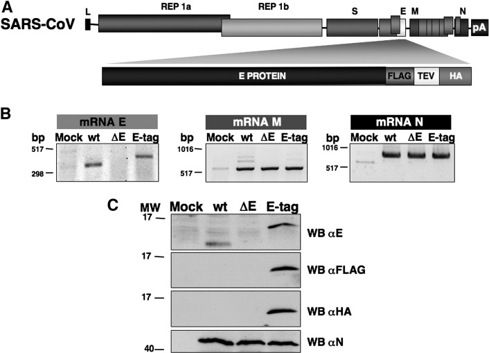
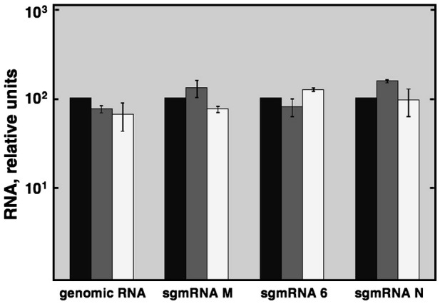
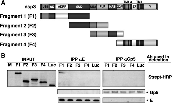
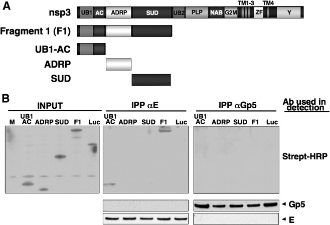
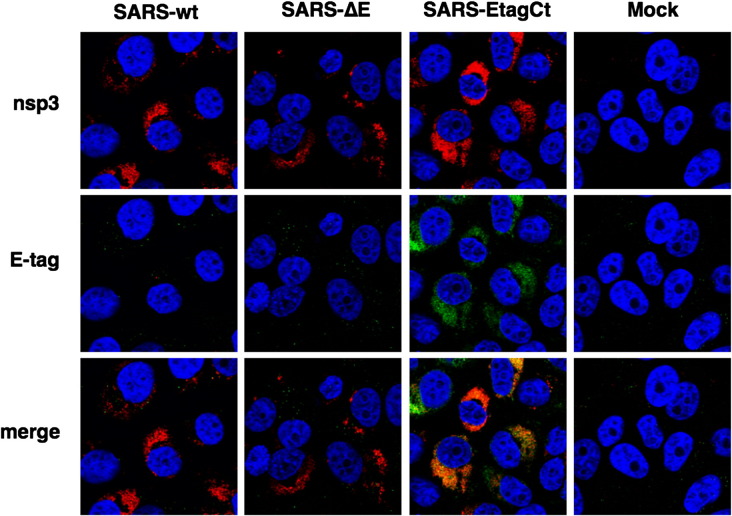
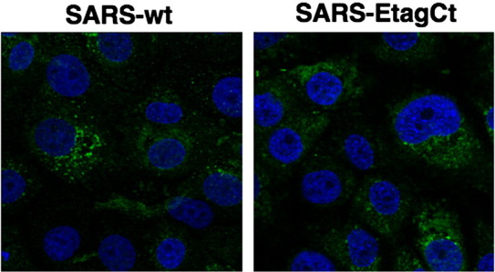
To analyze the proteins interacting with the severe acute respiratory syndrome coronavirus (SARS-CoV) envelope (E) protein, a SARS-CoV was engineered including two tags associated to the E protein. Using this virus, complexes of SARS-CoV E and other proteins were purified using a tandem affinity purification system. Several viral and cell proteins including spike, membrane, non-structural protein 3 (nsp3), dynein heavy chain, fatty acid synthase and transmembrane protein 43 bound E protein. In the present work, we focused on the binding of E protein to nsp3 in infected cells and cell-free systems. This interaction was mediated by the N-terminal acidic domain of nsp3. Moreover, nsp3 and E protein colocalized during the infection. It was shown that E protein was ubiquitinated in vitro and in cell culture, suggesting that the interaction between nsp3 and E protein may play a role in the E protein ubiquitination status and therefore on its turnover.


Figures

Fig. 1
Fig. 1
Generation of a recombinant SARS-CoV expressing a tagged E protein. (A) Scheme of recombinant virus expressing tagged E protein. L, leader sequence; REP, replicase gene; S, spike protein, E, envelope protein; M, membrane protein; N, nucleocapsid protein; pA, poly(A) tail; FLAG, FLAG epitope; TEV, tobacco etch virus protease site; and HA, influenza haemmaglutinin epitope. (B) Vero E6 cells were mock infected (Mock) or infected with the recombinant wild-type (WT), the rSARS-CoV-∆E (∆E) or rSARS-CoV-EtagCt (E-tag) viruses. Viral mRNA expression was analyzed by RT-PCR using the oligonucleotides specific for sgmRNAs of E, M and N genes. (C) Western blot analysis of infected cell lysates using E, FLAG, HA and N protein-specific antibodies followed by peroxidase-labelled goat anti-rabbit or anti-mouse antibodies.
Fig. 2
Fig. 2
Virus RNA synthesis in infected Vero E6 cells. Vero E6 cells were infected with rSARS-CoV-wt (black boxes), rSARS-CoV-∆E (grey boxes) or rSARS-CoV-EtagCt (white boxes) at an moi of 0.5. Total RNA was extracted at 16 h post-infection and the accumulation of viral genomic or subgenomic messenger RNA (sgmRNA) of genes M, 6 and N was quantified by Q-RT-PCR. Levels of viral RNAs are represented in comparison to reference levels from cells infected with SARS-CoV-wt. The experiment was performed three times and the data represent the average of triplicates. Standard deviation is indicated as error bars.
Fig. 3
Fig. 3
Growth kinetics of the recombinant viruses in monkey and human cells. Vero E6 (A), and Huh-7.5.1 (B) cells were infected at an moi of 0.5 with the recombinant wild-type virus (WT), the rSARS-CoV-∆E (∆E) or the rSARS-CoV-EtagCt (E-tag). At different times post-infection, virus titers were determined by plaque assay on Vero E6 cells. Error bars represent standard deviations of the mean of results from three experiments.
Fig. 4
Fig. 4
Identification of viral proteins interacting with SARS-CoV E protein. (A) Purification of proteins interacting with E protein using a tandem affinity purification method. Vero E6 extracts from mock-infected cells or cells infected with rSARS-CoV-wt (wt) or rSARS-CoV-EtagCt (E-tag) were used in a double affinity chromatography. Purified proteins were detected by staining gels with Coomassie blue dye. Bands were excised from gels and were identified by mass spectrometry. (B) Coimmunoprecipitation assays of nsp3 and E protein. Extracts from Vero E6 cells infected with rSARS-CoV-wt or rSARS-CoV-∆E were immunoprecipitated with nsp3, E or TGEV N protein-specific antibodies. Immunoprecipitations were analyzed by Western blot using E and nsp3 protein-specific antibodies followed by peroxidase-labelled goat anti-rabbit or anti-mouse antibodies.
Fig. 5
Fig. 5
Coimmunoprecipitation assays of nsp3 fragments. (A) Scheme of nsp3-derived fragments synthesized in TNT® coupled reticulocyte lysate systems (F1 to F4). The nsp3 domains are shown in the scheme: UB1, ubiquitin-like domain 1; AC, acidic hypervariable domain; ADRP, ADP-ribose-1″-phosphatase; SUD, SARS unique domain; UB2, ubiquitin-like domain 2; PLP, papain-like protease; NAB, group II-specific domain; G2M, group II-specific maker; TM, transmembrane motif; ZF, putative metal-binding region; and Y, Y region. Synthesized fragments were mixed with recombinant E or Gp5 proteins and then were used in immunoprecipitation assays using E or Gp5 protein-specific antibodies. Immunoprecipitations were analyzed by Western blot using E and Gp5 protein-specific antibodies followed by peroxidase-labelled goat anti-rabbit or anti-mouse antibodies. Luciferase (Luc) was used as a control.
Fig. 6
Fig. 6
Mapping the nsp3 region required to interact with E protein. (A) Scheme of nsp3-F1-derived fragments synthesized in TNT® coupled reticulocyte lysate systems (UB1-AC, ADRP and SUD). The nsp3 domains are shown in the scheme: UB1, ubiquitin-like domain 1; AC, acidic hypervariable domain; ADRP, ADP-ribose-1″-phosphatase; SUD, SARS unique domain; UB2, ubiquitin-like domain 2; PLP, papain-like protease; NAB, group II-specific domain; G2M, group II-specific marker; TM, transmembrane motif; ZF, putative metal-binding region; and Y, Y region. Synthesized fragments were mixed with recombinant E or Gp5 proteins and then were used in immunoprecipitation assays using E or Gp5 protein-specific antibodies. Immunoprecipitations were analyzed by Western blot using E and Gp5 protein-specific antibodies followed by peroxidase-labelled goat anti-rabbit or anti-mouse antibodies.
Fig. 7
Fig. 7
Colocalization of nsp3 and E proteins. Vero E6 cells grown on glass coverslips were infected with rSARS-CoV-wt, rSARS-CoV-∆E or rSARS-CoV-EtagCt at an moi of 0.5. At 15 h post-infection the cells were fixed with 8% paraformaldehyde. Cells were labelled with nsp3 (red) or HA (green) specific antibodies.
Fig. 8
Fig. 8
Ubiquitination of SARS-CoV E protein. Ubiquitin conjugation to E protein. Vero E6 cells were transiently transfected with the plasmid mixtures shown in the figure. The control plasmid pcDNA was used to equalize the total amount of transfected plasmid in all cases. 24 h after transfection, the cells were lysed by Laemmli lysis buffer followed by boiling for 5 min. The denatured lysates were analyzed by SDS-PAGE and Western blot with an antibody against E protein (A) or HA epitope (B). (C) In vitro ubiquitination of SARS-CoV E protein. Baculovirus purified E protein was incubated with mono-ubiquitin (mUb) or poly-ubiquitin (pUb) in the presence of the all the enzymes of the ubiquitination process. The reactions were resolved by SDS-PAGE and transferred to nitrocellulose. The ubiquitinated (Ub-E) and non-ubiquitinated E protein was detected by Western blot using a specific antibody against E protein.
Supplementary Fig. 1
Confocal microscopy analysis of cells infected with SARS-wt, and SARS-EtagCt. Vero E6 cells grown on glass coverslips were infected with rSARS-CoV-wt or rSARS-CoV-EtagCt at an moi of 0.5. At 15 h post-infection the cells were fixed with 8% paraformaldehyde. Cells infected with SARS-CoV-wt were labelled using a rabbit polyclonal antibody against E protein (left panel). Cells infected with SARS-CoV-EtagCt were labelled with HA specific antibody (right panel). Alexa 488-conjugated antibodies against the different species were used as secondary antibodies.

Similar articles

Cited by

References

    1. Almazan F., DeDiego M.L., Galan C., Escors D., Alvarez E., Ortego J., Sola I., Zuñiga S., Alonso S., Moreno J.L., Nogales A., Capiscol C., Enjuanes L. Construction of a SARS-CoV infectious cDNA clone and a replicon to study coronavirus RNA synthesis. J. Virol. 2006;80:10900–10906. - PMC - PubMed
    1. Barretto N., Jukneliene D., Ratia K., Chen Z., Mesecar A.D., Baker S.C. The papain-like protease of severe acute respiratory syndrome coronavirus has deubiquitinating activity. J. Virol. 2005;79:15189–15198. - PMC - PubMed
    1. Brierley I., Digard P., Inglis S.C. Characterization of an efficient coronavirus ribosomal frameshifting signal: requirement for an RNA pseudoknot. Cell. 1989;57:537–547. - PMC - PubMed
    1. Corse E., Machamer C.E. The cytoplasmic tails of infectious bronchitis virus E and M proteins mediate their interaction. Virology. 2003;312:25–34. - PMC - PubMed
    1. de Groot R.J., Ziebuhr J., Poon L.L., Woo P.C., Talbot P., Rottier P.J.M., Holmes K.V., Baric R., Perlman S., Enjuanes L., Gorbalenya A.E. International Committee on Taxonomy of Viruses, 2008.085-126V. 2008. Revision of the family Coronaviridae.

Publication types

MeSH terms