. 2008 Jul-Aug;2(4):239-66.
doi: 10.1016/j.jash.2007.12.002. Epub 2008 Jun 2.

Treating hypertension while protecting the vulnerable islet in the cardiometabolic syndrome

Affiliations

Treating hypertension while protecting the vulnerable islet in the cardiometabolic syndrome

Melvin R Hayden et al. J Am Soc Hypertens. 2008 Jul-Aug.

Abstract

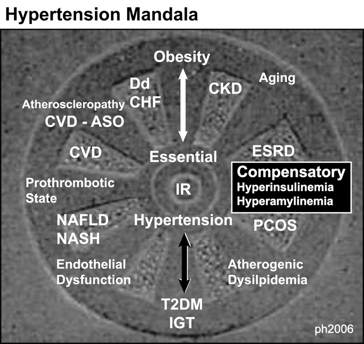
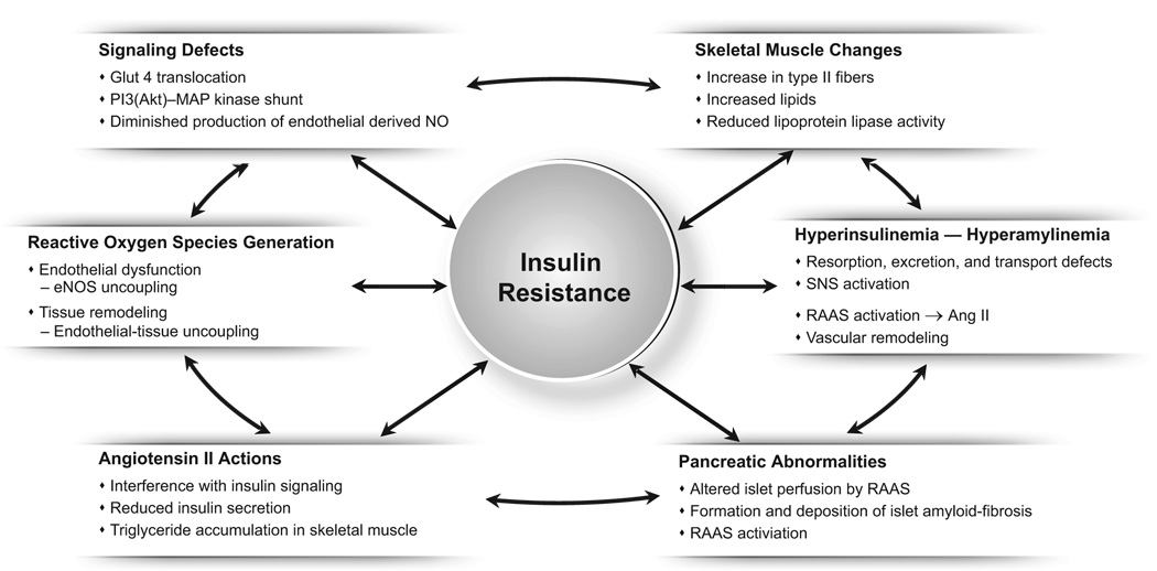
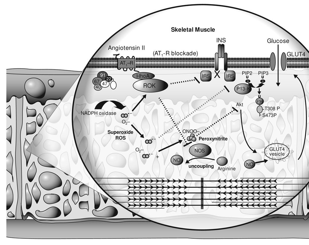
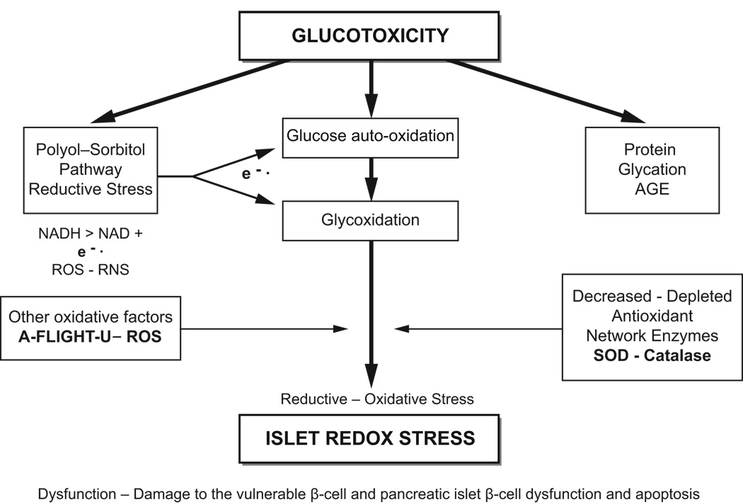
Hypertension, a multifactorial-polygenic disease, interacts with multiple environmental stressors and results in functional and structural changes in numerous end organs, including the cardiovascular system. This can result in coronary heart disease, stroke, peripheral vascular disease, congestive heart failure, end-stage renal disease, insulin resistance, and damage to the pancreatic islet. Hypertension is the most important modifiable risk factor for major health problems encountered in clinical practice. Whereas hypertension was once thought to be a medical condition based on discrete blood pressure readings, a new concept has emerged defining hypertension as part of a complex and progressive metabolic and cardiovascular disease, an important part of a cardiometabolic syndrome. The central role of insulin resistance, oxidative stress, endothelial dysfunction, metabolic signaling defects within tissues, and the role of enhanced tissue renin-angiotensin-aldosterone system activity as it relates to hypertension and type 2 diabetes mellitus are emphasized. Additionally, this review focuses on the effect of hypertension on functional and structural changes associated with the vulnerable pancreatic islet. Various classes of antihypertensive drugs are reviewed, especially their roles in delaying or preventing damage to the vulnerable pancreatic islet, and thus delaying the development of type 2 diabetes mellitus.


Figures

Figure 1
Figure 1
The hypertension mandala. Clustering of the cardiometabolic syndrome and cardiometabolic risk factors. IR is central to the development of hypertension and IGT–T2DM, along with multiple metabolic and clinical conditions surrounding the outer portions of this hypertension mandala. Obesity is placed at the top of the mandala because it is believed to be the driving force behind the subsequent clinical end-organ remodeling and disease. The central placement of hypertension was chosen because it represents the central importance regarding its treatment to prevent many of the associated clinical complications and specifically T2DM placed vertically below and opposite obesity. The interconnectedness between IR, hypertension, and T2DM is not to be underestimated. ASO, atheroscleropathy; CHF, congestive heart failure; CKD, chronic kidney disease; CVD, cardiovascular disease; Dd, diastolic dysfunction; ; ESRD, end-stage renal disease; IGT, impaired glucose tolerance; IR, insulin resistance; NAFLD, nonalcoholic fatty liver disease; NASH, nonalcoholic steatohepatitis; PCOS, polycystic ovarian syndrome; T2DM, type 2 diabetes mellitus.
Figure 2
Figure 2
Brief summary of the major metabolic and structural alterations in hypertension [4, 5, 7]. Ang II, angiotensin II; eNOS, endothelial nitric oxide synthase; Glut, glucose transporter; NO, nitric oxide; PI3(Akt)-MAP, phosphatidylinositol 3-kinase-mitogen activated protein; RAAS, renin angiotensin aldosterone system; SNS, sympathetic nervous system.
Figure 3
Figure 3
Counterregulatory roles of insulin and angiotensin II in glucose utilization: the role of oxidative stress [5]. There is increasing evidence of a strong association between hypertension, insulin resistance, and the development of T2DM. AT1-R, angiotensin II type 1 receptor; GLUT, glucose transporter; INS, insulin; IRS, insulin-receptor substrates; NADPH, nicotinamide adenine dinucleotide phosphate; NOS, nitric oxide synthase; NO, nitric oxide; PI3-K, phosphatidylinositol 3-kinase; ROK, Rho kinase; ROS, reactive oxygen species. Adapted from [5] with permission. American Journal of Physiology, Heart and Circulatory Physiology. JR Sowers. Copyright 2004 by American Physiological Society. Reproduced with permission of American Physiological Society in the format Journal via Copyright Clearance Center.
Figure 4
Figure 4
The role of glucotoxicity in the development of islet redox stress and pancreatic islet structural damage β-cell dysfunction [6]. A-FLIGHT-U, see Table 3 for explanation of this acronym; AGE, advanced glycosylation end products; NAD, nicotinamide adenine dinucleotide; NADH, nicotinamide adenine dinucleotide, (reduced); RNS, reactive nitrogen species; ROS, reactive oxygen species; SOD, super-oxide dismutase. Adapted from Hayden MR, Tyagi SC. J Pancreas 2002; 3: 86–108. [6].
Figure 5
Figure 5
The primary structure of human amylin (islet amyloid polypeptide) [55]. Reproduced from Hayden MR, et al. J Pancreas 2005; 6: 287–302 [55]).
Figure 6
Figure 6
The progression from unfolded or misfolded amylin to the formation of insoluble amyloid fibrils [55]. ER, endoplasmic reticulum; UPR, unfolded protein response. Reproduced from Hayden MR, et al. J Pancreas 2005; 6: 287–302 [55].
Figure 7
Figure 7
Islet structural changes associated with amylin-derived islet amyloid deposition. Panel 7A: Hematoxylin and eosin staining of a single human islet from a middle-aged patient with T2DM. Note the interbeta and pericapillary hyaline changes associated with islet amyloid deposition. Note the presence of islet amyloid surrounding the islet capillary. This critical location may serve as a barrier to docking of the insulin secretory granules as depicted in the TEM panel 7D. Magnification × 80. Panel 7B: A hypocellular islet (due to islet amyloid deposition) from the transgenic (Sprague-Dawley transfected with the human amylin gene) novel 8-month-old HIP transgenic rat model of T2DM. This Voerhoff Van Gieson–stained islet demonstrates peri-islet and perivascular fibrosis of the islet (arrows). Magnification × 40. Panel 7C: This same islet stained with Congo red and under crossed polarized light. Depicts islet amyloid appearing white in this black-and-white image. Note the novel findings of islet amyloid present in the peri-islet vessels. Amylin-derived islet amyloid in the perivascular region has not been previously demonstrated. Magnification × 40. Panel 7D: Demonstrates a TEM image of an islet from a 4-month-old transgenic HIP rat model of T2DM. It is interesting to note that the ISGs(*) seem to abut the pericapillary islet amyloid deposits (#) and indicate impaired ISG trafficking and docking with the capillary endothelial cell, which helps explain the impaired glucose tolerance and impaired first-phase insulin secretion. To our knowledge, this is the first image to demonstrate structurally this impaired trafficking and docking of the ISGs. The progressive deposition of islet amyloid seems to parallel the progressive nature of T2DM. Magnification ×4000. Eβ-C, endothelial beta-cell (representative of endothelial tissue uncoupling); EC, endothelial cell; hIAPP, human islet amyloid polypeptide; HIP, human islet-amyloid polypeptide c; ISG, insulin secretory granule; RBC, red blood cell within the islet capillary; T2DM, type 2 diabetes mellitus; TEM, transmission electron microscope.
Figure 8
Figure 8
The RAAS cascade. This image demonstrates the 3 critical steps in the production and blockade of Ang II. The renin enzyme, ACE, and the AT1 receptor. The current medications in use are the ACE inhibitors and ARBs, and this image portrays the emerging medication aliskiren. The 3 classes of medications will soon be the -rens, the –prils, and the -sartans. It will be interesting to see if the new renin inhibitor aliskiren will delay or prevent the development of T2DM in patients with hypertension compared with placebo or other antihypertensive medications. ACE, angiotensin-converting enzyme; ARBs, angiotensin receptor blockers; RAAS, renin-angiotensin aldosterone system; T2DM, type 2 diabetes mellitus.
Figure 9
Figure 9
Renin inhibitor–binding pockets on the active site of renin [135, 136]. Panel A represents the ribbon and band structure with the medication aliskiren overlying the binding pocket S1/S3 and extension into the hydrophobic S3 subpockets, which allows for the prerequisite higher affinity binding necessary for the desired clinical response. Panel B represents the model used to portray this binding pocket site within the renin molecule. Reprinted from Chemistry Biology, volume 7, Raheul J et al. Structure-based drug design. Pp 493–504. Copyright 2000, with permission from Elsevier.

Similar articles

Cited by

References

    1. Chobanian AV, Bakris GL, Black HR, Cushman WC, Green LA, Izzo JL, Jr, et al. The Seventh Report of the Joint National Committee on Prevention, Detection, Evaluation, and Treatment of High Blood Pressure: the JNC 7 report. JAMA. 2003;289:2560–2572. - PubMed
    1. Klag MJ, Whelton PK, Randall BL, Neaton JD, Brancati FL, Ford CE, et al. Blood pressure and end-stage renal disease in men. N Engl J Med. 1996;334:13–18. - PubMed
    1. Stamler J, Vaccaro O, Neaton JD, Wentworth D. Diabetes, other risk factors, and 12-yr cardiovascular mortality for men screened in the Multiple Risk Factor Intervention Trial. Diabetes Care. 1993;16:434–444. - PubMed
    1. Hayden MR, Sowers JR. Hypertension in type 2 diabetes mellitus. Insulin. 2006;1:22–37.
    1. Sowers JR. Insulin resistance and hypertension. Am J Physiol Heart Circ Physiol. 2004;286:H1597–H1602. - PubMed

LinkOut - more resources