Development of cardiomyopathy and atrial tachyarrhythmias associated with activating autoantibodies to beta-adrenergic and muscarinic receptors
- PMID: 20409953
- PMCID: PMC3116651
- DOI: 10.1016/j.jash.2008.10.004
Development of cardiomyopathy and atrial tachyarrhythmias associated with activating autoantibodies to beta-adrenergic and muscarinic receptors
Abstract
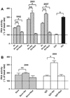
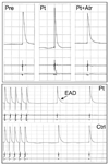
A 71-year-old male with well-controlled hypertension developed atrial tachyarrhythmias in 2002 and a restrictive cardiomyopathy in 2006 to 2007. Sera from 1992, 2001, and 2006 to 2008 demonstrated activating autoantibodies against beta-adrenergic (AAbetaAR) and M2 muscarinic receptors (AAM2R). These sera have been characterized for bioactivity using in vitro assays of cardiac contractility and automaticity using a canine cardiac Purkinje fiber assay as well as protein kinase assay activation in H9c2 cells. These assays demonstrated concurrent positive betaAR and inhibitory M2R effects that were blocked by nadolol and atropine, respectively. In a canine pulmonary vein atrial sleeve preparation, sera diluted 1:100 produced atrial hyperpolarization that was blocked by atropine. Atrial tachyarrhythmias developed in 2002 in the presence of a persistent bradycardia. Serial echocardiograms demonstrated progressive diastolic dysfunction in the absence of cardiac hypertrophy between 2006 and 2007. A dual-chamber pacemaker was installed with combined betaAR (nadolol) and M2<3R (oxybutynin) blockade, resulting in marked suppression of atrial ectopy and improved diastolic function. The estimated pulmonary artery pressure decreased and exercise tolerance returned. Blood pressure has remained normal with beta-blockade. AAbetaAR and AAM2R prospectively influenced atrial and ventricular function in this patient, and specific receptor blockade was associated with improved cardiac function.
Conflict of interest statement
Conflict of interest: none.
Figures


Similar articles
-
Opposing cardiac effects of autoantibody activation of β-adrenergic and M2 muscarinic receptors in cardiac-related diseases.Int J Cardiol. 2011 May 5;148(3):331-6. doi: 10.1016/j.ijcard.2009.11.025. Epub 2010 Jan 6. Int J Cardiol. 2011. PMID: 20053466 Free PMC article.
-
Activating autoantibodies to the beta-1 adrenergic and m2 muscarinic receptors facilitate atrial fibrillation in patients with Graves' hyperthyroidism.J Am Coll Cardiol. 2009 Sep 29;54(14):1309-16. doi: 10.1016/j.jacc.2009.07.015. J Am Coll Cardiol. 2009. PMID: 19778674 Free PMC article.
-
Autoantibody activation of beta-adrenergic and muscarinic receptors contributes to an "autoimmune" orthostatic hypotension.J Am Soc Hypertens. 2012 Jan-Feb;6(1):40-7. doi: 10.1016/j.jash.2011.10.003. Epub 2011 Nov 30. J Am Soc Hypertens. 2012. PMID: 22130180 Free PMC article.
-
Cardiomyopathy With Restrictive Physiology in Sickle Cell Disease.JACC Cardiovasc Imaging. 2016 Mar;9(3):243-52. doi: 10.1016/j.jcmg.2015.05.013. Epub 2016 Feb 17. JACC Cardiovasc Imaging. 2016. PMID: 26897687 Free PMC article. Review.
-
Autoantibodies against the beta- and muscarinic receptors in cardiomyopathy.Herz. 2000 May;25(3):261-6. doi: 10.1007/s000590050017. Herz. 2000. PMID: 10904849 Review.
Cited by
-
Drug-like actions of autoantibodies against receptors of the autonomous nervous system and their impact on human heart function.Br J Pharmacol. 2012 Jun;166(3):847-57. doi: 10.1111/j.1476-5381.2012.01828.x. Br J Pharmacol. 2012. PMID: 22220626 Free PMC article. Review.
-
Receptor autoantibodies: Associations with cardiac markers, histology, and function in human non-ischaemic heart failure.ESC Heart Fail. 2023 Apr;10(2):1258-1269. doi: 10.1002/ehf2.14293. Epub 2023 Jan 30. ESC Heart Fail. 2023. PMID: 36717981 Free PMC article.
-
Agonistic autoantibodies as vasodilators in orthostatic hypotension: a new mechanism.Hypertension. 2012 Feb;59(2):402-8. doi: 10.1161/HYPERTENSIONAHA.111.184937. Epub 2012 Jan 3. Hypertension. 2012. PMID: 22215709 Free PMC article. Clinical Trial.
-
Unmasking features of the auto-epitope essential for β1 -adrenoceptor activation by autoantibodies in chronic heart failure.ESC Heart Fail. 2020 Aug;7(4):1830-1841. doi: 10.1002/ehf2.12747. Epub 2020 May 21. ESC Heart Fail. 2020. PMID: 32436653 Free PMC article.
-
The adaptive immune response to cardiac injury-the true roadblock to effective regenerative therapies?NPJ Regen Med. 2017 Jun 19;2:19. doi: 10.1038/s41536-017-0022-3. eCollection 2017. NPJ Regen Med. 2017. PMID: 29302355 Free PMC article. Review.
References
-
- Limas CJ, Goldenberg IF, Limas C. Autoantibodies against beta-adrenoceptors in human idiopathic dilated cardiomyopathy. Circ Res. 1989;64:97–103. - PubMed
-
- Magnusson Y, Wallukat G, Waagstein F, Hjalmarson A, Hoebeke J. Autoimmunity in idiopathic dilated cardiomyopathy. Characterization of antibodies against the beta 1-adrenoceptor with positive chronotropic effect. Circulation. 1994;89:2760–2767. - PubMed
-
- Rosenbaum MB, Chiale PA, Schejtman D, Levin M, Elizari MV. Antibodies to beta-adrenergic receptors disclosing agonist-like properties in idiopathic dilated cardiomyopathy and Chagas’ heart disease. J Cardiovasc Electrophysiol. 1994;5:367–375. - PubMed
-
- Luft FC, Dechend R, Dragun D, Müller D, Wallukat G. Agonistic antibodies directed at cell surface receptors and cardiovascular disease. J Am Soc Hypertens. 2008;2:8–14. - PubMed
-
- Baba A, Yoshikawa T, Fukuda Y, Sugiyama T, Shimada M, Akaishi M, et al. Autoantibodies against M2-muscarinic acetylcholine receptors: new upstream targets in atrial fibrillation in patients with dilated cardiomyopathy. Eur Heart J. 2004;25:1108–1115. - PubMed
Grants and funding
LinkOut - more resources
Full Text Sources

