Comparative Study
. 2010 Apr 21;30(16):5489-97.
doi: 10.1523/JNEUROSCI.6383-09.2010.

Molecular characterization of mutations that cause globoid cell leukodystrophy and pharmacological rescue using small molecule chemical chaperones

Affiliations
Comparative Study

Molecular characterization of mutations that cause globoid cell leukodystrophy and pharmacological rescue using small molecule chemical chaperones

Wing C Lee et al. J Neurosci. .

Abstract

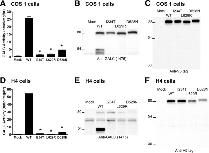
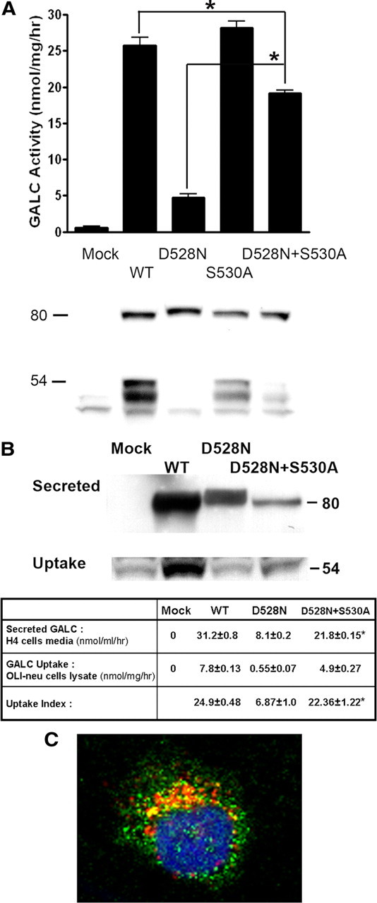
Globoid cell leukodystrophy (GLD) (Krabbe disease) is an autosomal recessive, degenerative, lysosomal storage disease caused by a severe loss of galactocerebrosidase (GALC) enzymatic activity. Of the >70 disease-causing mutations in the GALC gene, most are located outside of the catalytic domain of the enzyme. To determine how GALC mutations impair enzymatic activity, we investigated the impact of multiple disease-causing mutations on GALC processing, localization, and enzymatic activity. Studies in mammalian cells revealed dramatic decreases in GALC activity and a lack of appropriate protein processing into an N-terminal GALC fragment for each of the mutants examined. Consistent with this, we observed significantly less GALC localized to the lysosome and impairment in either the secretion or reuptake of mutant GALC. Notably, the D528N mutation was found to induce hyperglycosylation and protein misfolding. Reversal of these conditions resulted in an increase in proper processing and GALC activity, suggesting that glycosylation may play a critical role in the disease process in patients with this mutation. Recent studies have shown that enzyme inhibitors can sometimes "chaperone" misfolded polypeptides to their appropriate target organelle, bypassing the normal cellular quality control machinery and resulting in enhanced activity. To determine whether this may also work for GLD, we examined the effect of alpha-lobeline, an inhibitor of GALC, on D528N mutant cells. After treatment, GALC activity was significantly increased. This study suggests that mutations in GALC can cause GLD by impairing protein processing and/or folding and that pharmacological chaperones may be potential therapeutic agents for patients carrying certain mutations.


Figures

Figure 1.
Figure 1.
Expression of WT and mutant GALC in mammalian cells. A–F, Empty pcDNA4 vector (Mock), human wild-type GALC cDNA (WT), or GLD-causing GALC mutations (I234T, L629R, and D528N) were transiently transfected into COS-1 cells (A–C) or stably transfected into H4 cells (D–F). GALC enzymatic activity, measured by in vitro colorimetric assay, was significantly reduced in lysates from cells transfected with each mutant GALC, compared with WT GALC (*p < 0.05) (A, D). The precursor and processed forms of GALC were detected by Western blot using the CL1475 rabbit antibody against the N terminus of GALC (B, E), and the anti-V5 epitope tag antibody against the C terminus of GALC (C, F). Error bars indicate SEM.
Figure 2.
Figure 2.
Analysis of the secretion and endocytosis of WT and mutant GALC. Conditioned medium was collected from H4 cells stably transfected with WT GALC, mutant GALC (I234T, L629R, and D528N), or empty vector (Mock), and was then applied to OLI-neu oligodendrocyte precursor cells for 24 h to determine the efficiency of GALC uptake. A, Western blot (anti-V5 epitope tag) showing detectable levels of GALC protein in conditioned media from WT, I234T, and D528N GALC-transfected H4 cells. Secreted GALC was undetectable in conditioned media from empty vector (mock)-transfected and L629R GALC-transfected cells. B, Western blot (CL1021AP chicken anti-GALC) of lysates from recipient OLI-neu cells after incubation with conditioned medium from H4 cells expressing WT or mutant GALC. C, Summary of GALC activity determined by colorimetric assay from the H4 input media (A) and from the Oli-neu recipient lysates (B). Uptake index is defined as 100 times the ratio of the GALC activity measured in the cell lysate after uptake compared with the GALC activity in the corresponding input medium. The data in the table represent the mean values ± SE of triplicate samples in each group. GALC secretion and uptake were significantly decreased for each mutant compared with WT GALC (*p < 0.05).
Figure 3.
Figure 3.
Confocal immunocytofluorescent analysis of GALC in H4-GALC cells. H4 cells (mock), WT GALC, and mutant (I234T, L629R and D528N) GALC-expressing H4 cells were seeded at subconfluent density on glass coverslips 2 d before staining. The subcellular localization of GALC was analyzed by costaining of GALC (CL1475 rabbit anti-GALC antibody) and lamp 2 for lysosomes, costaining of GALC (anti-V5 antibody) and calnexin for ER, and costaining of GALC (anti-V5 antibody) and 58k Golgi protein for Golgi apparatus. GALC and the organelle markers were illustrated in green and red colors, respectively. Nuclei were labeled with DAPI and illustrated in blue color. The yellow color indicates colocalization of GALC with the organelle markers. All images were captured in 40× magnification using a Zeiss LSM 510 META confocal microscope.
Figure 4.
Figure 4.
Amino acid sequences near the D528N mutation in GALC. The aspartic acid residue (D) at position 528 was substituted by an asparagine residue (N), which creates an extra N-glycosylation site in the D528N GALC mutant. A double mutant, D528N+S530A, was generated to disrupt the extra N-glycosylation site created by the D528N mutation by replacing the serine residue at position 530 with an alanine residue. A construct containing the S530A mutation only was also generated for an assay control.
Figure 5.
Figure 5.
Western blot analysis of the hyperglycosylated D528N GALC mutant overexpressed in COS-1 cells. A–C, “Polymeric” GALC (A), monomeric GALC precursors (B), and C-terminal GALC fragments (C) were purified and analyzed without treatment (control) or after deglycosylation by Endo H or by PNG F.
Figure 6.
Figure 6.
Expression study of D528N mutant GALC in the presence or absence of glycosylation at position 528. Partial rescue in D528N GALC function, processing, secretion, endocytosis, and lysosomal localization was observed in the absence of the glycosylation motif that is introduced with the D528N mutation. A, GALC activity in lysates from COS-1 cells transfected with empty vector (mock), WT or mutant GALC (D528N, S530A or D528N+S530A) (top, graph). Data plotted represent the mean ± SE of at least triplicate samples in each group. GALC activity was significantly increased in cells transfected with the double mutant D528N+S530A, compared with the D528N mutant alone (*p < 0.05). The same lysates were analyzed by Western blot using the CL1475 rabbit anti-GALC antibody (bottom, immunoblot). B, H4 cells stably transfected with WT and mutant GALC (D528N or D528N+S530S) were used to determine the influence of glycosylation on GALC secretion and uptake. Conditioned medium from the transfected H4 cells was applied to OLI-neu cells for 24 h. Media containing the secreted GALC protein (B, top immunoblot) and lysates from recipient OLI-neu cells (B, bottom immunoblot) were analyzed by Western blot using the anti-V5 antibody and the chicken anti-GALC antibody, CL1021AP, respectively. GALC activities from the input cell medium and from the recipient OLI-neu cell lysate were determined and summarized in table format (B, table). Uptake index is defined as 100 times the ratio of the GALC activity measured in the cell lysate after uptake compared with the GALC activity in the corresponding input medium. The data in the table represent the mean ± SE of triplicate samples in each group. *p < 0.05, comparison between the D528N GALC and the D528N+S530A GALC within the same row. C, The subcellular localization of the D528N+S530A mutant GALC expressed in H4 cells was examined by confocal microscopy. Localization of GALC in lysosomes was analyzed by double-immunofluorescent staining using CL1475 rabbit antibody against GALC and anti-lamp 2 to label lysosomes, respectively. GALC is in green. Lamp 2 is in red. Nucleus (DAPI) is in blue. Magnification, 40×. The images are representative of a field (n = 3) of one of three independent experiments.
Figure 7.
Figure 7.
Activity and processing of GALC proteins in H4 cells under subphysiological temperature. WT and mutant GALC (D528N, I234T, L629R, and D528N+S530A) transfected H4 cells were cultured at 37 or 30°C for 72 h. A, GALC enzymatic activity in lysates was determined by in vitro colorimetric assay. Data shown represent the mean ± SE of at least triplicate samples in each group (*p < 0.05). B, Processed N-terminal GALC fragments (50–54 kDa) from cell lysate were analyzed by Western blot using the chicken anti-GALC antibody, CL1021AP.
Figure 8.
Figure 8.
Effect of α-lobeline on human GALC in vitro and in H4 cells expressing WT and mutant GALC (I234T, L629R, and D528N). A, The inhibition of recombinant GALC by α-lobeline was analyzed using an in vitro colorimetric enzymatic activity assay. B, C, H4 cells expressing WT GALC (B) or D528N GALC (C) were treated with different concentrations of α-lobeline (60–240 μm) for 72 h. GALC enzymatic activity in the lysates (black bars) and in the culture media (white bars) was analyzed. Data shown represent the mean ± SE of at least triplicate samples in each group. Statistical analysis was performed by one-way ANOVA test, followed by the Tukey's post hoc test (*p < 0.05).

Similar articles

Cited by

References

    1. Berson JF, Frank DW, Calvo PA, Bieler BM, Marks MS. A common temperature-sensitive allelic form of human tyrosinase is retained in the endoplasmic reticulum at the nonpermissive temperature. J Biol Chem. 2000;275:12281–12289. - PubMed
    1. Butters TD. Gaucher disease. Curr Opin Chem Biol. 2007;11:412–418. - PubMed
    1. Chen YQ, Wenger DA. Galactocerebrosidase from human urine: purification and partial characterization. Biochim Biophys Acta. 1993;1170:53–61. - PubMed
    1. Chen YQ, Rafi MA, de Gala G, Wenger DA. Cloning and expression of cDNA encoding human galactocerebrosidase, the enzyme deficient in globoid cell leukodystrophy. Hum Mol Genet. 1993;2:1841–1845. - PubMed
    1. De Gasperi R, Gama Sosa MA, Sartorato EL, Battistini S, MacFarlane H, Gusella JF, Krivit W, Kolodny EH. Molecular heterogeneity of late-onset forms of globoid-cell leukodystrophy. Am J Hum Genet. 1996;59:1233–1242. - PMC - PubMed

Publication types

MeSH terms