Comparative Study
. 2010 Apr 21;30(16):5617-28.
doi: 10.1523/JNEUROSCI.5296-09.2010.

Nerve growth factor-regulated emergence of functional delta-opioid receptors

Affiliations
Comparative Study

Nerve growth factor-regulated emergence of functional delta-opioid receptors

Bihua Bie et al. J Neurosci. .

Abstract

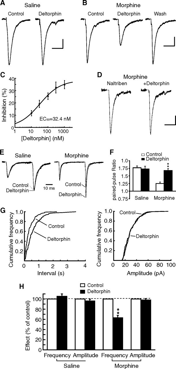
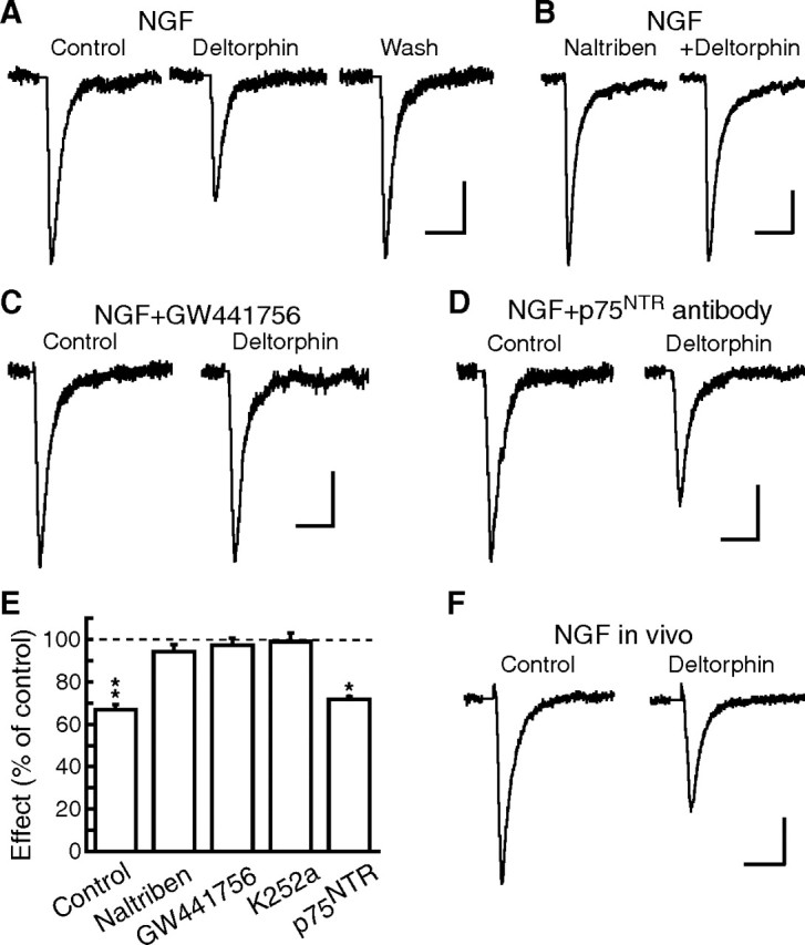
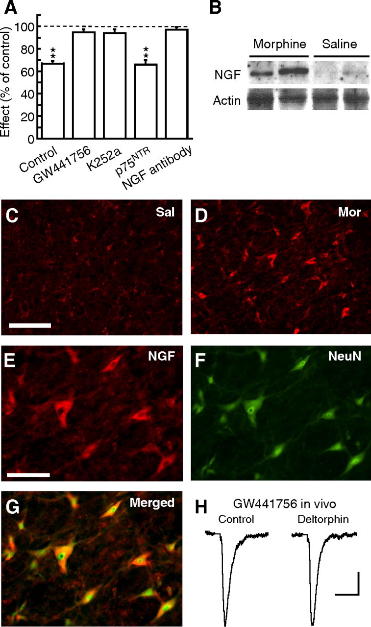
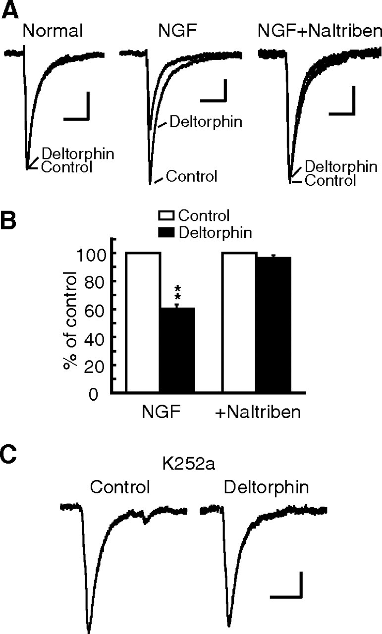
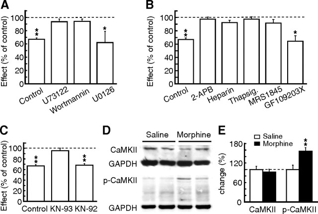
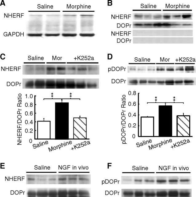
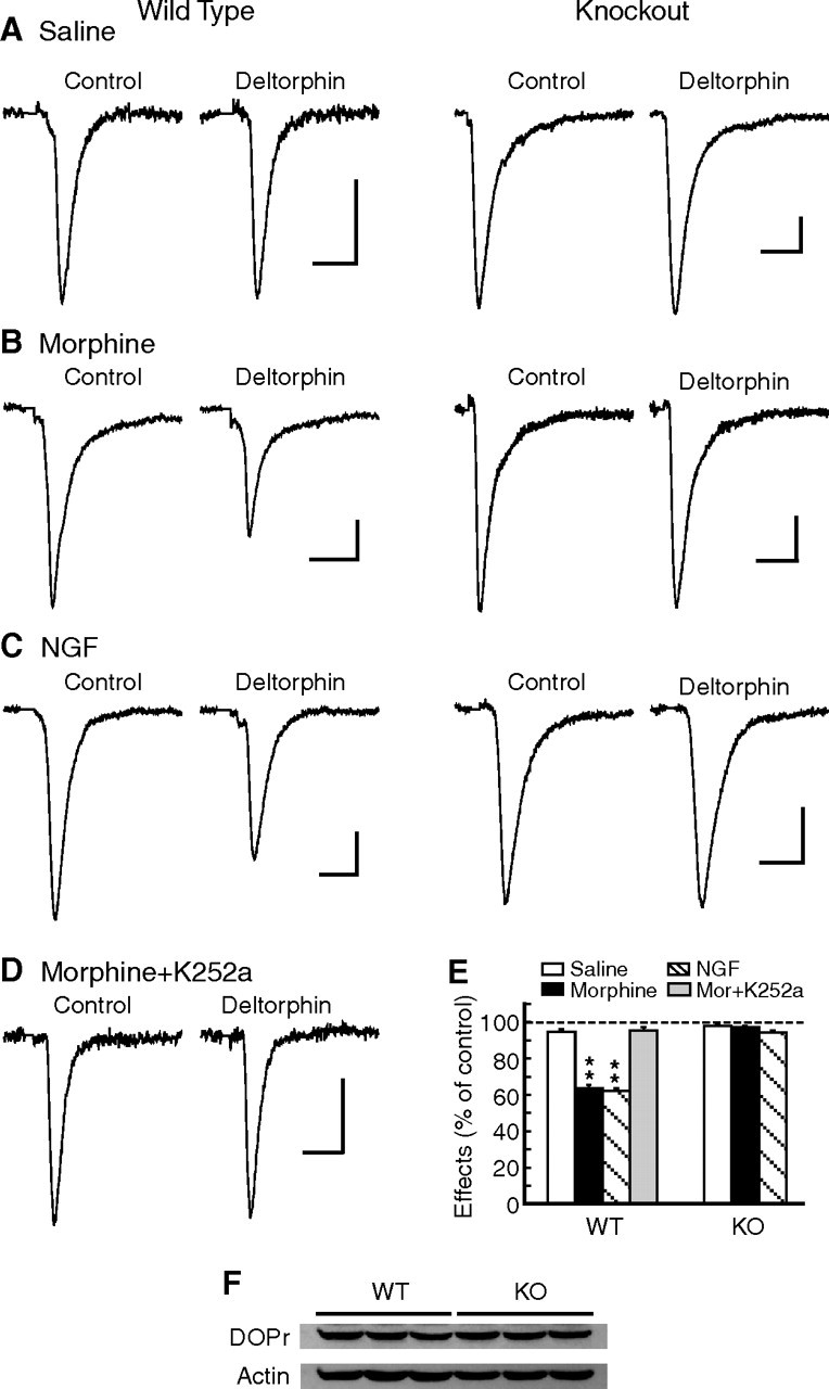
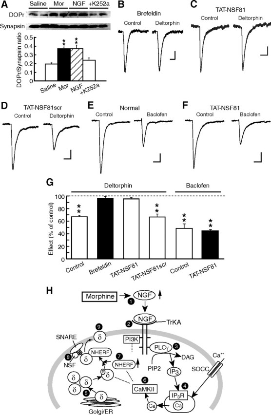
Sorting of intracellular G-protein-coupled receptors (GPCRs) either to lysosomes for degradation or to plasma membrane for surface insertion and functional expression is a key process regulating signaling strength of GPCRs across the plasma membrane in adult mammalian cells. However, little is known about the molecular mechanisms governing the dynamic process of receptor sorting to the plasma membrane for functional expression under normal and pathological conditions. In this study, we demonstrate that delta-opioid receptor (DOPr), a GPCR constitutively targeted to intracellular compartments, is driven to the surface membrane of central synaptic terminals and becomes functional by the neurotrophin nerve growth factor (NGF) in native brainstem neurons. The NGF-triggered DOPr translocation is predominantly mediated by the signaling pathway involving the tyrosine receptor kinase A, Ca(2+)-mobilizing phospholipase C, and Ca(2+)/calmodulin-dependent protein kinase II. Importantly, it requires interactions with the cytoplasmic sorting protein NHERF-1 (Na(+)/H(+) exchange regulatory factor-1) and N-ethyl-maleimide-sensitive factor-regulated exocytosis. In addition, this NGF-mediated mechanism is likely responsible for the emergence of functional DOPr induced by chronic opioids. Thus, NGF may function as a key molecular switch that redirects the sorting of intracellularly targeted DOPr to plasma membrane, resulting in new functional DOPr on central synapses under chronic opioid conditions.


Figures

Figure 1.
Figure 1.
Chronic morphine induces new functional DOPr on presynaptic terminals. A, B, Representative glutamatergic EPSCs in control, in the presence of DOPr agonist deltorphin II (1 μm), and after wash in a brainstem neuron of the NRM from a saline-treated rat (A) and from a morphine-treated rat (B). C, A dose–response curve for the deltorphin inhibition of EPSCs in brainstem neurons (n = 7–9 cells at each dose) from morphine-treated rats. Error bars here and in all following figures are SEMs. D, EPSCs in the presence of the DOPr antagonist naltriben (50 nm) and after addition of deltorphin in a neuron from a morphine-treated rat. E, Representative EPSC pairs superimposed and scaled to the amplitude of the first EPSC in a neuron from a saline- and a morphine-treated rat. F, Group data of the deltorphin effect on the paired-pulse ratios of EPSCs in neurons from the two rat groups (n = 10 in each group). G, Distribution graphs of miniature EPSC frequencies and amplitudes in a neuron from a morphine-treated rat. H, Summarized deltorphin effects on the frequency and amplitude of miniature EPSCs in neurons of saline-treated rats (n = 10) and morphine-treated rats (n = 11). **p < 0.01. Calibration: 50 pA and 10 ms.
Figure 2.
Figure 2.
NGF induces new functional DOPr on glutamate synapses. A, Representative glutamate EPSCs before (control), during and after (wash) application of deltorphin (1 μm) from an NRM neuron in a normal slice taken from a naive rat and treated in vitro with NGF for a long period (4 h). B, EPSCs in naltriben without and with deltorphin in an NGF-treated normal slice. C, D, EPSCs in a normal slice treated with NGF plus the TrKA receptor inhibitor GW441756 (100 nm, C) or plus an anti-p75NTR receptor antibody (D). E, Group data of the deltorphin effects on EPSCs in normal slices treated with long-period NGF alone (control, n = 9), in the presence of naltriben, or treated with NGF plus an inhibitor as indicated. F, Deltorphin inhibition of EPSCs from a naive rat treated with repeated NGF in vivo. *p < 0.05, **p < 0.01. Calibration: 50 pA and 10 ms.
Figure 3.
Figure 3.
NGF is responsible for morphine induction of functional DOPr. A, Summarized deltorphin effects on EPSCs in slices of morphine-treated rats in control or after treatment in vitro with the indicated inhibitor. B, Representative Western blot lanes of NGF and actin proteins in NRM tissues taken from saline- and morphine-treated rats. C, D, Microscopic images of immunofluorescence labeling for NGF in NRM sections from saline (Sal)- and morphine (Mor)-treated rats. Calibration: 100 μm. E–G, Images of labeling for NGF (red) and for neurons with the neuronal marker NeuN (green), and a merged image in NRM slices from a morphine-treated rat. Calibration: 20 μm. H, EPSCs from a rat treated with morphine plus GW441756 in vivo. **p < 0.01. Calibration: 50 pA and 10 ms.
Figure 4.
Figure 4.
NGF mediates morphine induction of DOPr on GABA synapses. A, Superimposed GABA-mediated IPSCs in control and in deltorphin (1 μm) in NRM neurons in a normal slice and in normal slices treated with NGF (100 ng/ml) alone or with NGF plus naltriben (50 nm) for 4 h in vitro. B, Group data of the deltorphin effects after treatment of normal slices with NGF or NGF plus naltriben. C, Representative GABA IPSCs in slices from morphine-treated rats and treated with the TrK receptor inhibitor K252a (200 nm) for 4 h in vitro. Note that use of KCl in the intrapipette solution with a holding potential of −60 mV resulted in a downward direction of the IPSCs. **p < 0.01. Calibration: 50 pA and 20 ms.
Figure 5.
Figure 5.
Morphine induction of DOPr involves the PLC–CaMKII pathway. A–C, Group data of deltorphin effects on EPSCs in slices from morphine-treated rats in control or treated in vitro with the indicated inhibitor for a long period. D, Representative lanes of CaMKII, p-CaMKII, and GAPDH proteins in NRM tissues from saline- and morphine-treated rats. E, Summarized Western blot results of percentage changes in CaMKII and p-CaMKII proteins normalized to GAPDH. Thapsig, Thapsigargin. *p < 0.05, **p < 0.01.
Figure 6.
Figure 6.
Chronic morphine increases interaction between DOPr and NHERF-1 proteins. A, Western blot lanes of NHERF-1 and GAPDH proteins in NRM tissues from saline- and morphine-treated rats. B, DOPr immunoprecipitates (IP) immunoblotted for NHERF-1 and for DOPr in NRM tissues from saline- and morphine-treated rats in control (top two panels) and after preincubation of the DOPr antibody with a DOPr-blocking peptide (bottom two panels). C, D, Similar IP data of NHERF-1 and DOPr (C) and DOPr immunoprecipitates with immunoblotted phosphorylated DOPr (pDOPr) and total DOPr (D) in tissues from saline-treated rats and rats treated with morphine without or with K252a cotreatment in vivo. Also shown are changes in the ratios of NHERF-1 over DOPr and pDOPr over total DOPr proteins in the indicated three groups of rats. E, F, Similar DOPr precipitates immunoblotted for NHERF-1 and DOPr (E) and for pDOPr and total DOPr (F) in NRM tissues from naive rats treated with saline or NGF in vivo. Mor, Morphine. **p < 0.01.
Figure 7.
Figure 7.
NHERF-1 is required for morphine and NGF induction of functional DOPr. A–D, Representative glutamate EPSCs in control and in deltorphin (1 μm) in an NRM neuron from WT mice (left column) and NHERF-1 KO mice (right column) treated with saline (A), morphine (B), from normal slices treated with long-period NGF (C), or from a K252a-treated slice of a morphine-treated WT mouse (D). E, Summarized data of the deltorphin effects in the four treatment groups of WT and KO mice. F, Western blot lanes of DOPr and actin proteins in NRM tissues from WT (n = 5) and NHERF-1 KO mice (n = 6). **p < 0.01. Calibration: 50 pA and 10 ms.
Figure 8.
Figure 8.
Morphine induction of DOPr involves DOPr translocation to surface membrane of synaptic terminals. A, Western blot lanes of DOPr protein and the synaptic terminal marker synaptophysin (synapsin), and their ratio changes in NRM synaptosomes from rats treated with saline, morphine, NGF, or morphine plus K252a in vivo. B, Representative EPSCs in a slice from a morphine-treated rat and treated in vitro with long-period brefeldin A. C, D, Deltorphin actions on EPSCs in slices from morphine-treated rats and treated in vitro with the exocytosis-inhibiting peptide TAT-NSF 81 (C) or with the corresponding but scrambled peptide TAT-NSF81scr (D) for 3 h. E, F, Effects of the GABAB receptor agonist baclofen (10 μm) on EPSCs in NRM slices from morphine-treated rats without (normal) or with the TAT-NSF81 treatment in vitro. G, Group data showing the effects of deltorphin and baclofen in NRM slices of morphine-treated rats in control or after long-period treatment with the agents indicated. **p < 0.01. Calibration: 50 pA and 10 ms. H, Diagram of the proposed signaling pathways for DOPr trafficking on a central synaptic terminal. Chronic morphine upregulates NGF (1), which activates the TrKA receptor (2) and its coupled PLCγ pathway (3) and PI3K pathway (downstream components to be investigated). PLCγ activation leads to production of IP3 and increased intracellular Ca2+ through Ca2+ release from the IP3 receptor (IP3R)-controlled Ca2+ store (4) and by Ca2+ influx through SOCC. DOPr is normally packed and transported through the ER/Golgi network and constitutively targeted to an intracellular pool (5). Increased intracellular Ca2+ activates CaMKII (6), which would lead to downstream changes in the expression of DOPr and NHERF-1 genes and proteins through yet unknown mechanisms of transcription and translation (7), resulting in an increased interaction between upregulated NHERF-1 and phosphorylated DOPr, and exocytotic translocation of intracellular DOPr to surface membrane. Interaction of DOPr-containing vesicles with the SNARE complex, whose function is regulated by NSF (8), causes membrane fusion for exocytosis and surface expression of functional DOPr (9).

Similar articles

Cited by

References

    1. Bie B, Peng Y, Zhang Y, Pan ZZ. cAMP-mediated mechanisms for pain sensitization during opioid withdrawal. J Neurosci. 2005;25:3824–3832. - PMC - PubMed
    1. Bie B, Zhu W, Pan ZZ. Rewarding morphine-induced synaptic function of delta-opioid receptors on central glutamate synapses. J Pharmacol Exp Ther. 2009;329:290–296. - PMC - PubMed
    1. Cahill CM, Holdridge SV, Morinville A. Trafficking of delta-opioid receptors and other G-protein-coupled receptors: implications for pain and analgesia. Trends Pharmacol Sci. 2007;28:23–31. - PubMed
    1. Chao MV, Bothwell M. Neurotrophins: to cleave or not to cleave. Neuron. 2002;33:9–12. - PubMed
    1. Chen YL, Law PY, Loh HH. NGF/PI3K signaling-mediated epigenetic regulation of delta opioid receptor gene expression. Biochem Biophys Res Commun. 2008;368:755–760. - PMC - PubMed

Publication types

LinkOut - more resources