Comparative Study
. 2010 Apr 21;30(16):5635-43.
doi: 10.1523/JNEUROSCI.6154-09.2010.

GSK3 beta regulates myelin-dependent axon outgrowth inhibition through CRMP4

Affiliations
Comparative Study

GSK3 beta regulates myelin-dependent axon outgrowth inhibition through CRMP4

Yazan Z Alabed et al. J Neurosci. .

Abstract

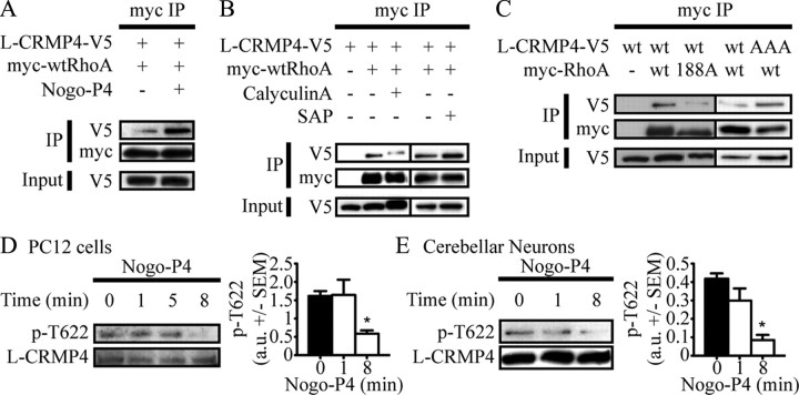
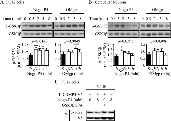
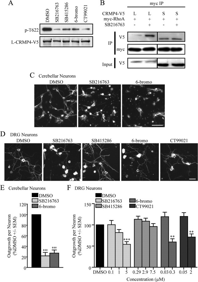
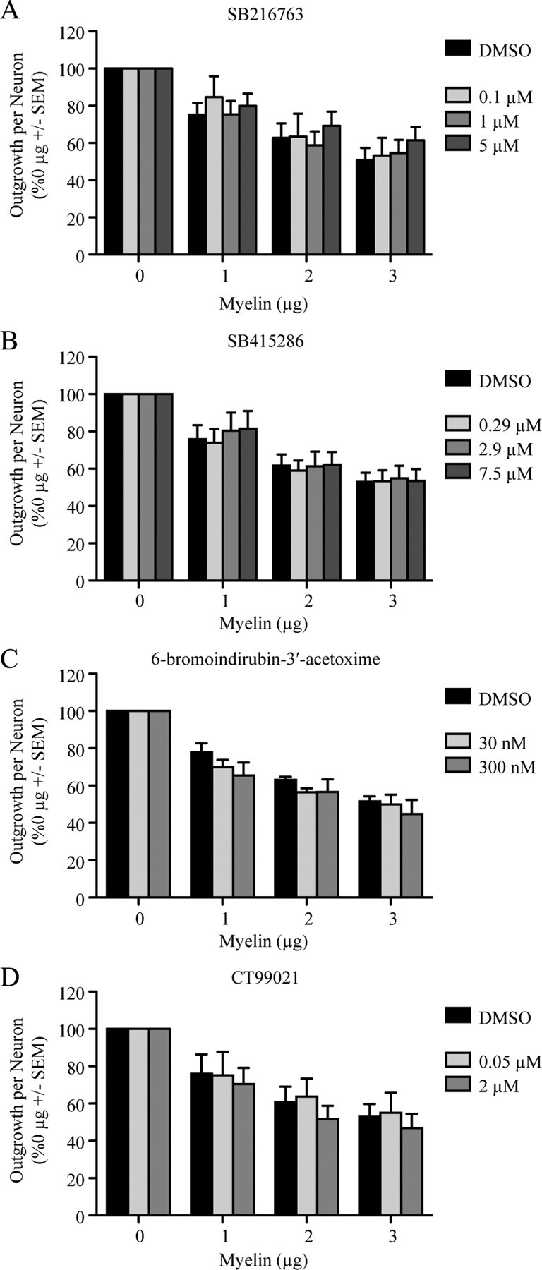
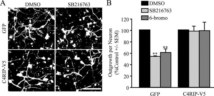
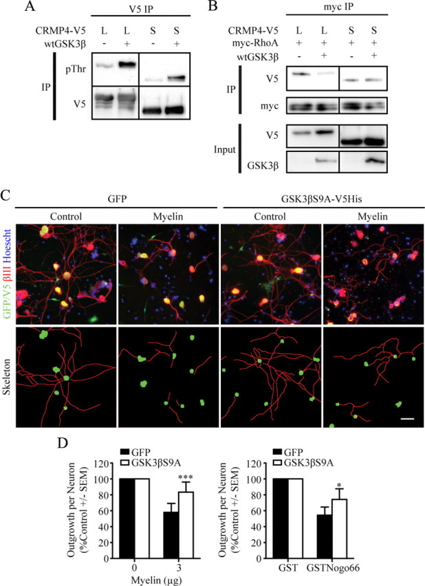
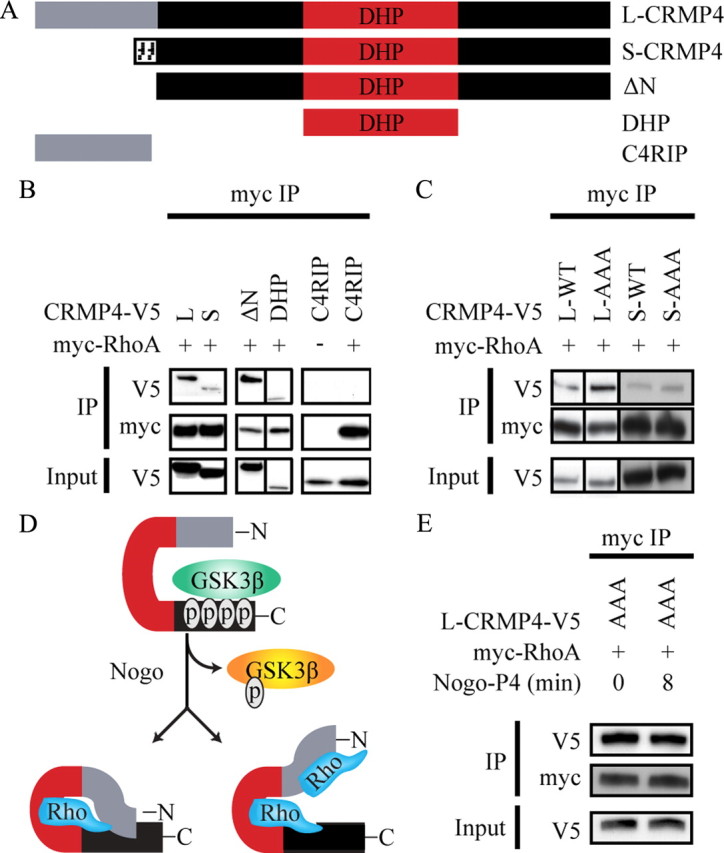
Myelin-associated inhibitors (MAIs) contribute to failed regeneration in the CNS. The intracellular signaling pathways through which MAIs block axonal repair remain largely unknown. Here, we report that the kinase GSK3beta is directly phosphorylated and inactivated by MAIs, consequently regulating protein-protein interactions that are critical for myelin-dependent inhibition. Inhibition of GSK3beta mimics the neurite outgrowth inhibitory effect of myelin. The inhibitory effects of GSK3beta inhibitors and myelin are not additive indicating that GSK3beta is a major effector of MAIs. Consistent with this, overexpression of GSK3beta attenuates myelin inhibition. MAI-dependent phosphorylation and inactivation of GSK3beta regulate phosphorylation of CRMP4, a cytosolic regulator of myelin inhibition, and its ability to complex with RhoA. Introduction of a CRMP4 antagonist attenuates the neurite outgrowth inhibitory properties of GSK3beta inhibitors. We describe the first example of GSK3beta inactivation in response to inhibitory ligands and link the neurite outgrowth inhibitory effects of GSK3beta inhibition directly to CRMP4. These findings raise the possibility that GSK3beta inhibition will not effectively promote long-distance CNS regeneration following trauma such as spinal cord injury.


Figures

Figure 1.
Figure 1.
L-CRMP4 dephosphorylation is stimulated by Nogo-P4 and regulates L-CRMP4–RhoA binding. A–C, PC12 cells (A) or HEK293T cells (B, C) were cotransfected with myc-RhoA constructs and L-CRMP4–V5 and subjected to immunoprecipitation with anti-myc antibody. A, PC12 cells were stimulated for 8 min with Nogo-P4 peptide. B, Cells were treated for 30 min with 200 nm calyculin A before lysis or cell lysates were treated for 30 min with 10 U SAP at 34°C before immunoprecipitation. D, E, PC12 cells (D) or cerebellar neurons infected with HSV-L-CRMP4–V5 (E) were stimulated for 1–8 min with Nogo-P4 peptide. Cell lysates were separated by SDS-PAGE and total and phosphorylated L-CRMP4 was detected by immunoblot. Quantifications in arbitrary units are from three experiments and are normalized for total L-CRMP4 loading. *p < 0.05 by paired t tests, two-tailed, compared with 0 min. a.u., Arbitrary units; IP, immunoprecipitation.
Figure 2.
Figure 2.
MAI-dependent phosphorylation of GSK3β results in L-CRMP4 dephosphorylation. A, B, PC12 cells (A) or cerebellar neurons (B) were stimulated for 0.5–8 min with Nogo-P4 peptide or OMgp. Membrane fractions were separated by SDS-PAGE and total and phosphorylated GSK3β was detected by immunoblot. Quantifications are from at least seven experiments and are normalized for total GSK3β protein loading. *p < 0.05, **p < 0.01, and ***p < 0.001 by Dunnett's post-test compared with 0 min following one-way RM ANOVA. ANOVA p values are indicated above the graphs. C, PC12 cells were transiently transfected with L-CRMP4–V5 and GSK3βS9A for 24 h and then treated with Nogo-P4 peptide for 8 min. L-CRMP4–V5 was immunoprecipitated and separated by SDS-PAGE, and total and phosphorylated L-CRMP4 was detected by immunoblot. a.u., Arbitrary units; IP, immunoprecipitation.
Figure 3.
Figure 3.
Pharmacologic inhibition of GSK3β enhances L-CRMP4–RhoA binding and inhibits neurite outgrowth. A, HSV-L-CRMP4–V5-infected cerebellar neurons were treated with 5 μm SB216763, 7.5 μm SB415286, 300 nm 6-bromoindirubin-3′-acetoxime, or 2 μm CT99021 for 18 h. Cell lysates were subjected to immunoprecipitation with anti-V5 antibody and analyzed by immunoblot with anti-pT622 to detect L-CRMP4 phosphorylation and anti-V5 to detect total L-CRMP4. Densitometry from two experiments shows a statistically significant reduction in phospho-L-CRMP4 signal normalized to total CRMP4; the one-way repeated-measures ANOVA p value is 0.002 with Tukey's post-tests, indicating that values for all inhibitors are significantly different from those with DMSO (p < 0.01). B, myc-RhoA and CRMP4–V5 constructs were transiently coexpressed in PC12 cells. To inhibit GSK3β, cells were treated overnight with 5 μm SB216763. Cell lysates were subjected to immunoprecipitation (IP) with anti-myc antibody and analyzed by immunoblot with anti-V5 or anti-myc antibodies. L, Long; S, short. C, D, Dissociated rat cerebellar neurons (C) or DRG neurons (D) were treated overnight with 5 μm SB216763, 7.5 μm SB415286, 300 nm 6-bromoindirubin-3′-acetoxime, or 2 μm CT99021, fixed, and stained with anti-βIII tubulin antibody to visualize neurite outgrowth. Scale bar, 50 μm. E, F, Quantification of cerebellar (E) or DRG (F) outgrowth/cell. Values are normalized to baseline outgrowth with DMSO vehicle control for each experiment. Determinations are from at least three experiments performed in duplicate. **p < 0.01 and ***p < 0.001 by Dunnett's post-tests following one-way RM ANOVA. 6-bromo, 6-Bromoindirubin-3′-acetoxime.
Figure 4.
Figure 4.
Effects of myelin and GSK3β inhibitors on neurite outgrowth inhibition are not additive. A–D, Quantification of DRG neurite outgrowth/cell from neurons plated on myelin and treated with vehicle control (DMSO) and increasing doses of SB216763 (A), SB415286 (B), 6-bromoindirubin-3′-acetoxime (C), or CT99021 (D). Values are normalized to outgrowth with no myelin. Determinations are from at least four experiments performed in duplicate. For DMSO-treated neurons, p < 0.001 for all doses of myelin compared with no myelin by Dunnett's post-tests following one-way RM ANOVA. Two-way ANOVA shows no significant differences in outgrowth on myelin between cultures treated with DMSO and those treated with any GSK inhibitor.
Figure 5.
Figure 5.
Pharmacologic inhibition of GSK3β inhibits neurite outgrowth in an L-CRMP4-dependent manner. A, HSV-GFP- or HSV-C4RIP-infected dissociated DRG neurons were grown overnight in the presence of 5 μm SB216763, fixed, and stained with anti-β-III tubulin antibody. Scale bar, 100 μm. B, Quantification of DRG neurite outgrowth/cell from HSV-GFP- or HSV-C4RIP-infected neurons treated overnight with 5 μm SB216763 or 300 nm 6-bromoindirubin-3′-acetoxime (6-bromo). Values are normalized to baseline outgrowth with DMSO vehicle control. Determinations are from three experiments performed in duplicate. **p < 0.01 by Student's t test compared with DMSO.
Figure 6.
Figure 6.
GSK3β overexpression attenuates myelin inhibition. A, Lysates from 293T cells transfected with GSK3β and CRMP4–V5 constructs were subjected to immunoprecipitation with an anti-V5 antibody and probed with anti-phosphothreonine or anti-V5 antibody. B, Lysates from 293T cells transfected with myc-RhoA, GSK3β, and CRMP4–V5 constructs were subjected to immunoprecipitation with an anti-myc antibody and probed with anti-V5, anti-myc, and anti-GSK3β antibodies. C, HSV-GFP- or GSK3βS9A-V5-infected dissociated DRGs were grown overnight on myelin, fixed, and stained with anti-β-III tubulin antibody (red) and Hoescht nuclear stain (blue). The green image reflects GFP signal from the control virus or anti-V5 staining detecting GSK3βS9A overexpression. Skeletonized output images of HSV-GFP- or GSK3βS9A-V5-positive neuronal cell bodies (green) and their associated neurites (red) used for quantification are shown in the bottom row. Scale bar, 50 μm. D, Quantification of DRG neurite outgrowth/cell from HSV-GFP- or GSK3βS9A-infected DRG neurons grown on myelin or GST-Nogo-66 substrates. Determinations are from three experiments. *p < 0.05 and ***p < 0.001 by Bonferroni's post-tests following two-way RM ANOVA. IP, Immunoprecipitation; L, long; S, short.
Figure 7.
Figure 7.
L-CRMP4 conformation is phospho dependent and regulates RhoA recruitment. A, Schematic representation of full-length and mutant CRMP4 constructs. B, C, HEK293T cells were transiently cotransfected with myc-RhoA and CRMP-V5 constructs. Lysates were subjected to immunoprecipitation with anti-myc antibody and immunoblotted with anti-myc or anti-V5 antibodies. D, A model of how L-CRMP4 binding to RhoA may be regulated by a phosphorylation-dependent conformational change in L-CRMP4. Dephosphorylation of the carboxy terminal region of L-CRMP4 may enhance the folding interaction between the amino and carboxy termini of the protein and modify the protein conformation to favor binding to RhoA or may change the conformation of the N terminus of L-CRMP4, exposing a second RhoA binding site. E, PC12 cells were transiently cotransfected with myc-RhoA and CRMP4 AAA-V5. Cells were stimulated with Nogo-P4 peptide and lysates were subjected to immunoprecipitation with anti-myc antibody and immunoblotted with anti-myc or anti-V5 antibodies. IP, Immunoprecipitation; L, long; S, short.

Similar articles

Cited by

References

    1. Alabed YZ, Grados-Munro E, Ferraro GB, Hsieh SH, Fournier AE. Neuronal responses to myelin are mediated by ROCK. J Neurochem. 2006;96:1616–1625. - PubMed
    1. Alabed YZ, Pool M, Ong Tone S, Fournier AE. Identification of CRMP4 as a convergent regulator of axon outgrowth inhibition. J Neurosci. 2007;27:1702–1711. - PMC - PubMed
    1. Bain J, McLauchlan H, Elliott M, Cohen P. The specificities of protein kinase inhibitors: an update. Biochem J. 2003;371:199–204. - PMC - PubMed
    1. Bain J, Plater L, Elliott M, Shpiro N, Hastie CJ, McLauchlan H, Klevernic I, Arthur JS, Alessi DR, Cohen P. The selectivity of protein kinase inhibitors: a further update. Biochem J. 2007;408:297–315. - PMC - PubMed
    1. Byk T, Dobransky T, Cifuentes-Diaz C, Sobel A. Identification and molecular characterization of Unc-33-like phosphoprotein (Ulip), a putative mammalian homolog of the axonal guidance-associated unc-33 gene product. J Neurosci. 1996;16:688–701. - PMC - PubMed

Publication types

MeSH terms

LinkOut - more resources