Comparative Study
. 2010 Apr 21;30(16):5781-91.
doi: 10.1523/JNEUROSCI.4947-09.2010.

Presynaptic and postsynaptic mechanisms of synaptic plasticity and metaplasticity during intermediate-term memory formation in Aplysia

Affiliations
Comparative Study

Presynaptic and postsynaptic mechanisms of synaptic plasticity and metaplasticity during intermediate-term memory formation in Aplysia

Igor Antonov et al. J Neurosci. .

Abstract

Synaptic plasticity and learning involve different mechanisms depending on the following: (1) the stage of plasticity and (2) the history of plasticity, or metaplasticity. However, little is known about how these two factors are related. We have addressed that question by examining mechanisms of synaptic plasticity during short-term and intermediate-term behavioral sensitization and dishabituation in a semi-intact preparation of the Aplysia siphon-withdrawal reflex. Dishabituation differs from sensitization in that it is preceded by habituation, and is thus a paradigm for metaplasticity. We find that whereas facilitation during short-term sensitization by one tail shock involves presynaptic covalent modifications by protein kinase A (PKA) and CamKII, facilitation during intermediate-term sensitization by four shocks involves both presynaptic (PKA, CaMKII) and postsynaptic (Ca(2+), CaMKII) covalent modifications, as well as both presynaptic and postsynaptic protein synthesis. The facilitation also involves presynaptic spike broadening 2.5 min after either one or four shocks, but not at later times. Dishabituation by four shocks differs from sensitization in several ways. First, it does not involve PKA or CaMKII, but rather involves presynaptic PKC. In addition, unlike sensitization with the same shock, dishabituation by four shocks does not involve protein synthesis or presynaptic spike broadening, and it also does not involve postsynaptic Ca(2+). These results demonstrate that not only the mechanisms but also the site of plasticity depend on both the stage of plasticity and metaplasticity during memory formation.


Figures

Figure 1.
Figure 1.
Behavioral pharmacology of sensitization and dishabituation in the siphon-withdrawal preparation. A, The preparation. B, Behavioral protocols. See the text for details. C, Examples of siphon withdrawal (SWR) before (PreTest) and 2.5 min after (PostTest) tail shock (Sensitization), 10 closely spaced siphon stimuli (Habituation), or habituation followed by tail shock (Dishabituation). D, Average results from experiments like the ones shown in C with the abdominal ganglion bathed in normal saline (Control), the PKA inhibitor KT5720, the CaMKII inhibitor KN93, the PKC inhibitor chelerythrine, or the protein synthesis inhibitor emetine, and no shock control. There were significant overall effects of group during sensitization following a single tail shock (F(2,15) = 8.04, p < 0.01, n = 6, 5, and 7), sensitization following four tail shocks (F(5,33) = 4.54, p < 0.01, n = 6, 7, 6, 6, 7, and 7), dishabituation following four tail shocks (F(4,26) = 4.19, p < 0.01, n = 6, 5, 6, 7, and 7), and a marginal effect during habituation following 10 closely spaced siphon stimuli (F(4,25) = 2.56, p < 0.10, n = 7, 5, 5, 7, and 6). The point at −2 min indicates the response to the 10th habituation stimulus. The amplitude of siphon withdrawal has been normalized to the average value on the three pretests in each experiment. The overall average pretest value was 2.4 mm, which was not significantly different in experiments with the different inhibitors by a one-way ANOVA. The average response to the tail shock was 5.5 mm. The error bars indicate SEMs.
Figure 2.
Figure 2.
Cellular mechanisms involved in sensitization. A, Examples of siphon withdrawal (SWR), evoked firing of an LFS siphon motor neuron, and the monosynaptic EPSP from an LE sensory neuron to the LFS neuron before (PreTest) and 2.5 min after (PostTest) four tail shocks (top) or no shock control (bottom). B, Average results from experiments like the ones shown in A with either a single shock (n = 6 for shock; n = 6 for no shock) or four shocks (n = 7 for shock; n = 6 for no shock). There was a significant overall effect of shock in each case. The overall average pretest values were 1.8 mm for siphon withdrawal, 15 spikes for evoked LFS firing, and 5.9 mV for the amplitude of the EPSP, which were not significantly different in experiments with shock and no shock. In this and Figs. 4, 5, and 6, *p < 0.05. +p < 0.05 one tail for the difference between shock and no shock.
Figure 3.
Figure 3.
Presynaptic and postsynaptic molecular mechanisms of the facilitation during sensitization. A, Average facilitation of the EPSP by one shock (colored symbols) and no shock controls (white symbols) following no injection (Control) or intracellular injection of a peptide inhibitor of PKA (PKAi), CaMKII (CamKi), or the Ca2+ chelator BAPTA into the sensory neuron (SN) or motor neuron (MN). There was a significant overall effect of group (F(4,52) = 2.75, p < 0.05, n = 12, 12, 13, 10, and 15), and a marginal effect for the group × shock interaction (F(4,52) = 2.45, p < 0.10). B, Average facilitation by four tail shocks following no injection (Control) or intracellular injection of a peptide inhibitor of PKA (PKAi), CaMKII (CamKi), PKC (PKCi), the protein synthesis inhibitor gelonin, or the Ca2+ chelator BAPTA into the SN or MN. There were significant overall effects of group (F(8,89) = 3.91, p < 0.01, n = 13, 10, 13, 13, 11, 12, 13, 11, and 11) and the group × shock interaction (F(8,89) = 2.15, p < 0.05). The overall average pretest value was 6.1 mV. The average response to the tail shock was 5.1 mm. These values were not significantly different in experiments with different injections. In this and Figs. 5 and 6, #p < 0.05 for the interaction between the inhibitor and shock at each test.
Figure 4.
Figure 4.
Changes in sensory neuron membrane properties during sensitization. A, Examples of evoked firing, membrane resistance, and action potential duration of an LE siphon sensory neuron before (PreTest) and 2.5 min after (PostTest) one tail shock (top) or no shock control (bottom). B, Average results from experiments like the ones shown in A with either a single shock (n = 6 for shock; n = 6 for no shock) or four shocks (n = 7 for shock; n = 6 for no shock). The overall average pretest values were 3.8 spikes for evoked LE firing and 2.3 ms for action potential duration, which were not significantly different in experiments with shock and no shock.
Figure 5.
Figure 5.
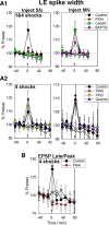
Molecular mechanisms of sensory neuron spike broadening and changes in the shape of the EPSP during sensitization. A1, Average LE spike broadening by one and four tail shocks (colored symbols) and no shock controls (white symbols) following no injection (Control) or intracellular injection of a peptide inhibitor of PKA (PKAi), CaMKII (CamKi), or the Ca2+ chelator BAPTA into the sensory neuron (SN) or motor neuron (MN). Results with one and four shocks were similar and have been pooled. There was a significant main effect of group (F(4,79) = 2.73, p < 0.05, n = 22, 18, 21, 19, and 19) and a marginal effect for the group × shock interaction (F(4,79) = 2.12, p < 0.10) at 2.5 min. A2, Average LE spike broadening by four tail shocks following no injection (Control) or intracellular injection of a peptide inhibitor of PKC (PKCi), PKA (PKAi), or the protein synthesis inhibitor gelonin into the sensory neuron or motor neuron (n = 13, 11,11, and 11). The overall average pretest value was 2.0 ms. B, Average ratio of the late (75 ms after peak) and peak amplitude of the EPSP in experiments with four tail shocks (colored symbols) and no shock controls (white symbols). There was a marginal main effect of group (F(1,19) = 3.16, p < 0.10, n = 13 and 10) and group × shock interaction (F(1,19) = 3.09, p < 0.10) at 2.5 min. The overall average pretest value was 0.31, not significantly different in experiments with PKAi injections.
Figure 6.
Figure 6.
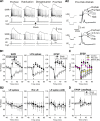
Cellular and molecular mechanisms involved in dishabituation. A, Examples of siphon withdrawal (SWR), evoked firing and membrane resistance of an LE siphon sensory neuron and an LFS siphon motor neuron, and the monosynaptic EPSP from the LE sensory neuron to the LFS motor neuron before (PreTest) and at the end of a series of 10 siphon stimuli [Habituation (Hab)], and 2.5 min [Dishabituation (Dishab)] and 77.5 min (PostTest) after four tail shocks (top) or no shock control (bottom). B, Average results from experiments like the ones shown in A (n = 7 for shock; n = 6 for no shock). B1, Siphon withdrawal, evoked LFS firing, and EPSP amplitude. There were significant effects of habituation and shock in each case. The average pretest values were 2.0 mm for siphon withdrawal, 15 spikes for evoked LFS firing, and 6.6 mV for the amplitude of the EPSP, which were not significantly different in experiments with shock and no shock. B2, Evoked LE firing, LE membrane resistance, and LE action potential duration. The average pretest values were 4.2 spikes for evoked LE firing and 2.4 ms for action potential duration, which were not significantly different in experiments with shock and no shock. C, Average depression and facilitation of the EPSP in experiments with tail shock (colored symbols) or no shock (white symbols) following no injection (Control) or intracellular injection of a peptide inhibitor of PKC (PKCi) into the sensory neuron or BAPTA into the motor neuron. There was a significant overall effect of group for the facilitation during dishabituation (F(2,29) = 4.40, p < 0.05, n = 13, 11, and 11). The overall average pretest value was 6.6 mV. The average response to the tail shock was 5.0 mm. These values were not significantly different in experiments with different injections. D, Average ratio of the late (75 ms after peak) and peak amplitude of the EPSP. The average pretest value was 0.56, which was not significantly different in experiments with shock and no shock.

Similar articles

Cited by

References

    1. Abraham WC, Bear MF. Metaplasticity: the plasticity of synaptic plasticity. Trends Neurosci. 1996;19:126–130. - PubMed
    1. Angers A, Fioravante D, Chin J, Cleary LJ, Bean AJ, Byrne JH. Serotonin stimulates phosphorylation of Aplysia synapsin and alters its subcellular distribution in sensory neurons. J Neurosci. 2002;22:5412–5422. - PMC - PubMed
    1. Antonov I, Kandel ER, Hawkins RD. The contribution of facilitation of monosynaptic PSPs to dishabituation and sensitization of the Aplysia siphon withdrawal reflex. J Neurosci. 1999;19:10438–10450. - PMC - PubMed
    1. Antonov I, Antonova I, Kandel ER, Hawkins RD. The contribution of activity-dependent synaptic plasticity to classical conditioning in Aplysia. J Neurosci. 2001;21:6413–6422. - PMC - PubMed
    1. Antonov I, Antonova I, Kandel ER, Hawkins RD. Activity-dependent presynaptic facilitation and Hebbian LTP are both required and interact during classical conditioning in Aplysia. Neuron. 2003;37:135–147. - PubMed

Publication types

MeSH terms