Comparative Study
. 2010 Apr 21;30(16):5792-801.
doi: 10.1523/JNEUROSCI.5659-09.2010.

Target-dependent regulation of neurotransmitter specification and embryonic neuronal calcium spike activity

Affiliations
Comparative Study

Target-dependent regulation of neurotransmitter specification and embryonic neuronal calcium spike activity

Qian Xiao et al. J Neurosci. .

Abstract

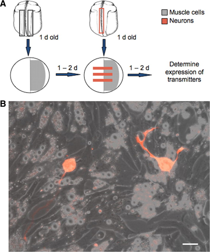
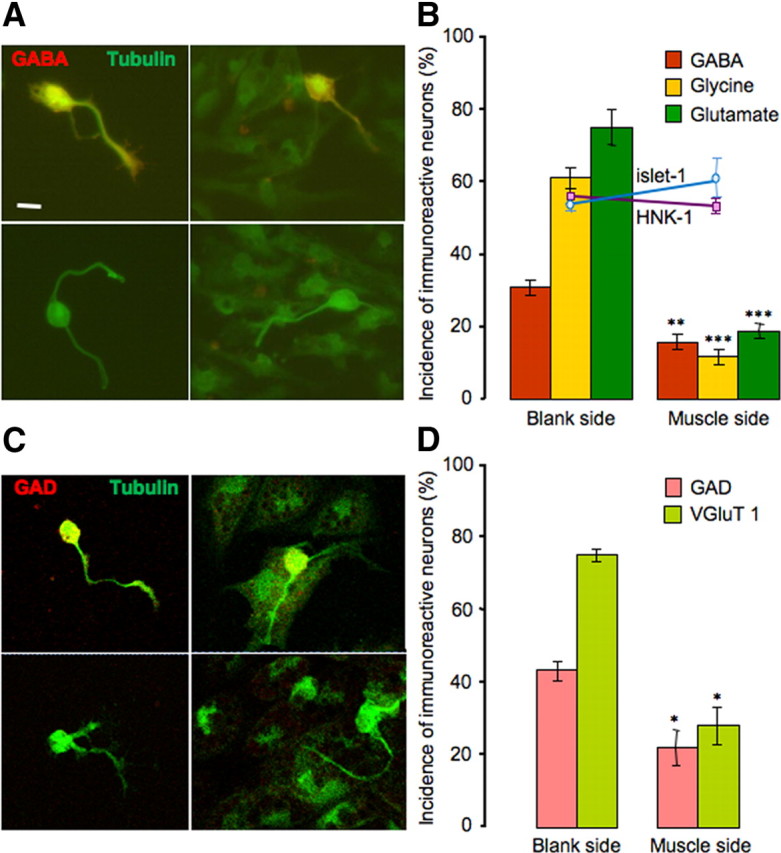
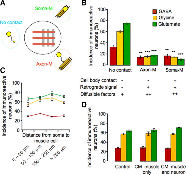
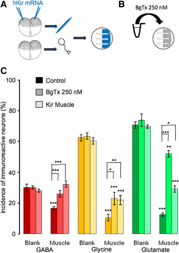
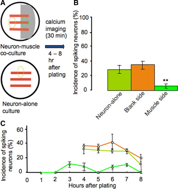
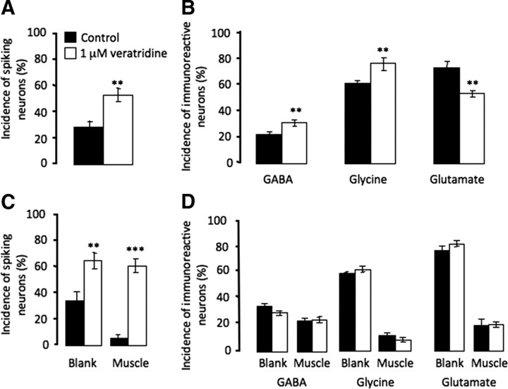
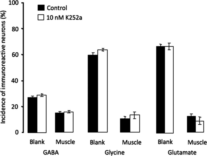
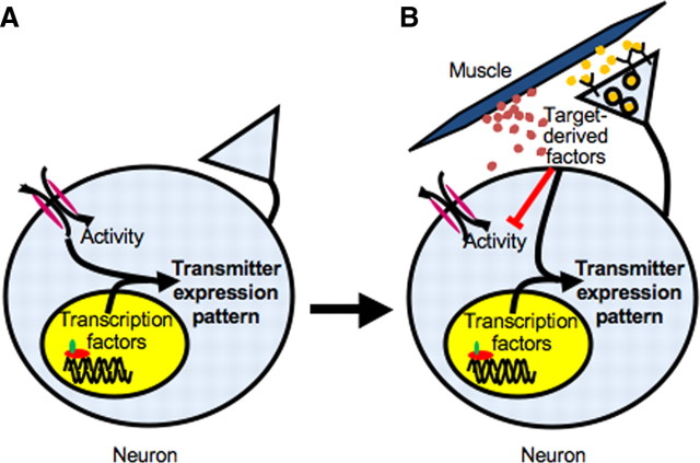
Neurotransmitter specification has been shown to depend on genetic programs and electrical activity; however, target-dependent regulation also plays important roles in neuronal development. We have investigated the impact of muscle targets on transmitter specification in Xenopus spinal neurons using a neuron-muscle coculture system. We find that neuron-muscle contact reduces the number of neurons expressing the noncholinergic transmitters GABA, glycine, and glutamate, while having no effect on the incidence of ChAT expression. We show that muscle activity is necessary for target-dependent reduction of noncholinergic transmitter expression. In addition, we demonstrate that coculture with muscle cells suppresses early spontaneous calcium spike activity in neurons and the presence of muscle cells abolishes activity-dependent transmitter specification. The results indicate that target-dependent regulation can be crucial in establishing neurotransmitter phenotypes and altering early neuronal excitability.


Figures

Figure 1.
Figure 1.
Neuron–muscle coculture system. A, Diagram illustrating the procedure for establishing neuron–muscle cocultures. Areas in which muscle cells are plated are shown in gray and areas in which neurons are located are shown in red. B, Neurons growing on muscle cells. Neurons were dissociated from rhodamine dextran-injected embryos. Scale bar, 10 μm.
Figure 2.
Figure 2.
Muscle cells suppress noncholinergic neurotransmitter expression. A, Immunostaining of neurons for GABA (red) and tubulin (green) on the blank side (left) and the muscle side (right). Top, Positive staining for GABA; bottom, negative staining. B, Noncholinergic transmitter expression is reduced on the muscle side (**p < 0.01, ***p < 0.001, compared with the blank side; n > 8 cultures per transmitter, >100 neurons per culture). HNK-1 and islet-1 expression does not differ between the two sides of the culture; the y-axis applies to the incidence of HNK-1, islet-1, and transmitter expression. Error bars indicate SEM. Scale bar, 10 μm. The Mann–Whitney U test was used to determine statistical significance. C, Immunostaining of neurons for GAD (red) and tubulin (green) on the blank side (left) and the muscle side (right). Top, Positive staining for GAD; bottom, negative staining. D, GAD and VGluT1 expression is reduced on the muscle side (*p < 0.05, compared with the blank side; n > 5 cultures per transmitter marker, >100 neurons per culture). Error bars indicate SEM. Scale as for B. The Mann–Whitney U test was used to determine statistical significance.
Figure 3.
Figure 3.
Neuronal ChAT expression is independent of muscle regulation. In situ hybridization for ChAT on the blank side (left) and the muscle side (right; neurons circled). Top, Positive staining; bottom, negative staining. Scale bar, 10 μm.
Figure 4.
Figure 4.
Cell contacts and not diffusible factors regulate muscle-dependent suppression of noncholinergic neurotransmitter expression. A, Schematic view of the lattice neuron–muscle coculture and groups of neurons with different interactions with muscles. B, Neurons with or without cell body interaction with muscle show similar levels of reduction in incidence of noncholinergic transmitter expression, when compared with the no-contact group. There is no significant difference in noncholinergic transmitter expression between axon-M and soma-M groups (**p < 0.01, ***p < 0.001; n > 5 cultures per condition per transmitter, >100 neurons per culture). C, The incidence of expression of the three noncholinergic transmitters in the no-contact group from B (GABA in red, glycine in yellow, and glutamate in green) does not change as a function of distance from neuronal soma to muscle cells (>100 neurons scored at each distance for each transmitter). D, The incidence of expression of the three noncholinergic transmitters does not differ between the control condition and conditioned medium condition (muscle alone or muscle cocultured with neurons) (n > 5 cultures per condition per transmitter, >100 neurons per culture). Error bars indicate SEM. The Kruskal–Wallis test and Conover post hoc test were used to determine statistical significance.
Figure 5.
Figure 5.
Rescue of noncholinergic neurotransmitter expression by blockade of muscle activity. A, Diagram illustrating hKir mRNA injection and neuron–muscle coculture with silenced muscle and wild type neurons. B, Diagram showing method of pharmacological blockade of muscle activity by α-bungarotoxin. C, Complete rescue of GABA expression and partial rescue of glycine and glutamate expression are achieved by either BgTx incubation or Kir expression. Lower asterisks indicate a significant difference between the bar beneath and its counterpart on the blank side. Upper asterisks indicate a significant difference between the bars (n > 5 cultures per condition per transmitter, >100 neurons per culture; *p < 0.05, **p < 0.01, ***p < 0.001). Error bars indicate SEM. The Kruskal–Wallis test and Conover post hoc test were used to determine statistical significance.
Figure 6.
Figure 6.
Muscle cells suppress early neuronal spontaneous calcium spike activity. A, Diagram showing areas of interest in which neurons were imaged. B, Neurons grown on muscle cells exhibit a significantly lower incidence of spiking at 4–8 h in vitro (n = 10 cultures, >50 neurons per condition; **p < 0.01). C, Neurons grown on muscle cells exhibit reduced incidence of spiking during each hour after plating (n > 10 cultures, >50 neurons per hour). A higher percentage of neurons exhibit calcium spikes from 4 to 8 h on the blank side of cocultures and in neuron-alone cultures. Imaging at earlier times was not performed with the two control groups due to lack of morphological distinction between neurons before axon outgrowth and the presence of other cell types. Error bars indicate SEM. The Kruskal–Wallis test and Conover post hoc test were used to determine statistical significance.
Figure 7.
Figure 7.
Muscle cells abolish activity-dependent neurotransmitter specification induced by veratridine. A, Veratridine increases the incidence of spiking neurons in neuron-alone cultures. B, In neuron-alone cultures, veratridine increases the incidence of neurons expressing the inhibitory transmitters GABA and glycine and reduces incidence of neurons expressing the excitatory transmitter glutamate. C, Veratridine increases the incidence of spiking neurons in neuron–muscle cocultures, both on the blank side and the muscle side. D, In neuron–muscle cocultures, veratridine fails to induce changes in either inhibitory or excitatory transmitter expression (n > 5 cultures per condition per transmitter, >100 neurons per culture; **p < 0.01, ***p < 0.001). Error bars indicate SEM. The Mann–Whitney U test (A–C) and the Kruskal–Wallis test (D) were used to determine statistical significance.
Figure 8.
Figure 8.
Blockade of trk receptors does not rescue expression of noncholinergic transmitters. K252a (10 nm) was applied from 5 to 24 h. No significant difference in expression of GABA, glycine, or glutamate was observed between the blank and muscle sides of the dish (n > 5 cultures per condition per transmitter, >100 neurons per culture). Higher concentrations (up to 10 μm) were also ineffective. Error bars indicate SEM. The Kruskal–Wallis test was used to determine statistical significance.
Figure 9.
Figure 9.
Model of neurotransmitter specification by transcription factors, activity, and target-derived factors. A, Early in development, when neurons have not yet established contact with their targets, spontaneous calcium spike activity and transcription factors work together to specify their transmitter expression profile. B, After neurons contact their targets, target-derived factors, including both retrograde factors (yellow dots) and diffusible factors (red dots), suppress expression of noncholinergic transmitters, block spontaneous calcium activity, and disrupt evoked activity-dependent transmitter specification. These factors work in combination with transcription factors to further refine and lock in the transmitter expression phenotype.

Similar articles

Cited by

References

    1. Barnett J, Baecker P, Routledge-Ward C, Bursztyn-Pettegrew H, Chow J, Nguyen B, Bach C, Chan H, Tuszynski MH, Yoshida K. Human beta nerve growth factor obtained from a baculovirus expression system has potent in vitro and in vivo neurotrophic activity. Exp Neurol. 1990;110:11–24. - PubMed
    1. Berg DK, Hall ZW. Increased extrajunctional acetylcholine sensitivity produced by chronic acetylcholine sensitivity produced by chronic post-synaptic neuromuscular blockade. J Physiol. 1975;244:659–676. - PMC - PubMed
    1. Borodinsky LA, Spitzer NC. Activity-dependent neurotransmitter-receptor matching at the neuromuscular junction. Proc Natl Acad Sci U S A. 2007;104:335–340. - PMC - PubMed
    1. Borodinsky LA, Root CM, Cronin JA, Sann SB, Gu X, Spitzer NC. Activity-dependent homeostatic specification of transmitter expression in embryonic neurons. Nature. 2004;420:523–530. - PubMed
    1. Brosenitsch TA, Katz DM. Expression of Phox2 transcription factors and induction of the dopaminergic phenotype in primary sensory neurons. Mol Cell Neurosci. 2002;20:447–457. - PubMed

Publication types

Substances

LinkOut - more resources