. 2010 Jun 15;21(12):2034-44.
doi: 10.1091/mbc.e10-02-0158. Epub 2010 Apr 21.

Insulin-regulated aminopeptidase is a key regulator of GLUT4 trafficking by controlling the sorting of GLUT4 from endosomes to specialized insulin-regulated vesicles

Affiliations

Insulin-regulated aminopeptidase is a key regulator of GLUT4 trafficking by controlling the sorting of GLUT4 from endosomes to specialized insulin-regulated vesicles

Ingrid Jordens et al. Mol Biol Cell. .

Abstract

Insulin stimulates glucose uptake by regulating translocation of the GLUT4 glucose transporter from intracellular compartments to the plasma membrane. In the absence of insulin GLUT4 is actively sequestered away from the general endosomes into GLUT4-specialized compartments, thereby controlling the amount of GLUT4 at the plasma membrane. Here, we investigated the role of the aminopeptidase IRAP in GLUT4 trafficking. In unstimulated IRAP knockdown adipocytes, plasma membrane GLUT4 levels are elevated because of increased exocytosis, demonstrating an essential role of IRAP in GLUT4 retention. Current evidence supports the model that AS160 RabGAP, which is required for basal GLUT4 retention, is recruited to GLUT4 compartments via an interaction with IRAP. However, here we show that AS160 recruitment to GLUT4 compartments and AS160 regulation of GLUT4 trafficking were unaffected by IRAP knockdown. These results demonstrate that AS160 is recruited to membranes by an IRAP-independent mechanism. Consistent with a role independent of AS160, we showed that IRAP functions in GLUT4 sorting from endosomes to GLUT4-specialized compartments. This is revealed by the relocalization of GLUT4 to endosomes in IRAP knockdown cells. Although IRAP knockdown has profound effects on GLUT4 traffic, GLUT4 knockdown does not affect IRAP trafficking, demonstrating that IRAP traffics independent of GLUT4. In sum, we show that IRAP is both cargo and a key regulator of the insulin-regulated pathway.


Figures
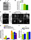
Figure 1.
Figure 1.
IRAP is essential for basal GLUT4 intracellular retention. (A) Representative immunoblot analysis of the indicated differentiation markers in cell extracts of control 3T3-L1 adipocytes and IRAP-KD adipocytes at day 5 after differentiation. (B) Quantification of the expression of the indicated differentiation markers normalized to actin as the ratio IRAP-KD adipocytes-to-control adipocytes. Each data point represents the average ± SEM of 5–7 independent experiments. (C) Epifluorescence images detecting endogenous GLUT4 in basal control and IRAP-KD adipocytes. Bar, 10 μm. (D) Epifluorescence images of basal control and IRAP-KD adipocytes expressing HA-GLUT4-GFP. Surface GLUT4 (PM) and endogenous IRAP were detected by indirect immunofluorescence of anti-HA and anti-IRAP, respectively. Arrows indicate HA-GLUT4-GFP–expressing cells. Bar, 10 μm. (E) PM-to-total HA-GLUT4-GFP ratios of control, IRAP-KD, and IRAP-KD adipocytes reexpressing IRAP, or IRAP-TR in basal and insulin-stimulated state. Each data point represents the average ± SEM of 8–20 experiments. The data from the individual experiments were normalized to basal IRAP-KD adipocytes reexpressing IRAP (rescue basal). Insulin-stimulated cells were incubated with 1 nM insulin for 30 min at 37°C. IRAP or IRAP-TR expression was verified by indirect immunofluorescence in saponin-permeabilized cells (***p < 0.0001 compared with control; ns, nonsignificant, paired Student's t test). (F) PM-to-total transferrin receptor (TR) ratios of IRAP-KD adipocytes or IRAP-KD adipocytes reexpressing IRAP in basal and insulin-stimulated state. Each data point represents the average ± SEM of 2–3 experiments (ns, nonsignificant; paired Student's t test). The data from the individual experiments are normalized to basal IRAP-KD adipocytes (Basal KD). IRAP expression was verified by indirect immunofluorescence.
Figure 2.
Figure 2.
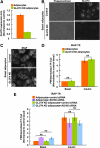
IRAP traffics independent of GLUT4. (A) Quantification of the expression of endogenous GLUT4 detected by indirect immunofluorescence in control and GLUT4-KD adipocytes in arbitrary units (A.U.). Each data point represents the average ± SEM; n = > 50 cells. (B) Phase-contrast and epifluorescence images of basal control and GLUT4-KD adipocytes. Endogenous GLUT4 was detected by indirect immunofluorescence with GLUT4 antibodies. Bar, 10 μm. (C) Epifluorescence images of basal control and GLUT4-KD adipocytes. Endogenous IRAP was detected by indirect immunofluorescence with IRAP antibodies. Bar, 10 μm. (D) PM-to-total IRAP-TR ratios of control and GLUT4-KD adipocytes in basal and insulin-stimulated states. Each data point represents the average ± SEM of three experiments. The data from the individual experiments were normalized to basal control adipocytes. Insulin-stimulated cells were incubated with 1 nM insulin for 30 min at 37°C. ns, nonsignificant; paired Student's t test. (E) Control and GLUT4-KD adipocytes were transfected with either control siRNA or AS160 siRNA. Data points represent PM-to-total IRAP-TR distributions in basal and insulin-stimulated state of three independent experiments ± SEM. Insulin-stimulated cells were incubated with 1 nM insulin for 30 min at 37°C. ns, nonsignificant; paired Student's t test.
Figure 3.
Figure 3.
Effects of IRAP-KD and IRAP overexpression on HA-GLUT4-GFP exocytosis and internalization. HA-GLUT4-GFP exocytosis in (A) basal control adipocytes, IRAP-KD adipocytes, IRAP-KD adipocytes reexpressing IRAP or IRAP-TR, and (B) control adipocytes and control adipocytes expressing IRAP or IRAP-TR. Data from at least five different experiments was averaged and plotted as the ratio internalized-to-total HA-GLUT4-GFP versus time ± SEM. Each data point from the individual experiments was normalized to the control plateau. In this assay the time to achieve the plateau reflects the exocytosis rate constant. (C) PM-to-total HA-GLUT4-GFP ratios of control adipocytes and adipocytes overexpressing IRAP in basal and insulin-stimulated state. Each data point represents the average ± SEM of 4–10 experiments (***p < 0.001 compared with basal control; paired Student's t test). The data from the individual experiments are normalized to basal control. IRAP expression was verified by indirect immunofluorescence. (D) Internalization of HA-GLUT4-GFP in basal control adipocytes, IRAP-KD adipocytes and control adipocytes expressing IRAP or IRAP-TR. IRAP coexpression with HA-GLUT4-GFP was verified by indirect immunofluorescence.
Figure 4.
Figure 4.
IRAP and AS160 function at different steps in GLUT4 intracellular retention. (A) Transient AS160 knockdown in basal IRAP-KD adipocytes has additive effects on PM GLUT4 levels. Control and IRAP-KD adipocytes were transfected with either control siRNA or AS160 siRNA. Data points represent the average basal PM-to-total HA-GLUT4-GFP distributions of four independent experiments ± SEM (*p < 0.05 compared with control siRNA, **p < 0.001 compared with control siRNA; paired Student's t test). The data from the individual experiments are normalized to IRAP-KD adipocytes reexpressing IRAP transfected with control siRNA (rescue). IRAP coexpression with HA-GLUT4-GFP was verified by indirect immunofluorescence. (B) IRAP-KD has no effect on the membrane localization of FLAG-AS160. Control and IRAP-KD adipocytes coexpressing HA-GLUT4-GFP and FLAG-AS160 were permeabilized, or not, before fixation. FLAG-AS160 was revealed by indirect immunofluorescence using rabbit anti-FLAG. Shown is membrane-bound fraction of FLAG-AS160 as the ratio of FLAG-AS160 fluorescence in permeabilized cells to nonpermeabilized cells. Each data point is the average of three independent experiments ± SEM (ns, nonsignificant; paired Student's t test). (C) IRAP is not essential for AS160 localization to GLUT4-positive compartments. GLUT4-containing compartments were isolated by immunoabsorption of HA-GLUT4-GFP using beads conjugated to anti-GFP and immunoblotted for endogenous AS160. Shown is the quantification of the fraction of total AS160 in the immunoabsorption (IA) of three independent experiments in control and IRAP KD adipocytes. Each data point of the individual experiments was normalized to the ratio in control adipocytes. (ns, nonsignificant; paired Student's t test).
Figure 5.
Figure 5.
The intracellular distribution of GLUT4 in IRAP-KD adipocytes and the effect of IRAP-KD on the behavior of mutant GLUT4. (A) Schematic overview of the specialized GLUT4 pathway versus the general TR pathway and the steps at which IRAP could function. In basal cells GLUT4 is distributed to the specialized GLUT4 compartments and the general TR-positive endosomes. IRAP-KD can affect either the sorting of GLUT4 from the TR pathway to the GSV (step 1) regulated by the TELE motif, or the GLUT4 storage compartments (step 2) regulated by the FQQI motif, or affect the slow recycling specialized pathway (step 3), which is under control of AS160. (B) Distribution of HA-GLUT4-GFP between endosomes and GSV is affected by IRAP-KD. Each data point represents the average ± SEM of four independent experiments (**p < 0.01 compared with control; paired Student's t test). (C) The behaviors of the FQQI5–8 and TELE487–490 HA-GLUT4-GFP mutants in control and IRAP-KD adipocytes. PM-to-total HA-GLUT4-GFP WT, F5A and E488A,E490A distributions in basal control, IRAP-KD or IRAP-KD adipocytes reexpressing IRAP. Each data point represents the average ± SEM of 4–7 independent experiments (**p < 0.01 compared with own control adipocytes, *p < 0.05 compared with F5A control adipocytes, #p < 0.05 compared with WT control, ##p < 0.01 compared with WT control; paired Student's t test). The data from the individual experiments are normalized to WT HA-GLUT4-GFP in IRAP KD reexpressing IRAP adipocytes (WT rescue). IRAP expression was verified by indirect immunofluorescence. (D) The increase in basal PM GLUT4 in IRAP-KD adipocytes is insensitive to AS160-4P. PM-to-total distributions of HA-GLUT4-GFP in IRAP-KD adipocytes expressing IRAP or AS160-4P in basal and insulin-stimulated state. Shown is the average of four independent experiments ± SEM (*p < 0.05 compared with rescue basal; unpaired Student's t test). The data of the individual experiments is normalized to basal IRAP-KD reexpressing IRAP adipocytes (rescue basal). IRAP and AS160 expression were verified by indirect immunofluorescence.

References

    1. Abel E. D., Graveleau C., Betuing S., Pham M., Reay P. A., Kandror V., Kupriyanova T., Xu Z., Kandror K. V. Regulation of insulin-responsive aminopeptidase expression and targeting in the insulin-responsive vesicle compartment of glucose transporter isoform 4-deficient cardiomyocytes. Mol. Endocrinol. 2004;18:2491–2501. - PubMed
    1. Albiston A. L., et al. Evidence that the angiotensin IV (AT(4)) receptor is the enzyme insulin-regulated aminopeptidase. J. Biol. Chem. 2001;276:48623–48626. - PubMed
    1. Albiston A. L., Mustafa T., McDowall S. G., Mendelsohn F. A., Lee J., Chai S. Y. AT4 receptor is insulin-regulated membrane aminopeptidase: potential mechanisms of memory enhancement. Trends Endocrinol. Metab. 2003;14:72–77. - PubMed
    1. Albiston A. L., Peck G. R., Yeatman H. R., Fernando R., Ye S., Chai S. Y. Therapeutic targeting of insulin-regulated aminopeptidase: heads and tails? Pharmacol. Ther. 2007;116:417–427. - PubMed
    1. Antonescu C. N., Foti M., Sauvonnet N., Klip A. Ready, set, internalize: mechanisms and regulation of GLUT4 endocytosis. Biosci. Rep. 2009;29:1–11. - PubMed

Publication types

MeSH terms