. 2010 Jun 15;21(12):2078-86.
doi: 10.1091/mbc.e09-11-0926. Epub 2010 Apr 21.

Neuralized promotes basal to apical transcytosis of delta in epithelial cells

Affiliations

Neuralized promotes basal to apical transcytosis of delta in epithelial cells

Najate Benhra et al. Mol Biol Cell. .

Abstract

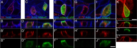
Notch receptors mediate short-range signaling controlling many developmental decisions in metazoans. Activation of Notch requires the ubiquitin-dependent endocytosis of its ligand Delta. How ligand endocytosis in signal-sending cells regulates receptor activation in juxtaposed signal-receiving cells remains largely unknown. We show here that a pool of Delta localizes at the basolateral membrane of signal-sending sensory organ precursor cells in the dorsal thorax neuroepithelium of Drosophila and that Delta is endocytosed in a Neuralized-dependent manner from this basolateral membrane. This basolateral pool of Delta is segregated from Notch that accumulates apically. Using a compartimentalized antibody uptake assay, we show that murine Delta-like 1 is similarly internalized by mNeuralized2 from the basolateral membrane of polarized Madin-Darby canine kidney cells and that internalized ligands are transcytosed to the apical plasma membrane where mNotch1 accumulates. Thus, endocytosis of Delta by Neuralized relocalizes Delta from the basolateral to the apical membrane domain. We speculate that this Neuralized-dependent transcytosis regulates the signaling activity of Delta by relocalizing Delta from a membrane domain where it cannot interact with Notch to another membrane domain where it can bind and activate Notch.


Figures

Figure 1.
Figure 1.
Apical-basal localization of Notch and Dl in the pupal notum. (A, D, and G) Schematic representation of apical to basal sections of epidermal, pI (SOP), pIIb, and pIIa cells from the pupal notum. Adherens junctions are depicted in orange and Sanpodo (Spdo) localization is schematized in blue. (B–I“') Localization of NICD (green in B–I'), Dl (red in B–I, gray in B”–I“) and Spdo (blue in B–I, B”'–I“') in epidermal and interphasic pI cells (B–C”'), pI at prometaphase (E–F“'), and pIIa/pIIb cells (H–I”'). B–B“', E–E”' and H–H“' show apical confocal sections. C–C”', F–F“', and I–I”' show basal confocal sections taken at the level of the nuclei. Sensory organ cells (pI, pIIb, and pIIa) were identified using Spdo (blue). Anterior is left. Bar, 5 μm (B–I”').
Figure 2.
Figure 2.
Dl accumulates at the basolateral plasma membrane in neur mutant cells. (A–H') Localization of Dl (A–H', red in A–E and F–H, gray in C'–E' and F'–H') and Notch (I–P', red in I–M and I–P and gray in K'–M' and N′–P') in neurIF65 mutant (F–H' and N–P') versus control sensory organ cells (C–E' and K–M'). A, C, C', F, F', I, K, K', N, and N′ show confocal sections at the apical cortex taken at the level of the adherens junctions (E-Cad; green), and B, D, D', G, G', J, L, L', O, and O' show confocal sections taken at the level of the nuclei (basolateral cortex). (E, E', H, H') Panels are orthogonal sections of the cells shown in D, D', G, G', respectively. (M, M', P, P') Panels are higher magnifications of the apical sections of the cells shown in K, K', N, N', respectively. In neurIF65 mutant cells, identified by the loss of nuclear GFP (A B, and F–H; green in B, G, and H), Dl localized at the level of E-Cad and at the basolateral cortex below E-Cad. By contrast, Notch was found at the apical cortex (I, N, N′, P, and P', red in I and N–P) and in intracellular dotted structures (red in O and O') but not at the basolateral cortex of neurIF65 mutant cells. A, B and I, J are lower magnifications of wild-type and mutant organs illustrated in C–H' and K–P', respectively. Arrowheads in E' and H' highlight Dl localizing at the basolateral membrane. Arrowheads in M' and P' are pointing to Notch localizing apically at the interface between sensory organ daughter cells. Sensory organ cells were identified using Spdo (blue in A–E, F–H, I–M, and N–P). Anterior is left. Bar, 25 μm (I and J), 15 μm (A, B), 5 μm (C–H', K–O'), and 2.5 μm (M–P').
Figure 3.
Figure 3.
Dl is internalized from the basolateral membrane in the pupal notum. (A–C”') Pulse-chase labeling experiments to monitor Dl internalization in living pupae. Dl internalization (red in A–C“' and separate channel in A”–C“) at the pIIa/pIIb cell stage was examined at three time points (0, 5, and 15 min). A–C show confocal sections at the apical cortex taken at the level of the adherens junctions labeled with E-Cad (green), whereas panels A'–C' show confocal sections taken at the level of the nuclei (basolateral cortex). Sensory organ cells were identified using Spdo (blue in A–C, A”'–C”', and D). At 0 min, Dl was detected at the basolateral plasma membrane of the pIIa cell as well as at the pIIa/pIIb interface, below E-Cad (green). After 5 min of internalization, clustered Dl still present at the plasma membrane and/or small endocytic vesicles containing Dl were detected in both pIIa and pIIb cells. At 15 min, Dl was predominantly detected in vesicular compartments within the pIIa and pIIb cells. Most of these vesicles were located in vicinity to E-Cad-positive junctions of the pIIb cell. A”'–C”' show confocal sections along the z-axis of images A–C', respectively. D–E”', distribution of Dl 15 min after internalization (D, D', E, E'; red in D, E) relative to Rab5-GFP (D, D'; green in D), Sec15-GFP (E, E”; green in E) and Rab11 (E, E“; blue in E). Sensory organ cells were identified using Spdo (blue in A–C', A”', C”'–D and E). Anterior is left. Bars, 10 μm.
Figure 4.
Figure 4.
Apical-basal localization of Notch and Dl in polarized MDCK cells. Distribution of a VSV-G–tagged version of wild-type murine Dll-1 (A–F”, red) or K17R Dll11 a mutant version of Dll1 (G–J“) in polarized MDCK cells (phalloidin in blue) in the absence of mNeur2 (A–B”) or the presence of mNeur2 (C–D“, I–J”) or mNeur2-ΔRF (E–F“) (in C–J”; mNeur2 was detected using the HA tag in green). B–B“, D–D”, F–F“, H–H”, and J–J“ show confocal Z-sections of images shown in A, C, E, G, and I, respectively. (K–L”) Distribution of murine Notch1 (green) in polarized MDCK cells (phalloidin in red). Apical is up. Bar, 10 μm (A–H“, J–J”).
Figure 5.
Figure 5.
Neur promotes basal to apical transcytosis of Dl in MDCK cells. (A–A“) Schematic representation of the cell surface accessibility assay (A and A') and transcytosis assay (A”). (B–F“) Polarized MDCK cells were transfected with VSV-G–tagged mouse Dll-1 (B–D”) or K17R Dll1 (E–F“) alone or together with wild type mNeur2 (C–C”, F–F“), or mNeur2-ΔRF (D–D”). They were then incubated with anti-VSV-G antibodies in the basal (A, B–F) or apical compartment (A', B'–F') at 4°C for 15 min. Alternatively, polarized MDCK cells were incubated with anti-VSG-G and anti-mouse Cy3 in the basal and apical compartment respectively for 120 min at 37°C (A“, B”–F“). Dll-1 was detected using rabbit anti-Dll-1 (green; phalloidin in blue). Orthogonal confocal sections taken along the z-axis are shown below images B–F”. The faint diffuse red staining seen in B“, D”, E“, and F” corresponds to background (laser intensity and detector sensitivity of the confocal were increased compared with C“). Bar, 10 μm (B–F') and 5 μm (B”–F“).
Figure 6.
Figure 6.
Quantitation of Dll-1 localization and transcytosis in polarized MDCK cells. (A) Steady-state distributions of Dll-1 (n = 48), Dll-1 + mNeur2 (n = 47), Dll-1+mNeur2-ΔRF (n = 47), K17R Dll-1 (K17R; n = 45), and K17R Dll1 + mNeur2 (n = 50). In this semiquantitative analysis of confocal images, basolateral staining refers to cells in which Dll1 was predominantly observed at the basolateral membrane (see Figure 4, A, B and E, F). In cells where Dll1 predominantly localized in intracellular compartment (see Figure 4, C, D and G–J'), staining was classified as intracellular. (B) Quantitation of apical and basolateral cell surface immunostaining of Dll-1 (n = 49 and n = 46, respectively), Dll-1 + mNeur2 (n = 68 and n = 60, respectively), Dll-1 + mNeur2-ΔRF (n = 54 and n = 48, respectively), K17R Dll1(n = 49 and n = 56, respectively), and K17R Dll1 + mNeur2 (n = 45 and n = 58, respectively) illustrated in Figure 5, B–F and B–F'. Numbers into brackets refer to the number of cells analyzed for apical and basolateral cell surface staining, respectively. Apical versus basolateral cell surface staining was done on separate transwell filters. In this quantitation the occurrence of the immunostaining (binary: yes or no) at the apical or basolateral membrane was assessed. All transfected cells exhibited a basolateral staining, including cells transfected with K17R Dll1 mutant that showed low plasma membrane staining. This apparent discrepancy is likely due to the fact that the anti-VSVG antibody is more sensitive than anti-Dll1. By contrast, apical staining was almost exclusively detected upon coexpression of mNeur2 and Dll-1 (80% of the cells expressing Dll1 showed a VSG-G staining at the apical surface). (C) Quantitation of basal to apical transcytosis of Dll-1 (n = 49), Dll-1 + mNeur2 (n = 90), Dll-1 + mNeur2-ΔRF (n = 54), K17R Dll1 (n = 56), and K17R Dll1 + mNeur2 (n = 58) as shown in Figure 5, A”–F“. In all cases, quantitation was performed on results obtained from at least three independent experiments. Experiments performed in parallel indicated that mNeur2 and Dll-1 constructs are typically coexpressed in 90% of transfected cells.

Similar articles

Cited by

References

    1. Apodaca G., Katz L. A., Mostov K. E. Receptor-mediated transcytosis of IgA in MDCK cells is via apical recycling endosomes. J. Cell Biol. 1994;125:67–86. - PMC - PubMed
    1. Bardin A. J., Schweisguth F. Bearded family members inhibit Neuralized-mediated endocytosis and signaling activity of Delta in Drosophila. Dev. Cell. 2006;10:245–255. - PubMed
    1. Carraway C. A., Carraway K. L. Sequestration and segregation of receptor kinases in epithelial cells: implications for ErbB2 oncogenesis. Sci. STKE. 2007;2007:re3. - PubMed
    1. Chanet S., Vodovar N., Mayau V., Schweisguth F. Genome engineering-based analysis of Bearded family genes reveals both functional redundancy and a nonessential function in lateral inhibition in Drosophila. Genetics. 2009;182:1101–1108. - PMC - PubMed
    1. Commisso C., Boulianne G. L. The NHR1 domain of Neuralized binds Delta and mediates Delta trafficking and Notch signaling. Mol. Biol. Cell. 2007;18:1–13. - PMC - PubMed

Publication types

LinkOut - more resources