. 2010 Jul;25(4):381-8.
doi: 10.1093/mutage/geq017. Epub 2010 Apr 21.

XPA impacts formation but not proteasome-sensitive repair of DNA-protein cross-links induced by chromate

Affiliations

XPA impacts formation but not proteasome-sensitive repair of DNA-protein cross-links induced by chromate

Alma Zecevic et al. Mutagenesis. 2010 Jul.

Abstract

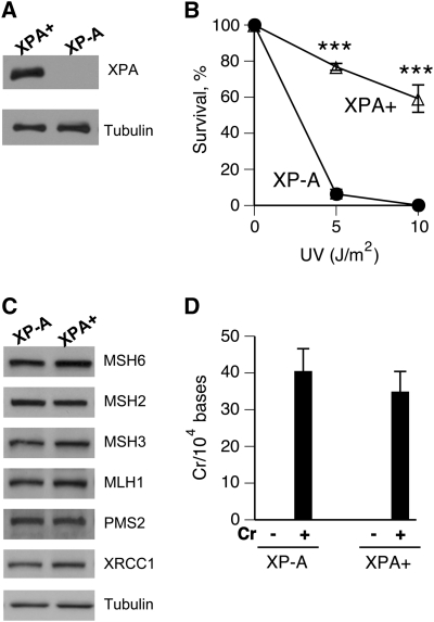
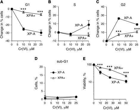
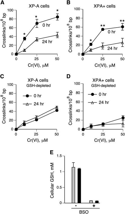
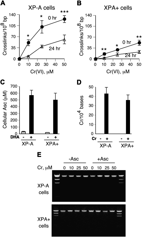
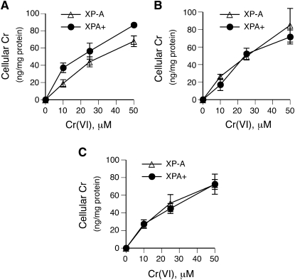
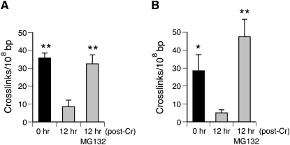
DNA-protein cross-links (DPCs) are caused by a large number of human carcinogens and anti-cancer drugs. However, cellular processes involved in decreasing a burden of these genotoxic lesions remain poorly understood. Here, we examined the impact of nucleotide excision repair (NER), which is a principal repair pathway for bulky DNA adducts, and the main cellular reducers on removal of chromium(VI)-induced DPC. We found that standard and ascorbate-restored cultures of isogenic XPA-null (NER deficient) and XPA-complemented human fibroblasts had very similar repair of Cr-DPC (60-65% average DPC removal after 24 h). However, XPA absence caused depletion of G1 and accumulation of G2 cells at low Cr(VI) doses, suggesting that Cr-DPC were not a significant cause of cell cycle perturbations. Interestingly, although pro-oxidant metabolism of Cr(VI) in glutathione-depleted cells generated significantly fewer DPC, they were repair resistant irrespective of the NER status of cells. Inhibition of proteasome activity by MG132 abolished DPC repair in both XPA-null and XPA-complemented cells. XPA loss caused two to three times higher initial DPC formation, demonstrating the importance of NER in removal of the precursor lesions. Our results indicate that human NER is not involved in removal of Cr-DPC containing non-histone proteins but it acts as a defence mechanism against these large lesions by preventing their formation. Therefore, individual differences in NER activity are expected to alter sensitivity but not persistence of DPC as a biomarker of hexavalent Cr.


Figures

Fig. 1
Fig. 1
Characterization of XP-A and XPA+ cells. (A) XPA western blot in extracts from XPA-null (XP-A cells) and XPA-complemented XP-A fibroblasts (XPA+ cells). (B) Clonogenic survival of XP-A and XPA+ fibroblasts treated with UVC (λmax = 254 nm). Data are means ± SDs from two independent experiments with three dishes per dose each. ***P < 0.001 relative to XP-A cells. (C) Western blots demonstrating expression of various DNA repair proteins in XP-A and XPA+ cells. (D) Cr–DNA binding in XP-A and XPA+ cells treated with 20 μM Cr(VI) for 3 h and collected immediately. The amount of DNA-bound Cr was determined by graphite furnace atomic absorption spectroscopy. Data are means ± SDs from four independent DNA samples.
Fig. 2
Fig. 2
Cell cycle changes and survival of Cr(VI)-treated XP-A and XPA+ cells. Cell cycle distribution was determined by FACS of propidium iodide-stained nuclei. Data are means ± SDs from three independent measurements. Statistics: *P < 0.05 and ***P < 0.001 relative to XP-A cells. (A) Changes in percentage of cells in G1 phase. (B) Changes in percentage of cells in S phase. (C) Changes in percentage of cells in G2/M phase. (D) Percentage of cells with subdiploid amount of DNA. (E) Cytotoxicity of Cr(VI) treatments at 48 h post-exposure (means ± SDs, n = 4).
Fig. 3
Fig. 3
Formation and repair of DPC in normal and GSH-depleted cells. DPC were measured by the K-SDS precipitation assay in cells collected immediately (0 h) and 24 h after 3-h long exposures to Cr(VI). Cr-induced DPC values are background subtracted and represent means ± SDs from three independent experiments in triplicates. Statistics: *P < 0.05 and **P < 0.01 relative to 24 h samples. (A) DPC levels in XP-A cells grown under standard tissue culture conditions. (B) DPC in XPA+ cells grown under standard conditions. (C) DPC in XP-A cells pre-treated with 0.1 mM BSO for 24 h prior to Cr(VI) treatments. (D) DPC in XPA+ cells pre-treated with 0.1 mM BSO for 24 h prior to Cr(VI) treatments. (E) Cellular levels of GSH in control and 0.1 mM BSO pre-treated cells (grey bars, XP-A cells; black bars, XPA+ cells). GSH concentrations were measured by HPLC detection of the specific GSH–monobromobimane conjugate.
Fig. 4
Fig. 4
Effects of Asc supplementation on DPC formation and repair. Cells were pre-loaded with 0.5 mM Asc during a 90-min long incubation with 1 mM DHA prior to Cr(VI) exposures. DPC data are means ± SDs from three independent experiments in triplicates. Statistics: *P < 0.05, **P < 0.01 and ***P < 0.001 relative to 24 h samples. (A and B) DPC in Asc-supplemented XP-A and XPA+ cells, respectively. (C) Cellular levels of Asc in control and 1 mM DHA-treated XP-A and XPA+ fibroblasts. Asc concentrations were measured by HPLC. (D) Cr–DNA binding in Asc-preloaded XP-A and XPA+ cells treated with 20 μM Cr(VI) for 3 h and collected immediately. (E) Ethidium bromide-stained agarose gels of DNA fragments generated during DPC measurements.
Fig. 5
Fig. 5
Cr(VI) uptake measurements. Data are means ± SDs from three independent samples. (A) Control cells. (B) Cells pre-treated with 0.1 mM BSO for 24 h. (C) Cells pre-incubated with 1 mM DHA for 90 min.
Fig. 6
Fig. 6
Inhibition of proteasomes suppresses DPC repair. Cells were exposed to Cr(VI) for 3 h and collected immediately (0 h) or following a 12-h recovery period in the presence or absence 0.5 μM MG132. Data are means ± SDs (n = 6). (A) DPC repair in XP-A cells. (B) DPC repair in XPA+ cells. Statistical significance relative to the 12-h samples without MG132: *P < 0.05 and **P < 0.01.

References

    1. Costa M. DNA-protein complexes induced by chromate and other carcinogens. Environ. Health Perspect. 1991;92:45–52. - PMC - PubMed
    1. Costa M, Zhitkovich A, Harris M, Paustenbauch D, Gargas M. DNA-protein crosslinks produced by various chemicals in cultured human lymphoma cells. J. Toxicol. Environ. Health. 1997;50:433–449. - PubMed
    1. Taioli E, Zhitkovich A, Toniolo P, Bernstein J, Blum R, Costa M. DNA-protein crosslinks as a biomarker of cis-platinum activity in cancer patients. Oncol. Rep. 1996;3:439–441. - PubMed
    1. Barker S, Weinfeld M, Murray D. DNA-protein crosslinks: their induction, repair, and biological consequences. Mutat. Res. 2005;589:111–135. - PubMed
    1. Minko IG, Zou Y, Lloyd RS. Incision of DNA-protein crosslinks by UvrABC nuclease suggests a potential repair pathway involving nucleotide excision repair. Proc. Natl Acad. Sci. USA. 2002;99:1905–1909. - PMC - PubMed

Publication types