. 2010 Oct;95(10):1722-9.
doi: 10.3324/haematol.2010.023358. Epub 2010 Apr 21.

Lack of expression of inhibitory KIR3DL1 receptor in patients with natural killer cell-type lymphoproliferative disease of granular lymphocytes

Affiliations

Lack of expression of inhibitory KIR3DL1 receptor in patients with natural killer cell-type lymphoproliferative disease of granular lymphocytes

Cristina Gattazzo et al. Haematologica. 2010 Oct.

Abstract

Background: Natural killer cell-type lymphoproliferative disease of granular lymphocytes is a disorder characterized by chronic proliferation of CD3(-)CD16(+) granular lymphocytes. By flow cytometry analysis, we previously demonstrated a dysregulation in killer immunoglobulin-like receptor (KIR) expression in natural killer cells from patients with this lymphoproliferative disease, the activating KIR receptors being mostly expressed. We also found that patients with natural killer cell-type lymphoproliferative disease of granular lymphocytes usually had KIR genotypes characterized by multiple activating KIR genes.

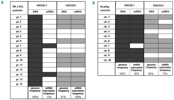
Design and methods: We investigated the mRNA levels of the KIR3DL1 inhibitory and the related KIR3DS1 activating receptors in 15 patients with natural killer cell-type lymphoproliferative disease of granular lymphocytes and in ten controls. These genes are usually expressed when present in the genome of the Caucasian population.

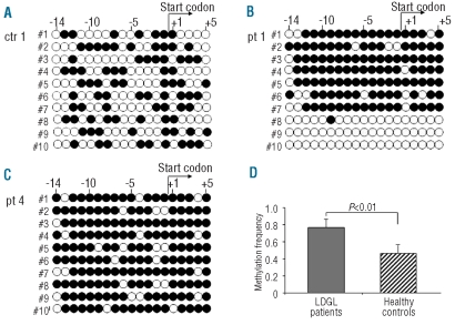
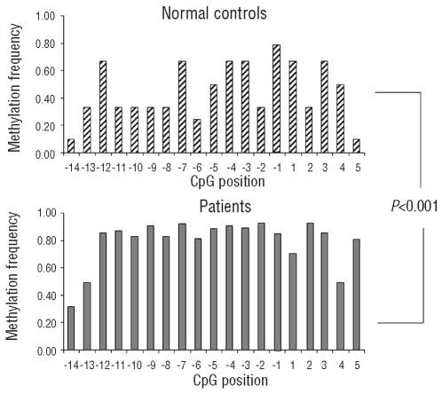
Results: We demonstrated the complete lack of KIR3DL1 expression in most of the patients analyzed, with the receptor being expressed in 13% of patients compared to in 90% of controls (P<0.01). Interestingly, studies of the methylation patterns of KIR3DL1 promoter showed a significantly higher methylation status (0.76 ± 0.12 SD) in patients than in healthy subjects (0.49±0.10 SD, P<0.01). The levels of expression of DNA methyl transferases, which are the enzymes responsible for DNA methylation, did not differ between patients and controls.

Conclusions: In this study we showed, for the first time, a consistent down-regulation of the inhibitory KIR3DL1 signal due to marked methylation of its promoter, thus suggesting that together with the increased expression of activating receptors, the lack of the inhibitory signal could also play a role in the pathogenesis of natural killer cell-type lymphoproliferative disease of granular lymphocytes.


Figures

Figure 1.
Figure 1.
Genotyping of KIR3DL1 and KIR3DS1 alleles and frequency of KIR3DL1/3DS1 mRNA expression in patients and normal controls. Genomic DNA was extracted from peripheral blood mononuclear cells of 15 patients and ten controls. The presence or absence of KIR3DL1/3DS1 gene was established by PCR-SSP using two different primer sets specific for both alleles. The figure (columns genome frequency) summarizes the KIR3DL1/3DS1 locus profiles observed in the patients (A) and controls (B). Filled boxes indicate the presence of KIR allele; open boxes indicate the absence. The difference of presence in the genome of KIR3DS1 between patients and controls was not statistically significant (P=0.28). For the investigation of KIR3DL1/DS1 expression total mRNA was isolated from sorted NK cells of patients and controls. Semi-quantitative PCR was carried out to determine the presence or absence of transcription for both KIR3DL1/3DS1 alleles (columns mRNA expression frequency). Amplification of β-actin was used as a control. The filled boxes indicate the corresponding expression of the mRNA, for both activating and inhibitory receptors in patients (A) and controls (B).
Figure 2.
Figure 2.
Distribution of CpG dinucleotides in the 5′ region of the KIR3D1 gene. The transcriptional start site is indicated by a square and the CpG dinucleotides are highlighted in gray. Analysis of the methylation patterns of the CpG island surrounding the transcriptional start site of this gene revealed that the methylation status consistently correlates with the transcriptional activity in primary NK cells. The primers used in this work to analyze the methylation pattern of this sequence are underlined.
Figure 3.
Figure 3.
Methylation of CpG dinucleotides correlates with transcriptional activity of the KIR3D1 gene in NK cells isolated from LDGL-patients. The figure reports the methylation pattern in the 5′ region of the KIR3D1 gene for one representative control (A) and two representative patients (B, C). The filled circles indicate methylated CpG dinucleotides, the empty circles unmethylated ones. The figure also shows the position of the start codon, the numbers indicate the CpG dinucleotides before and after the start codon in progressive order. The analysis of controls (5 subjects) was performed in the sorted CD3CD16+KIR3DL1/KIR3DS1 NK cells; instead in patients (5 subjects), the analysis was performed on sorted CD3 CD16+ NK cells. The methylation frequency, for each sequenced clone, was calculated as the number of methylated CpG dinucleotides over the total CpG dinucleotides present in the analyzed sequence. As shown by the numbers of the filled circles (A, B, C), methylation was increased in the patients with respect to the controls. The difference of median methylation frequency, considering each clone for the five patients and five controls, was statistically significant 0.76±0.12 SD versus 0.49±0.10SD, respectively P<0.01 (D).
Figure 4.
Figure 4.
The methylation frequency at individual CpG positions in normal controls and patients. The frequency for each CpG dinucleotide considered (from −14 to +5 with respect to the start codon) is reported as a median of the total sequenced clones for controls (upper panel) and patients (lower panel). The overall methylation frequency at individual positions was significantly higher in patients than in controls (P<0.001).

References

    1. Loughran TP., Jr Clonal diseases of large granular lymphocytes. Blood. 1993;82(1):1–14. - PubMed
    1. Oshimi K, Yamada O, Kaneko T, Nishinarita S, Iizuka Y, Urabe A, et al. Laboratory findings and clinical courses of 33 patients with granular lymphocyte-proliferative disorders. Leukemia. 1993;7(6):782–8. - PubMed
    1. Semenzato G, Zambello R, Starkebaum G, Oshimi K, Loughran TP., Jr The lymphoproliferative disease of granular lymphocytes: updated criteria for diagnosis. Blood. 1997;89(1):256–60. - PubMed
    1. Zambello R, Loughran TP, Jr, Trentin L, Pontisso P, Battistella L, Raimondi R, et al. Serologic and molecular evidence for a possible pathogenetic role of viral infection in CD3-negative natural killer-type lymphoproliferative disease of granular lymphocytes. Leukemia. 1995;9(7):1207–11. - PubMed
    1. Trinchieri G. Biology of natural killer cells. Adv Immunol. 1989;47:187–376. - PMC - PubMed

Publication types