. 2010 Aug 26;116(8):1321-8.
doi: 10.1182/blood-2009-07-229450. Epub 2010 Apr 21.

Differential ability of Tribbles family members to promote degradation of C/EBPalpha and induce acute myelogenous leukemia

Affiliations

Differential ability of Tribbles family members to promote degradation of C/EBPalpha and induce acute myelogenous leukemia

Priya H Dedhia et al. Blood. .

Abstract

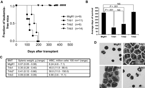
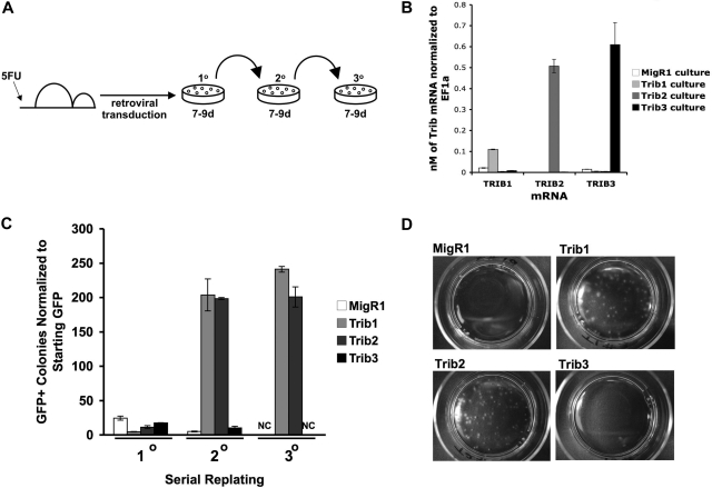
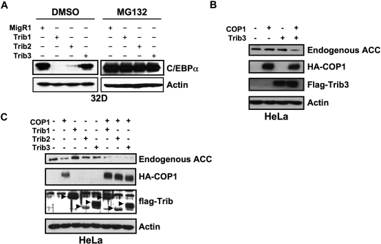
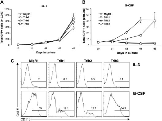
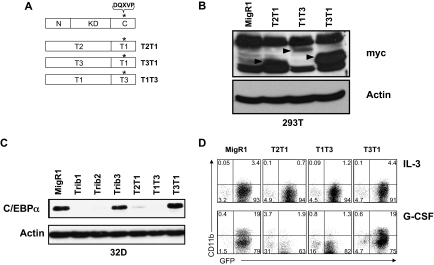
Trib1, Trib2, and Trib3 are mammalian homologs of Tribbles, an evolutionarily conserved Drosophila protein family that mediates protein degradation. Tribbles proteins function as adapters to recruit E3 ubiquitin ligases and enhance ubiquitylation of the target protein to promote its degradation. Increased Trib1 and Trib2 mRNA expression occurs in human myeloid leukemia and induces acute myeloid leukemia in mice, whereas Trib3 has not been associated with leukemia. Given the high degree of structural conservation among Tribbles family members, we directly compared the 3 mammalian Tribbles in hematopoietic cells by reconstituting mice with hematopoietic stem cells retrovirally expressing these proteins. All mice receiving Trib1 or Trib2 transduced hematopoietic stem cells developed acute myeloid leukemia, whereas Trib3 mice did not. Our previous data indicated that Trib2-mediated degradation of the transcription factor, CCAAT/enhancer-binding protein-alpha (C/EBPalpha), is important for leukemogenesis. Similar to Trib2, Trib1 induced C/EBPalpha degradation and inhibited its function. In contrast, Trib3 failed to inactivate or promote efficient degradation of C/EBPalpha. These data reveal that the 3 Tribbles homologs differ in their ability to promote degradation of C/EBPalpha, which account for their differential ability to induce leukemia.


Figures

Figure 1
Figure 1
Trib1 and Trib2, but not Trib3, induce AML. (A) Kaplan-Meier survival curve of mice receiving BM transplant with Trib1 (n = 11), Trib2 (n = 6), Trib3 (n = 14), or control MigR1 (n = 9) transduced cells. Results are derived from 2 independent experiments. (B) Average survival for indicated BM transplantation. P values determined by Student t test are indicated. NS indicates no significant difference. The difference between Trib1 and Trib2 yielded P > .1 (Student t test). (C) Average splenic weight and white blood cell count of MigR1, Trib1, Trib2, and Trib3 mice. *The splenic weights and white blood cell counts for the Trib2 mice are published. (D) Wright-Giemsa stain of BM cytospin from mice receiving the indicated BM transplantation.
Figure 2
Figure 2
Trib1 and Trib2, but not Trib3, confer serial plating activity in methylcellulose cultures. (A) Serial colony assays were performed as shown using primary BM cells transduced with retroviruses. (B) Primary methylcellulose cultures were sorted for GFP and used for RNA isolation. Absolute quantification of Trib1, Trib2, or Trib3 mRNA normalized to EF1α from sorted BM cells after 8 days of culture in methylcellulose. Values are mean (± SD) for triplicate samples. Data are representative of 2 independent experiments. (C) The colony numbers were counted during 3 rounds of serial culture and normalized to starting GFP. Values are mean (± SD) for triplicate samples. NC indicates no colonies. Data are representative of 3 independent experiments. (D) Plates from the third serial culture are shown.
Figure 3
Figure 3
Trib1 and Trib2, but not Trib3, induce efficient degradation of C/EBPα. (A) Western blot for C/EBPα in sorted 32D cells transduced with MigR1, Trib1, Trib2, or Trib3 and treated with either the proteasome inhibitor MG132 or the DMSO control. Endogenous C/EBPα expression was determined with an anti-C/EBPα antibody. β-Actin served as the protein loading control. Data are representative of 3 independent experiments. (B) Western blot for endogenous ACC in HeLa cells transfected with Trib3 and the E3 ubiquitin ligase COP1. (C) Western blot for endogenous ACC in HeLa cells transfected with the indicated Tribbles constructs and COP1. In panels B and C, ACC was detected with an anti-ACC antibody, COP1 was detected with an anti-HA antibody, Tribs were detected with an anti-FLAG antibody, and β-actin was the protein loading control. Data are representative of 2 independent experiments.
Figure 4
Figure 4
Trib1 and Trib2, but not Trib3, inhibit myeloid differentiation. The 32D cells were transduced with MigR1, Trib1, Trib2, or Trib3 and plated in IL-3 (A) or G-CSF (B) 48 hours after transduction. The number of GFP-expressing cells were determined at the indicated time points. Values are mean (± SD) for triplicate samples. (C) Flow cytometric analysis of CD11b expression, which indicates differentiation, is shown at day 3 after IL-3 or G-CSF treatment. Data are representative of 3 independent experiments.
Figure 5
Figure 5
The Trib3 C-terminus can functionally replace the Trib1 C-terminus. (A) Schematic of C-terminal domain swap mutants. *The COP1 binding site DQXVP. (B) Western blot for protein expression of indicated domain swap mutants in transfected 293T cells. The expressed proteins were detected with an anti-myc antibody (9E10) and are indicated with arrows. β-Actin was the protein loading control. (C) Western blot for C/EBPα in sorted 32D cells transduced with the indicated retrovirus. Endogenous C/EBPα expression was detected with an anti-C/EBPα antibody. β-Actin served as the protein loading control. (D) Flow cytometric analysis for CD11b in 32D cells transduced with indicated retrovirus, then plated in IL-3 or G-CSF for 3 days. Data are representative of 3 independent experiments.

References

    1. Grosshans J, Wieschaus E. A genetic link between morphogenesis and cell division during formation of the ventral furrow in Drosophila. Cell. 2000;101(5):523–531. - PubMed
    1. Mata J, Curado S, Ephrussi A, Rorth P. Tribbles coordinates mitosis and morphogenesis in Drosophila by regulating string/CDC25 proteolysis. Cell. 2000;101(5):511–522. - PubMed
    1. Rorth P, Szabo K, Texido G. The level of C/EBP protein is critical for cell migration during Drosophila oogenesis and is tightly controlled by regulated degradation. Mol Cell. 2000;6(1):23–30. - PubMed
    1. Seher TC, Leptin M. Tribbles, a cell-cycle brake that coordinates proliferation and morphogenesis during Drosophila gastrulation. Curr Biol. 2000;10(11):623–629. - PubMed
    1. Bowers AJ, Scully S, Boylan JF. SKIP3, a novel Drosophila tribbles ortholog, is overexpressed in human tumors and is regulated by hypoxia. Oncogene. 2003;22(18):2823–2835. - PubMed

Publication types

MeSH terms

Substances