. 2010 Aug 5;116(5):806-14.
doi: 10.1182/blood-2009-12-259267. Epub 2010 Apr 21.

Hematopoietic and nonhematopoietic cell tissue factor activates the coagulation cascade in endotoxemic mice

Affiliations

Hematopoietic and nonhematopoietic cell tissue factor activates the coagulation cascade in endotoxemic mice

Rafal Pawlinski et al. Blood. .

Abstract

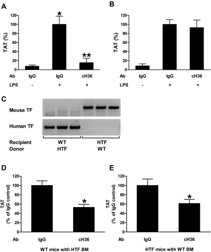
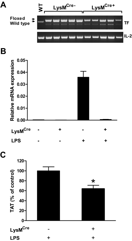
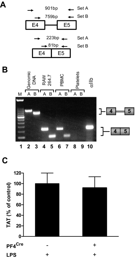
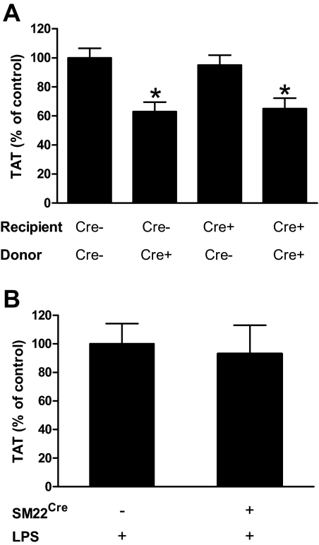
Tissue factor (TF) is the primary activator of the coagulation cascade. During endotoxemia, TF expression leads to disseminated intravascular coagulation. However, the relative contribution of TF expression by different cell types to the activation of coagulation has not been defined. In this study, we investigated the effect of either a selective inhibition of TF expression or cell type-specific deletion of the TF gene (F3) on activation of coagulation in a mouse model of endotoxemia. We found that inhibition of TF on either hematopoietic or nonhematopoietic cells reduced plasma thrombin-antithrombin (TAT) levels 8 hours after administration of bacterial lipopolysaccharide (LPS). In addition, plasma TAT levels were significantly reduced in endotoxemic mice lacking the TF gene in either myeloid cells (TF(flox/flox),LysM(Cre) mice) or in both endothelial cells (ECs) and hematopoietic cells (TF(flox/flox),Tie-2(Cre) mice). However, deletion of the TF gene in ECs alone had no effect on LPS-induced plasma TAT levels. Similar results were observed in mice lacking TF in vascular smooth muscle cells. Finally, we found that mouse platelets do not express TF pre-mRNA or mRNA. Our data demonstrate that in a mouse model of endotoxemia activation of the coagulation cascade is initiated by TF expressed by myeloid cells and an unidentified nonhematopoietic cell type(s).


Figures

Figure 1
Figure 1
Contribution of TF expressed by hematopoietic and nonhematopoietic cells to the activation of coagulation. (A) Effect of cH36 (50 μg/mL) or IgG (50 μg/mL) control antibody on LPS-induced plasma TAT levels in HTF mice (mean ± SEM for IgG, IgG/LPS and cH36/LPS groups were 2.6 ± 0.9, 34.2 ± 6.2 and 5.3 ± 3.4 ng/mL, respectively) 8 hours after LPS injection. (B) Effect of cH36 (50 μg/mL) or IgG (50 μg/mL) control antibody on LPS-induced plasma TAT levels in WT mice (mean ± SEM for IgG, IgG/LPS and cH36/LPS groups were 3.3 ± 1.7, 38.9 ± 4.2 and 36.1 ± 6.6 ng/mL, respectively) 8 hours after LPS injection (n = 3 for all treatment conditions). *Statistically significant difference (P < .05) between IgG- and IgG/LPS-treated group. **Statistically significant difference (P < .05) between IgG/LPS- and cH36/LPS-treated group. (C) PCR analysis of DNA isolated from blood cells from mice that underwent bone marrow transplantation to demonstrate reconstitution of the donor mice with recipient bone marrow 6 weeks after the transplantation. (D) Plasma TAT levels were analyzed in WT mice transplanted with bone marrow from HTF mice after treatment with IgG (n = 13; mean ± SEM 55.9 ± 5.7 ng/mL) or cH36 (n = 13; mean ± SEM 29.6 ± 3.6 ng/mL) 8 hours after LPS injection. *P < .05. (E) Plasma TAT levels were analyzed 8 hours after LPS injection in HTF mice transplanted with WT bone marrow after treatment with IgG (n = 12; mean ± SEM 46.6 ± 6.5 ng/mL) or cH36 (n = 13; mean ± SEM 28.5 ± 4.1 ng/mL;). *P < .001.
Figure 2
Figure 2
Analysis of TFflox/flox, LysMCre mice. (A) PCR analysis of mouse TF DNA isolate from blood cells demonstrates partial deletion of TF gene. Arrows indicate PCR products for wild type and floxed alleles. Note a nonspecific band below the TF band. The mouse interleukin-2 (IL-2) gene was used as a loading control. WT indicates DNA from a WT mouse. (B) Real-time PCR analysis of TF mRNA in blood cells isolated from TFflox/flox and TFflox/flox,LysMCre mice before (n = 2 per group) and after (n = 4 per group) LPS injection (2 hours). (C) Plasma TAT levels were analyzed in TFflox/flox (n = 35; mean ± SEM 19.4 ± 1.6 ng/mL) and TFflox/flox,LysMCre (n = 36; mean ± SEM 12.4 ± 1.4 ng/mL) mice 8 hours after LPS injection. *P < .01.
Figure 3
Figure 3
Mouse platelets do not express TF. (A) Diagram showing the position of 2 sets of primers used to detect TF pre-mRNA and mRNA. The expected size of PCR products for primer sets A and B are shown. (B) PCR analysis of TF was performed on either mouse genomic DNA or cDNA from LPS stimulated RAW 264.7 macrophages, LPS stimulated mouse peripheral blood leukocytes, or LPS stimulated mouse platelets. Lane 10 contains platelet cDNA amplified with primers for integrin αIIb. This gel is representative of 11 independent experiments. (C) Plasma TAT levels were analyzed in TFflox/flox (n = 13; mean ± SEM 18.9 ± 2.1 ng/mL) and TFflox/flox,PF4Cre (n = 13; mean ± SEM 17.5 ± 2.2 ng/mL) mice 8 hours after LPS injection.
Figure 4
Figure 4
Immunohistochemical analysis of TF expression. TF expression was analyzed in lungs of untreated TFflox/flox mice (A) or TFflox/flox (B) and TFflox/flox,Tie-2Cre (C) mice 8 hours after LPS injection. TF-positive cells are brown. ↑ indicates leukocytes and formula image 
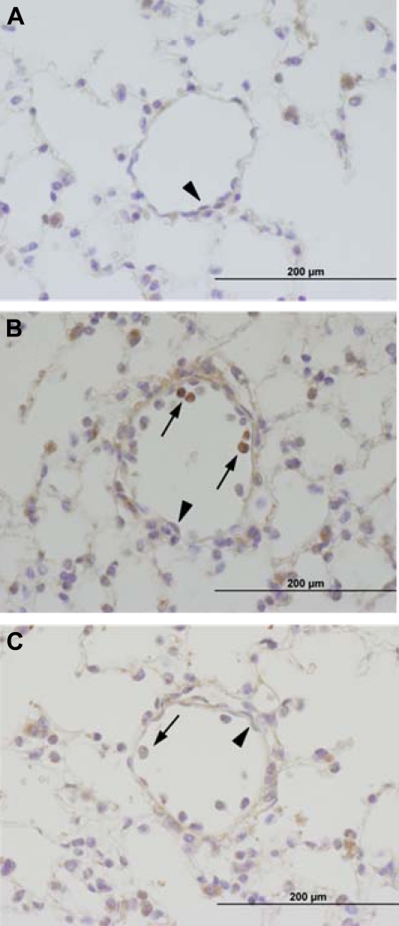
indicates ECs. Incubation of serial sections with control rat IgG demonstrated no staining (data not shown).
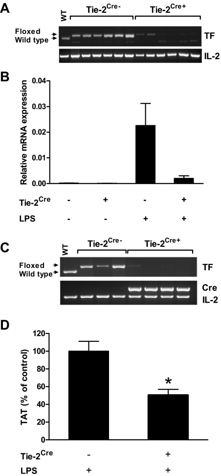
Figure 5
Figure 5
Analysis of TFflox/flox, Tie-2Cre mice. (A) PCR analysis of mouse TF DNA isolated from blood cells demonstrates deletion of the TF gene. Arrows indicate PCR products for wild type and floxed alleles. A nonspecific band is present below the TF band. The mouse IL-2 gene was used as a loading control. WT indicates DNA from a WT mouse. (B) Real-time PCR analysis of TF mRNA in blood cells isolated from TFflox/flox and TFflox/flox,Tie-2Cre mice before (n = 2 per group) and after (n = 4 per group) LPS injection (2 hours). (C) PCR analysis of mouse TF DNA isolated from ECs demonstrates deletion of the TF gene. Arrows indicate PCR products for wild type and floxed alleles. The mouse IL-2 gene was used as a loading control. Presence or absence of Cre recombinase (Cre) in different mouse groups is shown. Low levels of fibroblast contamination likely explain the residual TF-positive band in 1 of the 4 cell preparations from TFflox/flox,Tie-2Cre mice[b]. (D) Plasma TAT levels were analyzed in TFflox/flox (n = 19; mean ± SEM 15.2 ± 1.7 ng/mL) and TFflox/flox,Tie-2Cre (n = 21; mean ± SEM 7.7 ± 0.9 ng/mL) mice 8 hours after LPS injection. *P < .001.
Figure 6
Figure 6
Deletion of the TF gene in ECs or VSMCs has no effect on activation of coagulation. (A) Plasma TAT levels were analyzed 8 hours after LPS injection in TFflox/flox transplanted with TFflox/flox bone marrow (n = 18, mean ± SEM 35.9 ± 2.3 ng/mL), TFflox/flox transplanted with TFflox/flox,Tie-2Cre bone marrow (n = 12, mean ± SEM 22.7 ± 2.3 ng/mL), TFflox/flox,Tie-2Cre transplanted with TFflox/flox bone marrow (n = 13, mean ± SEM 34.1 ± 2.5 ng/mL) and TFflox/flox,Tie-2Cre transplanted with TFflox/flox,Tie-2Cre bone marrow (n = 13, mean ± SEM 23.4 ± 2.6 ng/mL). (B) Plasma TAT levels were analyzed 8 hours after LPS injection in TFflox/flox (n = 17; mean ± SEM 24.5 ± 3.5 ng/mL) and TFflox/flox,SM22Cre (n = 19; mean ± SEM 22.9 ± 3.4 ng/mL). In panel A the presence or absence of Cre recombinase in recipient and donor mice is indicated by Cre+ and Cre−. *P < .05 compared with control group expressing normal levels of TF (first bar in panel A).

Comment in

  • O tissue factor, where art thou?
    McVey JH. McVey JH. Blood. 2010 Aug 5;116(5):676-7. doi: 10.1182/blood-2010-05-283135. Blood. 2010. PMID: 20688964 No abstract available.

References

    1. Levi M. Disseminated intravascular coagulation: What's new? Crit Care Clin. 2005;21(3):449–467. - PubMed
    1. Taylor FB, Jr, Chang A, Ruf W, et al. Lethal E. coli septic shock is prevented by blocking tissue factor with monoclonal antibody. Circ Shock. 1991;33(3):127–134. - PubMed
    1. Taylor FB, Chang AC, Peer G, Li A, Ezban M, Hedner U. Active site inhibited factor VIIa (DEGR VIIa) attenuates the coagulant and interleukin-6 and -8, but not tumor necrosis factor, responses of the baboon to LD100 Escherichia coli. Blood. 1998;91(5):1609–1615. - PubMed
    1. Pawlinski R, Pedersen B, Schabbauer G, et al. Role of tissue factor and protease-activated receptors in a mouse model of endotoxemia. Blood. 2004;103(4):1342–1347. - PMC - PubMed
    1. Drake TA, Morrissey JH, Edgington TS. Selective cellular expression of tissue factor in human tissues. Implications for disorders of hemostasis and thrombosis. Am J Pathol. 1989;134(5):1087–1097. - PMC - PubMed

Publication types

MeSH terms