. 2010 Apr 21;2(28):28ra28.
doi: 10.1126/scitranslmed.3000664.

Lentiviral overexpression of GRK6 alleviates L-dopa-induced dyskinesia in experimental Parkinson's disease

Affiliations

Lentiviral overexpression of GRK6 alleviates L-dopa-induced dyskinesia in experimental Parkinson's disease

Mohamed R Ahmed et al. Sci Transl Med. .

Abstract

Parkinson's disease is caused primarily by degeneration of brain dopaminergic neurons in the substantia nigra and the consequent deficit of dopamine in the striatum. Dopamine replacement therapy with the dopamine precursor l-dopa is the mainstay of current treatment. After several years, however, the patients develop l-dopa-induced dyskinesia, or abnormal involuntary movements, thought to be due to excessive signaling via dopamine receptors. G protein-coupled receptor kinases (GRKs) control desensitization of dopamine receptors. We found that dyskinesia is attenuated by lentivirus-mediated overexpression of GRK6 in the striatum in rodent and primate models of Parkinson's disease. Conversely, reduction of GRK6 concentration by microRNA delivered with lentiviral vector exacerbated dyskinesia in parkinsonian rats. GRK6 suppressed dyskinesia in monkeys without compromising the antiparkinsonian effects of l-dopa and even prolonged the antiparkinsonian effect of a lower dose of l-dopa. Our finding that increased availability of GRK6 ameliorates dyskinesia and increases duration of the antiparkinsonian action of l-dopa suggests a promising approach for controlling both dyskinesia and motor fluctuations in Parkinson's disease.


Conflict of interest statement

Competing interests: The authors have declared no competing interests.

Figures

Fig. 1
Fig. 1
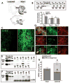
Lentivirus-induced GRK6 expression in the rat dopamine-depleted striatum. (A) Loss of dopaminergic innervation in the striatum and dopaminergic neurons in the substantia nigra was detected by immunohistochemistry for TH. ACsh, shell of the accumbens; ACc, core of the accumbens; SNc, substantia nigra pars compacta; SNr, substantia nigra pars reticulata; VTA, ventral tegmental area. (B) Quantification of the Western blots for TH in the lesioned (Les) and intact (Int) striatum in animals expressing GFP or GRK6. The inset shows a representative Western blot for TH. Numbers refer to individual animals. n = 8 in each group displayed in lower panel. (C) Photomicrograph of the intact striatum (left panel) and the striatum infected with the GRK6-GFP lentivirus (right panel) immunostained for GFP. (D) Expression of GRK6-GFP in striatal neurons. The upper row of images demonstrates the expression of GRK6 in medium spiny neurons by double staining for GFP and DARPP-32. The circle in (C) (right panel) indicates the approximate position where these photographs were taken. The middle row of images shows photomicrographs taken from similar positions in the control uninfected hemisphere [left image in (C)]. The lower row of images demonstrates the expression of GRK6 in cholinergic interneurons by double staining for GFP and choline acetyltransferase (ChAT). (E) Detection of the GRK6-GFP expression by Western blot. The expression of the GRK6 transgene in four animals in the intact and lesioned injected hemisphere detected with antibody to GFP is shown. The lower row shows expression of GFP in control animals. (F) Detection of the endogenous GRK6 and GRK6-GFP transgene expression by Western blot. Black arrowhead, GRK6A; white arrowhead, nonspecific band. (G) Quantification of the Western blot data for the expression of GRK6-GFP and endogenous GRK6A in the intact and lesioned hemisphere. Serial dilutions of purified human GRK6A were used as standards (15). The expression of endogenous GRK6A and total GRK6A (endogenous plus transgenic) in the intact and lesioned (infected) hemisphere was compared by paired t test. *P < 0.05 to the intact striatum for endogenous GRK6A; #P < 0.05 to the intact striatum, total GRK6A.
Fig. 2
Fig. 2
The expression of GRK6 in the lesioned striatum inhibits, and knockdown of GRK6 exacerbates, rotations and AIMs in the hemiparkinsonian rat. Data are shown as means ± SEM. (A) Overall frequency (for 1 hour) and peak frequency (5 min) of apomorphine-induced net contralateral rotations (contralateral - ipsilateral) in rats expressing GRK6 as compared to animals expressing GFP (control). *P < 0.05, **P < 0.01. (B) L-Dopa–induced rotations on repeated L-dopa treatment in rats expressing GRK6. *P < 0.05, between the GRK6 and control GFP groups, post hoc Student’s t test. (C) L-Dopa–induced rotations in rats expressing GRK6 presensitized with L-dopa for 5 days before virus injection. *P < 0.05, between the GRK6 and control GFP groups, post hoc unpaired Student’s t test. (D) Combined AIM scores per session in rats expressing GRK6 or GFP pretreated with L-dopa before the virus injection. *P < 0.05, between the GRK6 and GFP groups, Mann-Whitney test. (E) L-Dopa–induced rotation frequencies in rats injected with the GRK6 miRNA virus as compared to control (GFP). The animals were presensitized with L-dopa for 5 days before the virus injection. *P < 0.05, between the GRK6 miRNA and GFP groups, post hoc unpaired Student’s t test. (F) Combined AIM scores per session in rats with GRK6 knockdown as compared to control. Both groups were pretreated with L-dopa before the virus injection. *P < 0.05, between the GRK6 miRNA and GFP groups, Mann-Whitney test.
Fig. 3
Fig. 3
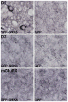
GRK6 promotes D1 dopamine receptor internalization in the lesioned striatum but does not affect D2 dopamine and mGluR5 glutamate receptor localization. In an area delineated by GFP immunohistochemistry, medium spiny neurons immunopositive for the D1 receptor showed plasma membrane and neuropil D1 receptor localization in GFP animals (top right) and prominent cytoplasmic localization in GRK6-expressing animals (top left; arrows). This effect was specific for the D1 receptor, as two other GPCRs, D2 receptor (middle) expressed in different medium spiny neurons and mGluR5 (lower) expressed in both D1- and D2-bearing neurons, remained cytoplasmic (D2, arrows) and both membranous and cytoplasmic (mGluR5, arrows) in both groups.
Fig. 4
Fig. 4
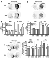
The expression of GRK6 in the lesioned striatum inhibits D1 receptor– and D2 receptor–mediated signaling. Rats lesioned with 6-OHDA and treated with L-dopa for 10 days expressed either GFP (control) or GRK6 in the lesioned striatum. (A) Representative autoradiograms showing the expression of prodynorphin mRNA. (B) Quantification of the prodynorphin in situ hybridization. Data are shown for the lesioned hemisphere as percentages of the intact hemisphere values (means ± SEM). *P < 0.001, **P < 0.01, as compared to the intact hemisphere by repeated-measures ANOVA; aP < 0.05, as compared to the L-dopa–treated GFP-expressing group (CO), Mann-Whitney test. (C) Representative autoradiograms showing the D3 receptor binding in the lesioned striatum. Note the reduction of the D3 receptor concentration in the shell (arrows) and core (approximate borders indicated by dashed lines) of the nucleus accumbens. Also note the up-regulation of the D3 receptor in the lesioned striatum caused by L-dopa and the reduction of the D3 receptor in the CPu of GRK6-expressing rats. (D) Quantification of the D3 receptor binding data (means ± SEM). *P < 0.001, **P < 0.01, as compared to the intact hemisphere by repeated-measures ANOVA; bP < 0.01, as compared to the L-dopa–treated GFP-expressing group, Mann-Whitney test. (E) Representative autoradiograms showing the expression of preproenkephalin mRNA in the lesioned striatum. (F) Quantification of the preproenkephalin in situ hybridization data (mean ± SEM). *P < 0.001, **P < 0.01, as compared to the intact hemisphere by repeated-measures ANOVA; aP < 0.05, as compared to the GFP-expressing group, Mann-Whitney test.
Fig. 5
Fig. 5
Extent of dopaminergic lesion and lentivirus-mediated expression of GRK6 in the motor striatum of MPTP-lesioned monkeys. (A and B) Representative examples and quantification (right panel) of DAT binding (A) and TH immunohistochemistry (B) in the motor striatum show marked dopamine denervation in both the caudate nucleus (CN) and the putamen (Pu) (37) with a faint signal around the globus pallidus (GPi), as reported (38). Both GFP and GRK6-GFP groups showed lesions of comparable extent [DAT: F(2,17) = 3828, P < 0.0001; TH: F(2,17) = 3331, P < 0.0001; *P < 0.001 versus control animals, one-way ANOVA followed by Bonferroni; means ± SEM]. (C) Representative GFP immunostaining at the most caudal level targeted in the striatum [globus pallidus pars externalis (GPe)] with a needle track. (D) Detection of the GRK6-GFP expression by Western blot. Samples of the monkey putamen were collected at the most rostral injection site (AC0) and 3 mm further rostrally to the injection site (AC+3). Total protein (5 μg) was loaded per lane, and GRK6-GFP and GFP were detected with mouse antibody to GFP (Clontech). Expression of the GRK6 and GFP transgenes in two GRK6-GFP–injected and two GFP-injected monkeys is shown. Recombinant proteins were loaded for comparison (right side of the blot). (E) Prodynorphin mRNA in situ detection in non–L-dopa–treated MPTP, L-dopa–treated MPTP, (L-dopa–treated MPTP) GFP, and (L-dopa–treated MPTP) GRK6-GFP monkeys. Quantitative analysis (right panel) showed that prodynorphin mRNA is reduced in the GRK6-GFP group to a level comparable to that of the non–L-dopa–treated situation [F(3,23) = 14.01, P < 0.0001; *P < 0.01 versus non–L-dopa–treated MPTP and (L-dopa–treated MPTP) GRK6-GFP monkeys, one-way ANOVA followed by Bonferroni correction; means ± SEM].
Fig. 6
Fig. 6
Virus-mediated GRK6 expression in the macaque motor striatum decreases LID intensity. (A and B) Only the median scores are shown, without ranges for readability. The dashed vertical line indicates the administration of L-dopa (L-dopa–carbidopa, 4:1; see Materials and Methods). (A) GRK6 expression had no impact on parkinsonian disability (PD) score at any time point. (D) Analysis of the area under the curve (AUC) of PD scores revealed a trend for a positive effect (mean ± SEM). The data for pretesting and posttesting sessions were analyzed separately by two-way repeated-measures ANOVA, with Group (GRK6 versus GFP) as the between-group factor and Session (before and after surgery) as the repeated-measures factor [Group effect: F(1,21) = 6.85, P = 0.027; Session effect: F(1,21) = 5.69, P = 0.04; Interaction: F(1,21) = 13.83, P = 0.004]. The significant interaction suggests that the GRK6 animals benefit longer from L-dopa than the GFP animals. (B) GRK6 expression reduced L-dopa–induced LID from 100 min until 160 min in comparison with the presurgery situation (median scores; *P < 0.05, Wilcoxon matched-pairs signed-ranks test). (E) The overall positive effect on LID severity is further exemplified by the AUC data [means ± SEM; Group effect: F(1,21) = 11.09, P = 0.008; Session effect: F(1,21) = 30.23, P = 0.0003; Interaction: F(1,21) = 11.09, P = 0.008], which show a significant difference in LID severity in GRK6 animals after surgery compared to their scores before surgery and to the GFP animals. *P < 0.05 versus all others. (C and F) Consequently, locomotor activity was lower in GRK6 animals [Group effect: F(1,21) = 31.82, P = 0.0003; Session effect: F(1,21) = 157.41, P < 0.0001; Interaction: F(1,21) = 206.75, P < 0.0001]. *P < 0.05 versus all others.
Fig. 7
Fig. 7
Virus-mediated GRK6 expression in the macaque motor striatum prolongs L-dopa action and reduces the dyskinesia elicited by D1 and D2 agonists. (A) Parkinsonian scores as function of time after administration of full or 50% L-dopa dose in GFP- or GRK6-expressing animals (#, scores not statistically different from baseline from 180 min onward in the GFP group treated with 50% dose). (C) AUC data of PD scores. The GFP group displayed worse PD scores with the 50% dose as compared to the full dose (*P < 0.05, Wilcoxon matched-pairs signed-ranks test), whereas the GRK6 group did not. (B) Dyskinesia scores as function of time after administration of full or 50% L-dopa dose in GFP- or GRK6-expressing animals. Dyskinesia is reduced with 50% dose in GFP from 140 min onward (blue arrow; *P < 0.05 to 100% dose, Wilcoxon matched-pairs signed-ranks test) and GRK6 from 120 min (red arrow). (D) Analysis of the AUC of LID scores shows significant reduction in LID in both groups (*P < 0.05, between 100 and 50% doses, Wilcoxon matched-pairs signed-ranks test). GRK6 animals display less severe LID than their GFP counterparts (#P < 0.05, Mann-Whitney test). (E) GRK6 animals show lower AUC of activity counts than the GFP group (#P < 0.05) with the full dose but the same AUC at 50% dose. Both groups show a significant reduction in activity counts at the 50% dose in comparison with their respective 100% dose (*P < 0.05). (F) GRK6 animals displayed reduced duration of antiparkinsonian effects of D1-and D2-selective agonists, with PD scores worsening from 120 min until 180 min (solid gray line) and from 120 min until 140 min (dashed gray line), respectively (median scores; #P < 0.05, Mann-Whitney test). (H) AUC analysis shows shorter effects of D1 and D2 agonists in GRK6 than in GFP animals [#P = 0.004 (D1 agonist) and 0.03 (D2 agonist), Mann-Whitney test]. (G) GRK6 animals displayed a reduced duration of D1 agonist– and D2 agonist–induced dyskinesia from 140 min until 160 min (solid gray line) and around 120 min (dashed gray line), respectively (median scores; #P < 0.05, Mann-Whitney test). (I) AUC analysis shows reduced duration of D1 agonist– and D2 agonist–induced dyskinesia in GRK6 animals [#P = 0.004 (D1 agonist) and 0.004 (D2 agonist), Mann-Whitney test]. (J) AUC of activity counts is reduced in GRK6 animals as compared to GFP animals after both D1 and D2 agonist administration (#P < 0.05, unpaired t test).

References

    1. Fahn S. How do you treat motor complications in Parkinson’s disease: Medicine, surgery, or both? Ann Neurol. 2008;64(Suppl 2):S56–S64. - PubMed
    1. Bezard E, Gross CE, Qin L, Gurevich VV, Benovic JL, Gurevich EV. L-DOPA reverses the MPTP-induced elevation of the arrestin2 and GRK6 expression and enhanced ERK activation in monkey brain. Neurobiol Dis. 2005;18:323–335. - PubMed
    1. Bychkov E, Ahmed MR, Dalby KN, Gurevich EV. Dopamine depletion and subsequent treatment with L-DOPA, but not the long-lived dopamine agonist pergolide, enhances activity of the Akt pathway in the rat striatum. J Neurochem. 2007;102:699–711. - PubMed
    1. Sgambato-Faure V, Buggia V, Gilbert F, Lévesque D, Benabid AL, Berger F. Coordinated and spatial upregulation of arc in striatonigral neurons correlates with L-dopa-induced behavioral sensitization in dyskinetic rats. J Neuropathol Exp Neurol. 2005;64:936–947. - PubMed
    1. Aubert I, Guigoni C, Håkansson K, Li Q, Dovero S, Barthe N, Bioulac BH, Gross CE, Fisone G, Bloch B, Bezard E. Increased D1 dopamine receptor signaling in levodopa-induced dyskinesia. Ann Neurol. 2005;57:17–26. - PubMed

Publication types

MeSH terms