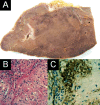
Symptomatic splenoma (hamartoma) of the spleen. A case report
- PMID: 20411063
- PMCID: PMC2843574
Symptomatic splenoma (hamartoma) of the spleen. A case report
Abstract
Hamartomas of the spleen (splenomas) are very rare benign tumors composed of an aberrant mixture of normal splenic elements. Herein we present a unique case of a symptomatic non-palpable splenoma in a 64-year-old female patient presented with anemia and thrombocytopenia and we describe imaging findings in ultrasound, computed tomography and magnetic resonance imaging. To our knowledge, this is the first case of a relatively small splenic hamartoma (35 mm at histopathology) associated with thrombocytopenia and anemia that resolved completely several months after splenectomy.
Keywords: hamartoma; spleen; splenoma.
Figures


References
-
- Silverman ML, Livolsi VA. Splenic Hamartoma. Am J Clin Pathol. 1978;70:224–229. - PubMed
-
- Lam KY, Yip KH, Peh WC. Splenic vascular lesions: unusual features and review of the literature. Aust N J Surg. 1999;69:422–425. - PubMed
-
- Lozzo RY, Haas JE, Chard RL. Symptomatic splenic hamartoma: a report of two cases and review of the literature. Pediatrics. 1980;66:261–265. - PubMed
-
- Darden JW, Teeslink R, Parrish A. Hamartoma of the spleen: a manifestation of tuberous sclerosis. Am Surg. 1975;41:564–566. - PubMed
-
- Huff DS, Lischner HW, Go HC, DeLeon GA. Unusual tumors in two boys with Wiskott-Aldrich-like syndrome. Lab Invest. 1979;40:305–306.
Publication types
LinkOut - more resources
Full Text Sources
