Review
. 2010 Jun;38(3):147-60.
doi: 10.1007/s00240-010-0271-8. Epub 2010 Apr 22.

Three pathways for human kidney stone formation

Affiliations
Review

Three pathways for human kidney stone formation

Fredric L Coe et al. Urol Res. 2010 Jun.

Abstract

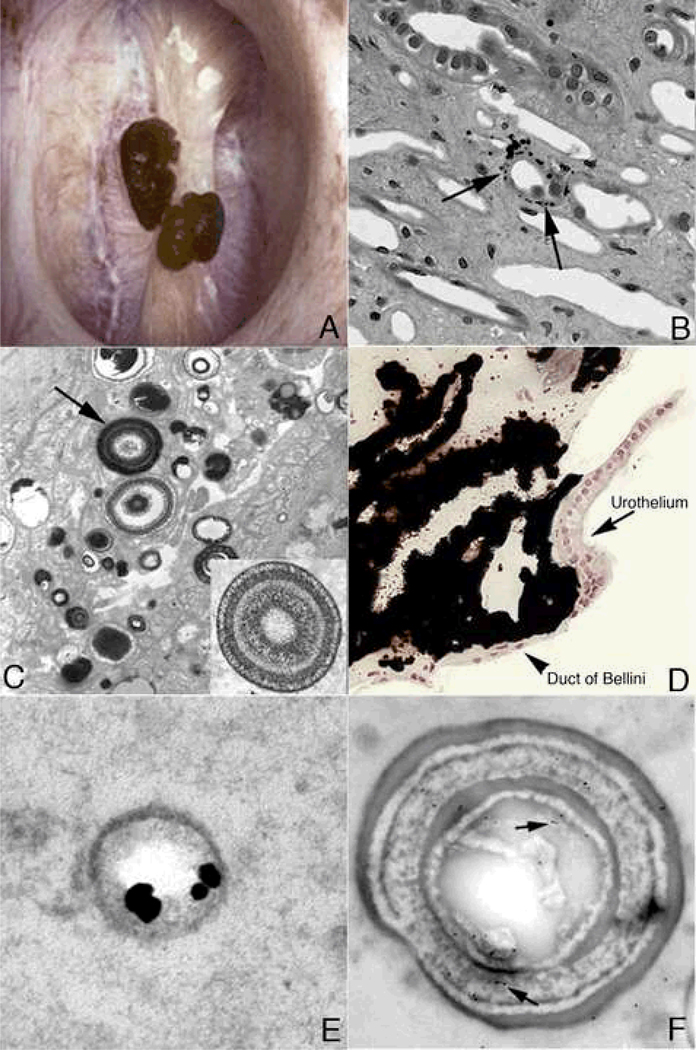
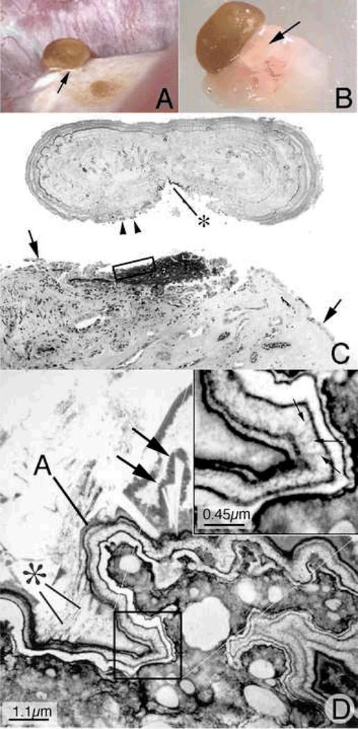
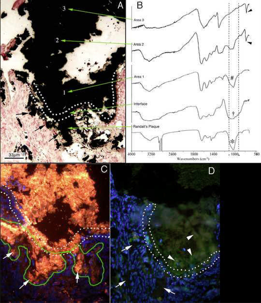
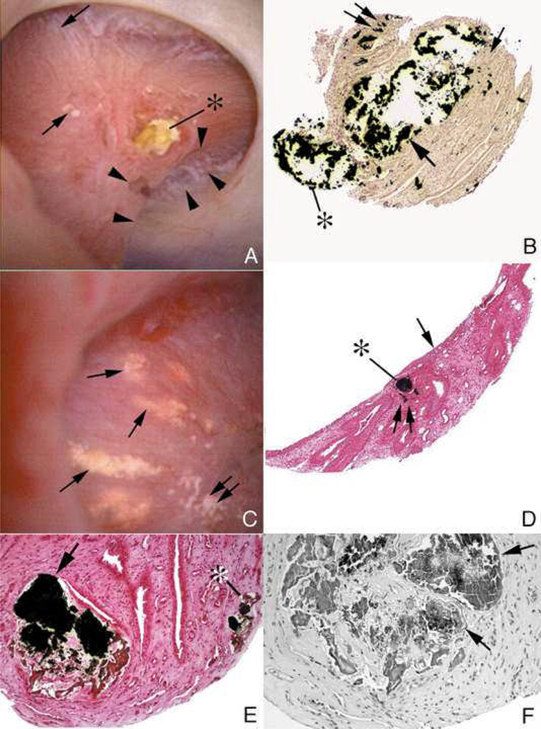
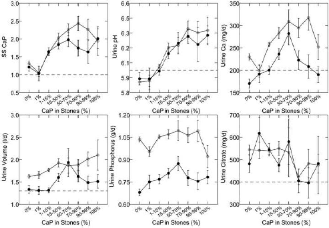
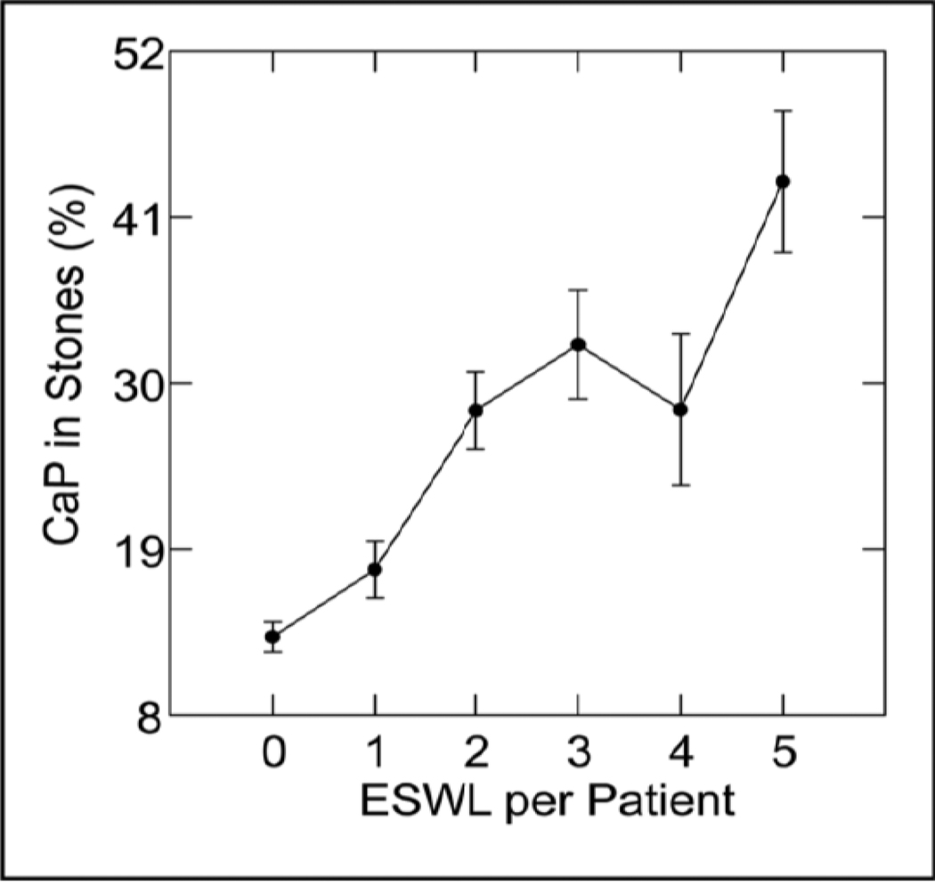
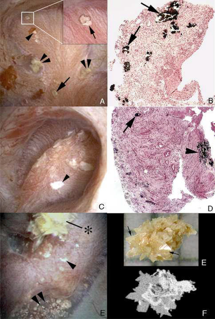
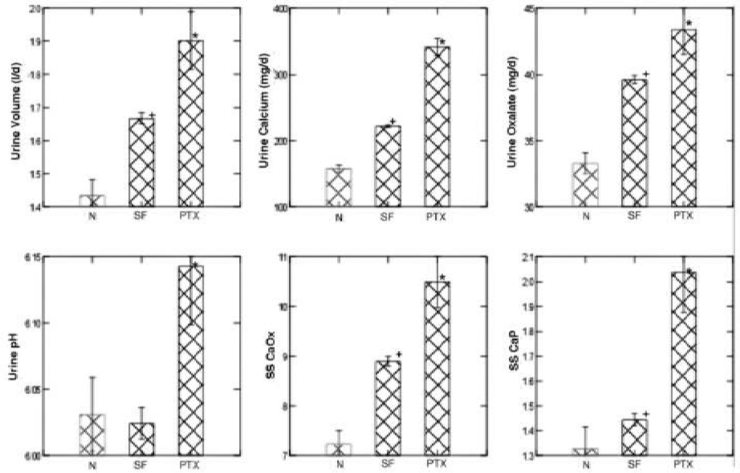
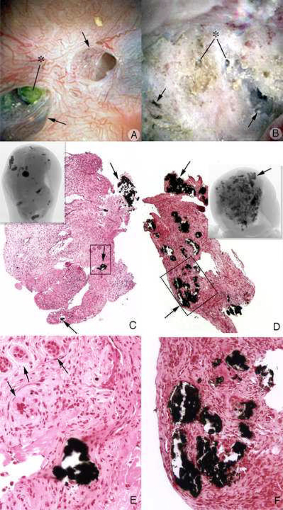
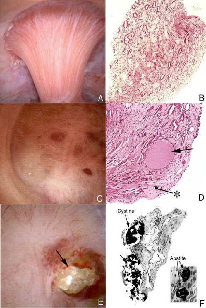
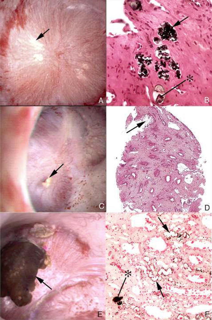
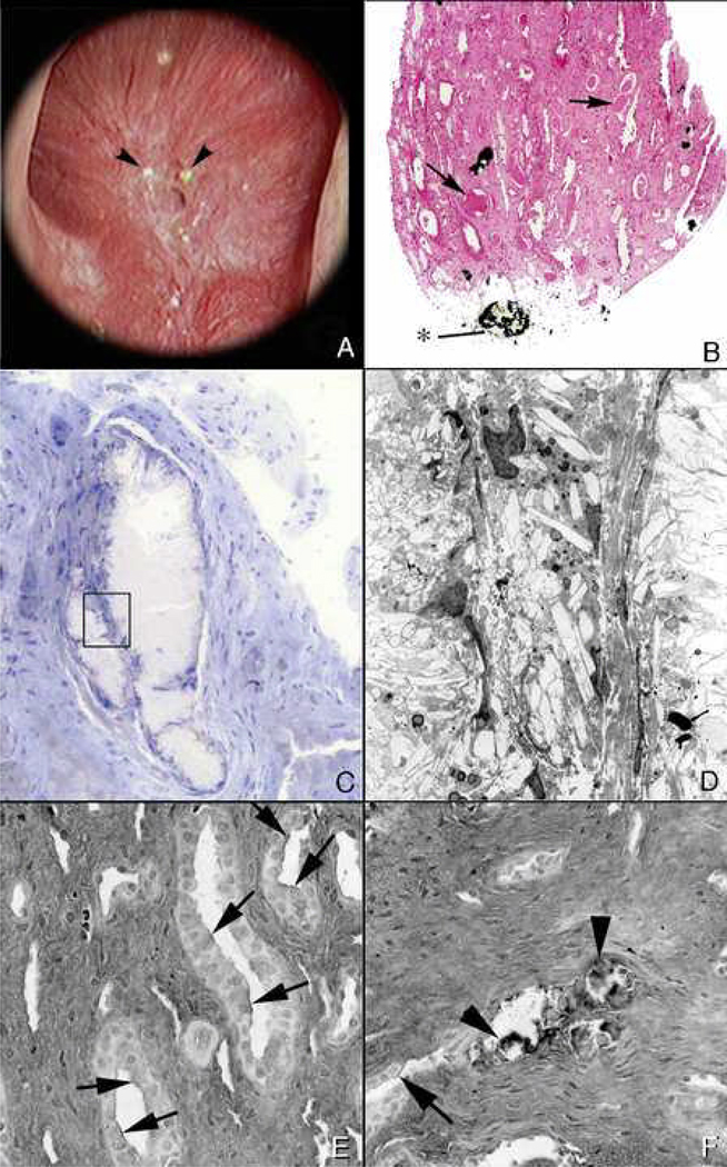
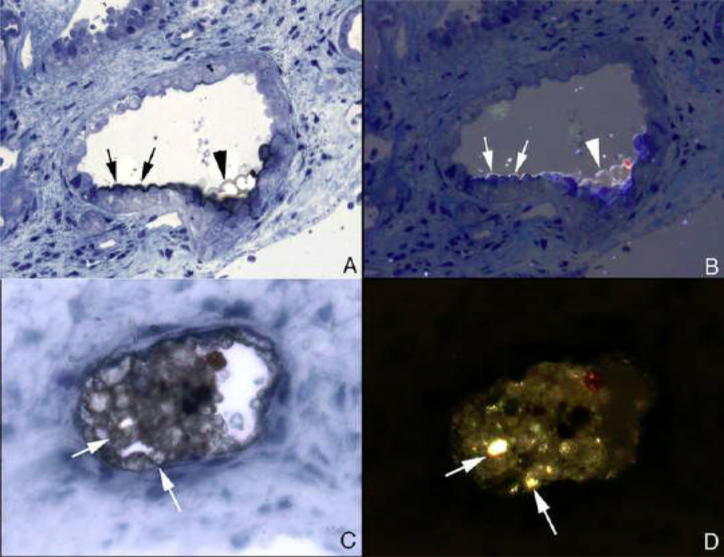
No single theory of pathogenesis can properly account for human kidney stones, they are too various and their formation is too complex for simple understanding. Using human tissue biopsies, intraoperative imaging and such physiology data from ten different stone forming groups, we have identified at least three pathways that lead to stones. The first pathway is overgrowth on interstitial apatite plaque as seen in idiopathic calcium oxalate stone formers, as well as stone formers with primary hyperparathyroidism, ileostomy, and small bowel resection, and in brushite stone formers. In the second pathway, there are crystal deposits in renal tubules that were seen in all stone forming groups except the idiopathic calcium oxalate stone formers. The third pathway is free solution crystallization. Clear examples of this pathway are those patient groups with cystinuria or hyperoxaluria associated with bypass surgery for obesity. Although the final products may be very similar, the ways of creation are so different that in attempting to create animal and cell models of the processes one needs to be careful that the details of the human condition are included.


Figures

Figure 1
Figure 1. Endoscopic and histologic images of Randall’s plaque in ICSF patients
Panel A shows two CaOx stones attached to the papilla tip at sites of Randall’s plaque as seen by endoscopy at the time of stone removal. On biopsy (panel B), tissue sections stained by the Yasue method reveals black, small, spherically shaped deposits (arrows) in the basement membrane of the thin loops of Henle, which appears to be the initiating site of plaque formation. By TEM, these deposits appear as multi-layered spheres with alternating light (crystal) and dark (matrix) bands. The smallest deposits were about 50 nm in diameter. Rather dense regions of interstitial plaque located near the papilla tip were commonly noted in ICSF patient (panel D) and at such sites crystal accumulated beneath the urothelium and around the distal ends of ducts of Bellini. Immunoelectron microscopic studies localized osteopontin immunogold particles (dark dots in panel E) at the crystal-matrix boundary whereas the third heavy chain of the inter-alpha trypsin molecule (arrows in panel F) was detected in the matrix layer.
Figure 2
Figure 2. Histologic examination of attached human kidney stone
During percutaneous nephrolithotomy for stone removal in a ICSF patient, two attached stones were noted on a single papillum (Panel A), a clear region of Randall’s plaque (arrow) is noted at the base of the larger of the two stones. Panel B shows the same stone and the underlying renal tissue with interposing region of Randall’s plaque (arrow) after biopsy removal of this entire complex. The entire biopsy specimen was subsequently decalcified and sectioned first for light microscopy (panel D). During the sectioning process the stone detached from its underpinning so the two images have been approximated in this figure. A large base of interstitial plaque, stained black here by the Yasue method, and framed in part by the rectangle was in continuity with the base of the stone that also reveals a small darkly stained regions (*). This region of plaque is completely devoid of its normal urothelial lining cells. Several attached epithelial cells (arrowheads) were seen at the periphery of the stone while arrows mark the site on the tissue where intact epithelial cells are seen again. The region in Figure 2C marked by the rectangle was prepared and sectioned for transmission electron microscopy (panel D). In this image, the region of interstitial plaque is at the lower right and the stone in the upper left. Between these two regions is a multilayered ribbon structure (labeled A) that forms a sharp boundary. This ribbon is formed by alternating layers of five thin black organic laminia and four, white crystalline laminia (see square and its enlarge in the insert at upper right of panel D). Small arrows in insert show perpendicularly oriented crystals in the white laminia. Various sized crystals (marked by * and large arrows) are seen embedded in the outer matrix layer of the ribbon-like structure.
Figure 3
Figure 3. Mineral analysis of attachment site of human kidney stone
A second stone was removed from another ICSF patient and this stone was processed so that the mineral composition of the stone-plaque interface retained intact so that a precise FTIR analysis could be performed across the interface. The double-doted line in panel A delineates the transition from interstitial plaque to stone. Sites of Randall’s plaque marked by arrows at the lower left side of the panel were confirmed to be composed of hydroxyapatite (lower green arrow in panel B). The region of the interface was composed of amorphous apatite. Region 1 (panel A) was hydroxyapatite while region 2 was a mixture of calcium oxalate and hydroxyapatite. Region 3, a site well within the stone was only calcium oxalate. Immunohistochemistry was used to determine the distribution of osteopontin (panel C) and Tamm-Horsfall protein (panel D) in sites of Randall’s plaque (outlined by a green line) in the tissue, through the interface region (outlined with the double dotted lines) and into the three areas of the stone. Panel C is a serial section to panel A. Note the presence of osteopontin (stained orange) in the interstitial plaque (arrows), interface and all three areas of the stone. Tamm-Horfall protein (stained green) was only found in the stone (arrowheads).
Figure 4
Figure 4. Urine Measurements in idiopathic calcium stone formers ICSF)
Urine molarity of calcium (Upper left and middle panels) among ICSF (Grey solid bars) exceed normals (crosshatch) in the intervals from lunch to supper (L to S), supper to home (S to H) and overnight (ON). Oxalate molarity (upper middle panel), urine pH (lower left panel), and urine volumes (lower middle panel) do not differ between groups. SS CaOx and CaP are higher among ICSF vs. normal at multiple periods (+, p<0.05; #, <0.01; *, p<0.001. Note that normals never exhibit SS values above 1 for CaP whereas values above 1 are common among ICSF.
Figure 5
Figure 5. Relationship between plaque abundance (Y-Axes of each panel) and urine measurements
Plaque area is proportional to urine calcium excretion and inverse to volume and pH (upper panels); multivariate scores using calcium and volume or all three variables (lower panels) account for much of the variation in plaque.
Figure 6
Figure 6. Evidence for reduced tubule calcium reabsorption in ICSF
In a CGRC controlled diet three meal day, serum Ultrafiltrate calcium (upper left panel) rose modestly and equally in ICSF (black circles) and normals (Grey circles) after breakfast (B to L) and remained high after lunch (L) and supper (S), but urine calcium rose (upper right panel) above normal throughout the day and overnight (ON) and tubule calcium reabsorption (lower right panel) fell far more in INSF; filtered loads of calcium (lower left panel) did not change in either group.
Figure 7
Figure 7. Relationship between delivery out of proximal tubule and urine calcium excretion
Distal calcium delivery from proximal tubule estimated by endogenous lithium clearance was higher in ICSF (Black circles) vs. normals (Grey circles) and highest urine calcium excretions corresponded with highest delivery. Each point is a complete study in a single individual.
Figure 8
Figure 8. Endoscopic and histologic observations of brushite stone formers
Brushite stone formers have unique papillary characteristics in that they possess white and yellow plaque as well as protruding plugs from ducts of Bellini. Although the papilla from brushite stone formers have sites of white plaque (arrows in panel A and double arrow in panel C), the most prominent features are mineral plugs protruding from dilated ducts of Bellini (* in panels A and B) and radially oriented sites of yellow plaque (single arrows in panel C) shown to be localized to inner medullary collecting ducts (*, panel D) just beneath the urothelium (arrow, panel D). Many of the papilla of brushite stone formers are deformed as noted by flattening, large dilated opening to ducts of Bellini (Panel A) and large pits (arrowheads in panel A). Histopathology detected grossly dilated inner medullary collecting ducts and ducts of Bellini filled with mineral deposits (arrows in panel B) shown to be apatite. These dilated tubules were surrounded by interstitial fibrosis. Regions of interstitial plaque were easily found (double arrows, panels B and D). Using light microscopy of 1 micron thick plastic sections of decalcified papillary biopsies, extensive regions of cellular damage with mineralization were seen in inner medullary collecting ducts (arrows, panels E and F) and loops of Henle (*, panel E) adjacent to normal appearing tubular segments (panel E). Extensive interstitial fibrosis is noted around these sites of tubular injury.
Figure 9
Figure 9. Urine measurements in ICSF with increasing stone calcium phosphate (CaP) percent
With increasing percent of CaP in analyzed stones (X-axes of all panels) SS CaP and urine pH (upper left and middle panels) rose progressively, and urine calcium excretion (upper right panel) in a less constant manner. Urine volume, and phosphate and citrate excretions (lower panels) showed no consistent relationship to stone CaP.
Figure 10
Figure 10. Stone CaP percent vs. extra-corporeal shock wave lithotripsies (ESWL)
Stone CaP% rose with numbers of ESWL procedures after correcting for sex, age, number of stones, and years of stone disease.
Figure 11
Figure 11. Endoscopic and histologic observations of stone formers with primary hyperparathyroidism
Endoscopic evaluation of papilla from stone formers with primary hyperparathyroidism shows the coexistence of attached stones (panel A, withinwhite box; , panel E) and plugging of ducts of Bellini (panel A, lower single arrow) on the same papilla. In additions, regions of white plaque (arrowhead, panels A, C and E) and yellow plaque double arrowheads, panels A and E) were also seen on the same papilla. Histopathology of the papillary biopsies showed extensive regions of intratubular plugging of inner medullary collecting ducts and ducts of Bellini (arrows, panels B and D) with areas of interstitial plaque (arrowhead, panel D). Extensive interstitial fibrosis surrounded the plugged tubular segments. Panel E shows an attached calcium oxalate stone (*) before while panel E shows it after detachment. Micro-CT analysis of this same stone (panel F) reveals a mixture of apatite (white regions) and calcium oxalate dihydrate (gray regions).
Figure 12
Figure 12. Urine abnormalities in primary hyperparathyroid stone formers (PTX)
Compared to ICSF and normals (SF and N on X axes of all panels) PTX showed higher urine volume, calcium and oxalate excretion, and especially urine pH and SS CaP. This was true even though the main bulk of stone mineral in this series was CaOx. *, differs from N and SF, p<0.01; +, differs from N, p<0.01.
Figure 13
Figure 13. Endoscopic and histologic observations of stone formers with renal tubular acidosis
Stone patients with renal tubular acidosis had normal papilla apart from an occasional dilated duct of Bellini (arrows, panel A) to severely flattened and fibrotic with pitted appearance (arrow, panel B). These papilla possess multiple dilated ducts of Bellini, some with protruding mineral plugs (*, panels A and B). Histopathlogy and micro-CT imagery shows a range of abnormalities. Plugging of inner medullary collecting ducts varied from minimal in number of tubules involved and size of deposits (arrows, panel C) with some interstitial fibrosis (arrows, panel E), to extensive plugging (arrows, panel D), loss of tubular cells and dense cuffs of fibrotic tissue (arrows, panel F).
Figure 14
Figure 14. Endoscopic and histologic observations of stone formers with cystinuria
Papillary morphology varied in these patients with some appearing normal (panel A) to distorted with flattening and greatly dilated opening of ducts of Bellini (panel C). Protruding plugs of cystine were noted in some dilated ducts (arrow, panel E). Histopathology confirmed the observations seen by endoscopy, in that, tissues form papillary biopsies appeared normal (panel B) to abnormal characterized by extensive inner medullary plugging with (panel F) and without mineral deposits (panel D). An occasional mineral plug was noted in loops of Henle (*, panel D). Intraluminal plugs of the ducts of Bellini were primarily cystine in nature while deposits in inner medullary and loops of Henle were always apatite (panel F).
Figure 15
Figure 15. Endoscopic and histologic observations of ileostomy patients with stones
Papillary morphology as observed by endoscopy varied from a normal conical shape (panel A) to flattened and retracted (panel C). Papilla show areas of white plaque (arrow, panel A), plugs in opening of ducts of Bellini, (arrow, panel C) and attached stones (arrow, panel E). Histopathology revealed intraluminal plugs of apatite in dilated inner medullary collecting ducts (arrows, panels B and F), those with a mixture of apatite and sodium acid urate/ammonium acid urate (*, panel B) while others had only sodium acid urate/ammonium acid urate (arrow, panel D). Some biopsy sections contained large amounts of interstitial plaque (arrows, panel F).
Figure 16
Figure 16. Endoscopic and histologic observations of stone patients with bypass surgery for obesity
Endoscopic observations revealed normal appearing papilla except for an occasional dilated opening of a duct of Bellini with adjacent nodular-appearing structures (arrowheads, panel A). Histopathogy shows an occasional intraluminal deposit in an inner medullary collecting duct and plugs in the ducts of Bellini (*, panel B). A few dilated inner medullary collecting ducts possessed cast material and regions of fibrosis (arrows, panel B). At a higher magnification (panel C), a site of intraluminal plugging reveals a loss of tubular lining cells and cuffs of interstitial fibrosis. A region within panel C (see rectangle) is further magnified in panel D with transmission electron microscopy. There is a complete loss of cellular detail. Biopsy sections from stone patients with bypass surgery for obesity were also stained for hyaluronan to detect sites of cellular injury. A number of inner medullary collecting ducts with (arrowheads, panel F) and without mineral deposits showed focal regions of hyaluronan staining of their apical cell membranes (arrows, panels E and F).
Figure 17
Figure 17. Birefringent and nonbirefringent crystals in inner medullary collecting ducts of stone patients with bypass surgery for obesity
Panels A and B show serial sections of a inner medullary collecting duct stained by the Yasue method and viewed direct (panel A) and with polarized light (panel B). The inner medullary collecting duct cells seen in panel A are coated with a thin black layer (arrows) that represent areas of calcium deposits. At the arrowhead is a thin region through apical cell surfaces in which the black overlay on the cells is particularly clear. By polarizing light (panel B) the overlay is birefringent suggesting the mineral is calcium oxalate while the rest of the nonbirefringent material is apatite. Panels C and D show another inner medullary collecting duct seen in serial sections by direct (panel C) and polarized light (panel D). The intraluminal plug that partial fills the tubular lumen was stained by Yasue and shows small sites of Yasue negative staining (arrows, panel C) which are birefringent with polarizing optics (arrows, panel D) suggesting the presences of two minerals as seen in panels A and B.

References

    1. Evan A, Lingeman J, Coe FL, Worcester E. Randall's plaque: pathogenesis and role in calcium oxalate nephrolithiasis. Kidney Int. 2006;69:1313–1318. - PubMed
    1. Matlaga BR, Williams JC, Jr, Kim SC, Kuo RL, Evan AP, Bledsoe SB, Coe FL, Worcester EM, Munch LC, Lingeman JE. Endoscopic evidence of calculus attachment to Randall's plaque. J Urol. 2006;175:1720–1724. - PubMed
    1. Randall R. Papillary pathology as precursor of primary renal calculus. Journal of Urology. 1940;44:580–589.
    1. Evan AP, Lingeman JE, Coe FL, Parks JH, Bledsoe SB, Shao Y, Sommer AJ, Paterson RF, Kuo RL, Grynpas M. Randall's plaque of patients with nephrolithiasis begins in basement membranes of thin loops of Henle. J Clin.Invest. 2003;111:607–616. - PMC - PubMed
    1. Evan AP, Coe FL, Rittling SR, Bledsoe SM, Shao Y, Lingeman JE, Worcester EM. Apatite plaque particles in inner medulla of kidneys of calcium oxalate stone formers: osteopontin localization. Kidney Int. 2005;68:145–154. - PubMed