. 2010 May 25;49(20):4327-38.
doi: 10.1021/bi901945w.

Heme binding to the Mammalian circadian clock protein period 2 is nonspecific

Affiliations

Heme binding to the Mammalian circadian clock protein period 2 is nonspecific

Michael V Airola et al. Biochemistry. .

Abstract

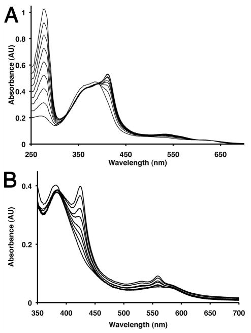
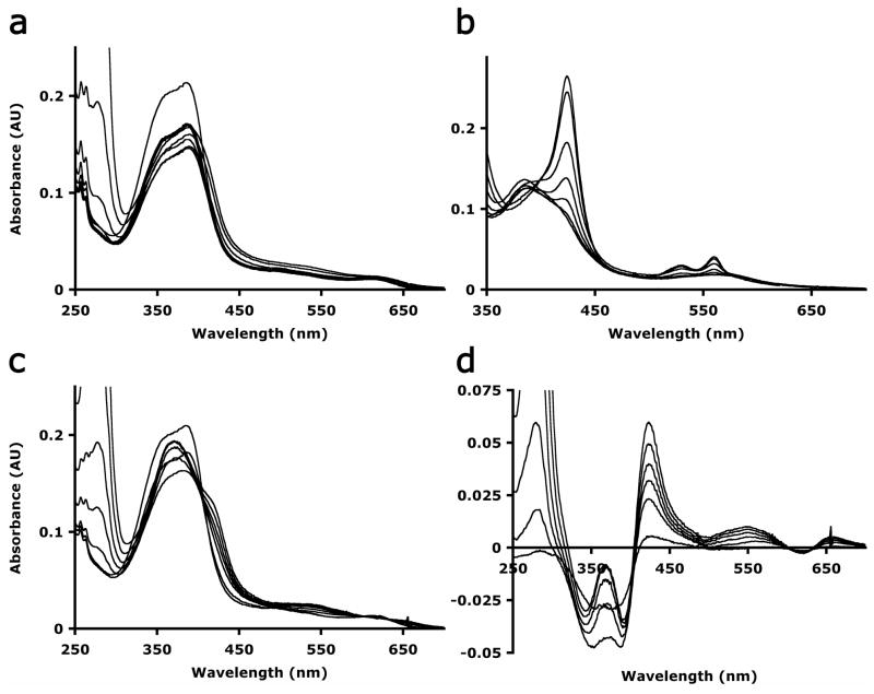
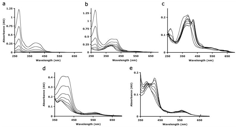
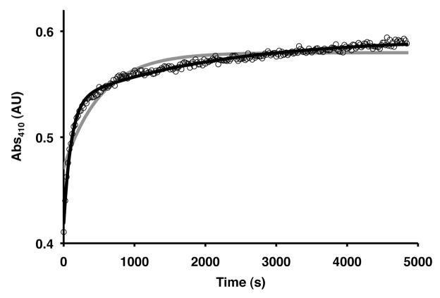
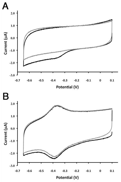
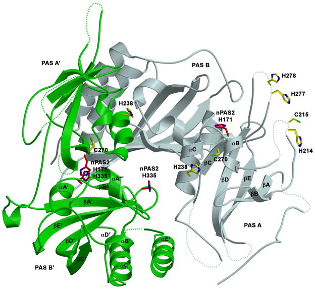
The mammalian circadian clock synchronizes physical and metabolic activity with the diurnal cycle through a transcriptional-posttranslational feedback loop. An additional feedback mechanism regulating clock timing has been proposed to involve oscillation in heme availability. Period 2 (PER2), an integral component in the negative feedback loop that establishes circadian rhythms in mammals, has been identified as a heme-binding protein. However, the majority of evidence for heme binding is based upon in vitro heme-binding assays. We sought to ascertain if these largely spectral assays could distinguish between specific and nonspecific heme interactions. Heme-binding properties by a number of other well-characterized proteins, all with no known biological role involving heme interaction, corresponded to those displayed by PER2. Site-directed mutants of putative heme-binding residues identified by MCD were unable to locate a specific heme-binding site on PER2. Protein film electrochemistry also indicates that heme binds PER2 nonspecifically on the protein surface. Our results establish the inability of qualitative in vitro assays to easily distinguish between specific and nonspecific heme binding. We conclude that heme binding to PER2 is likely to be nonspecific and does not involve the hydrophobic pocket within the PER2 PAS domains that in other PAS proteins commonly recognizes cofactors. These findings also question the significance of in vivo studies that implicate heme interactions with the clock proteins PER2 and nPAS2 in biological function.


Figures

FIGURE 1
FIGURE 1
PER2 binds heme in a manner dependent on redox state. (A) UV-Visible absorption spectra of PER2 (1-506) reconstituted with heme (5 μM, pH 7.5) before (-----) and after (- - -) treatment with dithionite. (B) UV-Visible absorption spectra of PAS A [PER2 (1-320)] reconstituted with heme (4 μM, pH 7.5) before and after treatment with dithionite. (C) Titration of ferric heme (10 μM) with PER2 (1-506) (1.5, 3, 5, 6.5, 8, 9.5, 11 μM). (D) Titration of ferrous heme (10 μM) with PER2 (1-506) (2.1, 4.2, 6.3, 8.4, 10.5, 12.6, 14.7, 16.8, 19.0, 21.1, 23.2, 25.3, 27.4, 29.5, 31.6, 33.7, 35.8, 37.9, 40.0, 43.9, 47.8, 59.5 μM).
FIGURE 2
FIGURE 2
Heme Binding Does Not Effect the Oligomeric State of PER2. The elution profiles of PER2 (1-506) with heme bound (black) and without heme bound (grey) are identical and indicate PER2 (1-506) forms a dimer with an apparent MW = 150 kDa (actual MW = 56 kDa).
FIGURE 3
FIGURE 3
Magnetic circular dichroism spectra of ferric PAS A, ferrous PAS A and ferrous-CO PAS A reveal ferric and ferrous hemes are bound through His-Cys and bis-His ligation respectively. A) MCD spectra of ferric PAS A (red) and ferric cytochrome P450-Camphor with imidazole bound (blue). B) MCD spectra of ferrous PAS A (red) and ferrous H93G horse heart myoglobin with bis-imidazole ligation (black). C) MCD spectra of ferrous-CO PAS A (red) and ferrous-CO wild type horse heart myoglobin (blue). All spectra are reported in ε(mM−1cm−1) units.
FIGURE 4
FIGURE 4
Secondary Structure Alignment of the PAS domains of mPER2 (PAS A: 186-319; PAS B: 325-435), dPER (PAS A: 235-377; PAS B: 385-498), nPAS2 (PAS A: 90-237; PAS B: 245-353), bjFixL (149-257), rmFixL (143-251), and ecDOS (18-126). Secondary structure information is derived from mPER2 (3GDI), dPER (1WA9), bjFixL (1DP9), rmFixL (1EW0), ecDOS (1VB6). Jpred3 (47) was used to predict the secondary structure of nPAS2. Alpha helices (red) and beta strands (blue) are colored accordingly. All cysteine and histidine residues in mPER2 are highlighted. All residues implicated in heme binding are highlighted and denoted with black triangles below. Alignments were generated with ClustalW and manually adjusted as presented.
FIGURE 5
FIGURE 5
Cysteine Modification Alters the Ferric But Not the Ferrous-Heme PER2 Spectra. (A) Titration of ferric heme (8.3 μM) with PER2 (129-506) modified with iodoacetamide (0, 6, 10, 14, 18, 22, 26, 30, 34 μM). Cysteine modification alters the position of the Soret peak shifting it to 410 nm compared to unmodified PER2 with a Soret peak at 422 nm. (B) Titration of ferrous heme (8.3 μM) with PER2 (129-506) modified with iodoacetamide (0, 4.1, 8.2, 12.4, 24.7, 37, 58, 78 μM) with a Soret peak growing in at 426 nm identical to unmodified PER2.
FIGURE 6
FIGURE 6
Heme binds to the chemotaxis kinase CheA (A) Titration of ferric heme (4 μM) with CheA (0, 0.23, 0.45, 1.1, 3.5, 11.6, 35 μM). (B) Titration of ferrous heme (4 μM) with CheA (0, 0.45, 2.3, 5.8, 11.6, 23, 46.5 μM). (C) Titration of ferric heme (4 μM) with CheA D371C variant (0, 0.5, 2.5, 6.4, 12.8, 25, 45 μM). (D) Difference spectra from panel C.
FIGURE 7
FIGURE 7
Addition of YtvA induces spectral changes to ferric and ferrous heme. (A) UV-Visible absorption spectra of YtvA (0, 1.2, 3, 6, 12, 24, & 42 μM) after photo bleaching. (B) Titration of ferric heme (4 μM) with YtvA (0, 1.2, 3, 6, 12, 24, & 42 μM). (C) Absorption spectra from titration of ferric heme with YtvA after subtraction of FMN absorbance. (D) Titration of ferrous heme (4 μM) with YtvA (0, 1.2, 3, 6, 12, 24, & 42 μM). (E) UV-Visible absorption spectra from titration of ferrous heme with YtvA after subtraction of FMN absorption.
FIGURE 8
FIGURE 8
Heme Dissociation from PER2 Occurs From Multiple Sites. Heme transfer from PER2 (1-320) to H64Y/V68F apomyoglobin was measured as an increase in the Absorbance at 410 nm in 0.2 M Na phosphate, pH 7.0 and 0.45 Sucrose at 25 °C. Rate constants were calculated by analyzing the data in terms of both single (grey) and bi-exponential processes (black) (Table 2). The data for PER2 and other proteins was best represented by a bi-exponential process indicating multiple binding sites with different affinities.
FIGURE 9
FIGURE 9
Electrochemistry of PER2-Heme Complex. A) Cyclic voltammogram of PER2-heme complex immobilized on glassy carbon electrode. B) Cyclic voltammogram of heme immobilized on glassy carbon electrode under the same conditions as (A). Black and grey traces denote the first and second potential sweeps. All scan rates were 100 mV/s.
FIGURE 10
FIGURE 10
mPER2 structure with location of possible heme binding residues. Ribbon representation of mPER2 PAS A/B dimer (3GDI): molecule 1 (PAS A and PAS B), molecule 2 (PAS A′ and PAS B′). Side chains are shown for residues H214, C215, H238, C270, H277, and H278 in mPER2 (yellow) and C170, H171, H335 in nPAS2 (red). H119 of nPAS2 is not shown but corresponds to C215 of PER2. Residues with missing density are depicted as a dotted line and were manually drawn. The secondary structure elements of the PAS fold are labeled for PAS A and PAS B′.

Similar articles

Cited by

References

    1. Reppert SM, Weaver DR. Coordination of circadian timing in mammals. Nature. 2002;418:935–941. - PubMed
    1. Harms E, Kivimae S, Young MW, Saez L. Posttranscriptional and posttranslational regulation of clock genes. Journal of Biological Rhythms. 2004;19:361–373. - PubMed
    1. Lowrey PL, Takahashi JS. Mammalian circadian biology: Elucidating genome-wide levels of temporal organization. Annual Review of Genomics and Human Genetics. 2004;5:407–441. - PMC - PubMed
    1. Young MW, Kay SA. Time zones: A comparative genetics of circadian clocks. Nature Reviews Genetics. 2001;2:702–715. - PubMed
    1. Hennig S, Strauss HM, Vanselow K, Yildiz O, Schulze S, Arens J, Kramer A, Wolf E. Structural and Functional Analyses of PAS Domain Interactions of the Clock Proteins Drosophila PERIOD and Mouse PERIOD2. Plos Biology. 2009;7:836–853. - PMC - PubMed

Publication types

MeSH terms