. 2010 Jul;101(7):1624-31.
doi: 10.1111/j.1349-7006.2010.01570.x. Epub 2010 Mar 19.

Subcellular distribution and expression of prenylated Rab acceptor 1 domain family, member 2 (PRAF2) in malignant glioma: Influence on cell survival and migration

Affiliations

Subcellular distribution and expression of prenylated Rab acceptor 1 domain family, member 2 (PRAF2) in malignant glioma: Influence on cell survival and migration

Tamás Borsics et al. Cancer Sci. 2010 Jul.

Abstract

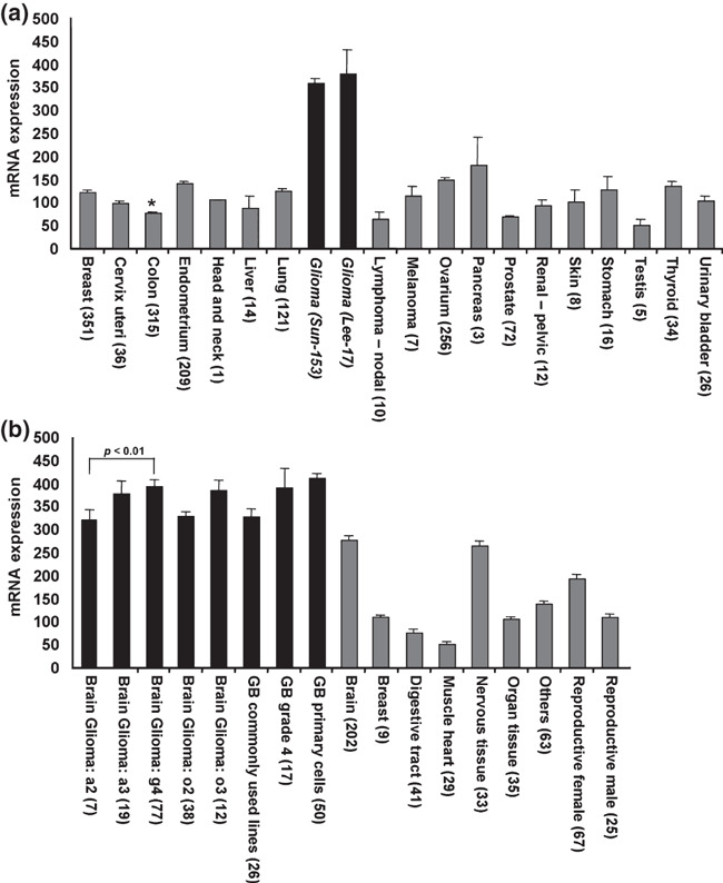
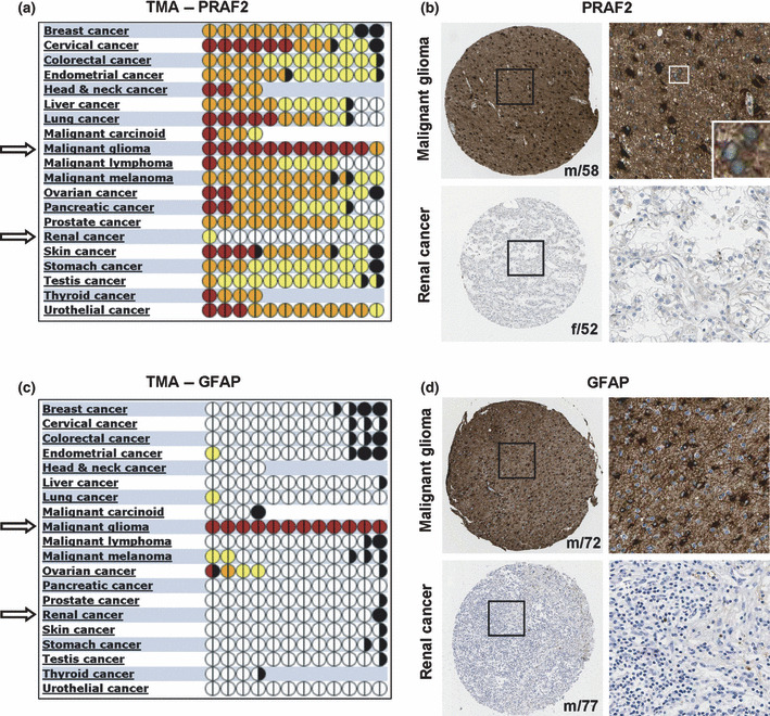
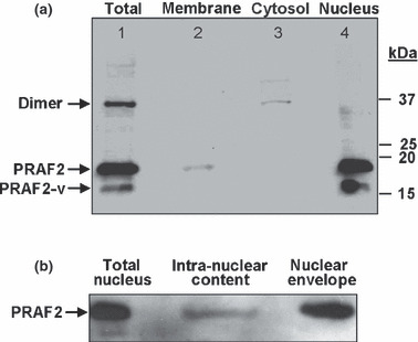
Our previous studies revealed that the expression of the 19-kDa protein prenylated Rab acceptor 1 domain family, member 2 (PRAF2) is elevated in cancer tissues of the breast, colon, lung, and ovary, when compared to noncancerous tissues of paired samples. PRAF2 mRNA expression also correlated with several genetic and clinical features and is a candidate prognostic marker in the pediatric cancer neuroblastoma. The PRAF2-related proteins, PRAF1 and PRAF3, play multiple roles in cellular processes, including endo/exocytic vesicle trafficking and glutamate uptake. PRAF2 shares a high sequence homology with these family members, but its function remains unknown. In this study, we examined PRAF2 mRNA and protein expression in 20 different human cancer types using Affymetrix microarray and human tissue microarray (TMA) analyses, respectively. In addition, we investigated the subcellular distribution of PRAF2 by immunofluorescence microscopy and cell fractionation studies. PRAF2 mRNA and protein expression was elevated in several cancer tissues with highest levels in malignant glioma. At the molecular level, we detected native PRAF2 in small, vesicle-like structures throughout the cytoplasm as well as in and around cell nuclei of U-87 malignant glioma cells. We further found that monomeric and dimeric forms of PRAF2 are associated with different cell compartments, suggesting possible functional differences. Importantly, PRAF2 down-regulation by RNA interference significantly reduced the cell viability, migration, and invasiveness of U-87 cells. This study shows that PRAF2 expression is elevated in various tumors with exceptionally high expression in malignant gliomas, and PRAF2 therefore presents a candidate molecular target for therapeutic intervention.


Figures

Figure 1
Figure 1
Prenylated Rab acceptor 1 domain family, member 2 (PRAF2) mRNA expression in malignant and healthy tissues. (a) PRAF2 mRNA level in a variety of human tumors. The expression of PRAF2 in two glioma tumor sets (black bars) was higher than in any other tumor tissue (gray bars). For comparison: average expressions of GAPDH and β‐actin in this dataset were 9240 and 11 890, respectively. *Expression data for other colon cancer sets are: 68.73 ± 6.63 for rectosigmoid tumors (n = 9) and 92.01 ± 10.25 (n = 38) for rectal tumors. Tumor sets are described in the Materials and Methods. (b) PRAF2 mRNA level is significantly higher in glioma (black bars) than in healthy tissues (gray bars) including the brain. Statistically significant differences in mRNA expression are shown above the bars. T‐tests were calculated in Excel. Datasets used: brain glioma Sun‐153, glioblastoma (GB) Lee‐101, healthy tissues Roth‐504. Error bars represent the SEM, numbers in parentheses refer to sample size.
Figure 2
Figure 2
Expression of prenylated Rab acceptor 1 domain family, member 2 (PRAF2) protein in tumor tissues. (a,c) Quantitative analysis of 220 tissue microarray (TMA) samples of various cancer types stained with PRAF2 antibody or astrocyte‐marker protein GFAP‐specific antibody. Each full circle represents one patient, and halves refer to staining performed in duplicates; color codes determine the strength/intensity of the immunohistochemical staining signal: white, no staining; yellow, light; orange, moderate; purple, strong; black, not evaluated. Tissue samples of malignant gliomas show extensive PRAF2 expression compared to other cancers. (b,d) Representative histological images of malignant glioma and renal cancer tissue sections stained with (b) anti‐PRAF2 or (d) anti‐GFAP antibody. In each case, a total of 12 tumor tissue samples were stained in duplicates (n = 12) and counterstained with hematoxylin. Patients’ gender and age are displayed in the lower right corner of each sample. f, female; m, male. Inset represents a higher magnification of PRAF2 perinuclear staining in malignant glioma.
Figure 3
Figure 3
Immunohistochemical staining of normal cerebral cortex tissue samples with antibodies directed against proteins prenylated Rab acceptor 1 domain family, member 2 (PRAF2), neuronal marker neurexin 2α, and glial marker glial fibrillary acidic protein (GFAP). (a) Cerebral cortex tissue stained with PRAF2 shows weak PRAF2 presence in glial cells (G) and high expression in neurons (N). (b) Neuronal marker protein neurexin 2α localized to pyramidal neurons in normal brain sections and showed a similar staining pattern as observed for PRAF2. (c) Distribution of the glial marker GFAP in the cerebral cortex. GFAP is strongly expressed in glial cells and is absent in neurons. Each image is representative of three independent experiments.
Figure 4
Figure 4
Subcellular localization of prenylated Rab acceptor 1 domain family, member 2 (PRAF2) in U‐251 malignant glioma cells. (a–d) PRAF2 (green) is expressed in punctuate structures throughout the cytoplasm and also in/around the nucleus (blue). The endoplasmic reticulum (ER) (yellow) and microtubules (red) are shown to further visualize the subcellular localization of PRAF2. (e) Superimposition by overlay of images (a b, and d). (f,g) Power images of (e), showing individual U‐251 cells. The images are representative of two independent experiments, and each experiment was performed in duplicate.
Figure 5
Figure 5
Subcellular distribution of native prenylated Rab acceptor 1 domain family, member 2 (PRAF2) in U‐87 malignant glioma cells. (a) Samples were prepared by gravity‐based fractional centrifugation of homogenized cells as described in the Materials and Methods. “Total” represents all cellular proteins prior to fractionation. While monomeric PRAF2 (19.3 kDa) and the alternatively spliced monomeric variant PRAF2‐v (16.7 kDa) are associated with membranous compartments, dimeric PRAF2 (37 kDa) is mainly detected in the membrane‐free cytosolic fraction. The specificity of all bands, including PRAF2‐v was confirmed by probing the stripped membrane with peptide‐blocked PRAF2 antibody (not shown). Molecular weights in kilodalton (kDa) are indicated. Lane 1, total cell lysate; lane 2, membranes; lane 3, cytosol; lane 4, nucleus. Data are representative of three independent experiments (n = 3). (b) Nuclear distribution of PRAF2 in U‐87 cells. Isolated nuclei were lysed, separated into intra‐nuclear and nuclear membrane fractions (envelope) and probed for PRAF2 by western blot analysis (n = 2).
Figure 6
Figure 6
Functional analysis of prenylated Rab acceptor 1 domain family, member 2 (PRAF2) knockdown in glioblastoma cells. (a) Stable transfection of U‐87 cells with shPRAF2 significantly reduced PRAF2 protein expression compared to controls (wild type and scrambled). Tubulin served as a loading control. (b) The cell viability was determined at 24, 48, and 72 h in triplicate wells in two separate experiments with similar results. (c,d) PRAF2 down‐regulation inhibits cell migration (c) and invasion (d). Five × 104 cells were seeded in the top chamber of Transwell plates. Serum‐induced migration/invasion was measured after 24 h. PRAF2 down‐regulation appeared to inhibit glioblastoma migration and invasion, compared to wild‐type and scrambled control glioblastoma cells. Three independent experiments were performed in triplicates and data are represented as mean ± SD (n = 9). WT, wild type.

References

    1. Stupp R, Mason WP, Van den Bent MJ et al. Radiotherapy plus concomitant and adjuvant temozolomide for glioblastoma. N Engl J Med 2005; 352: 987–96. - PubMed
    1. Bucci C, Chiariello M, Lattero D, Maiorano M, Bruni CB. Interaction cloning and characterization of the cDNA encoding the human prenylated rab acceptor (PRA1). Biochem Biophys Res Commun 1999; 258: 657–62. - PubMed
    1. Maier S, Reiterer V, Ruggiero AM et al. GTRAP3‐18 serves as a negative regulator of Rab1 in protein transport and neuronal differentiation. J Cell Mol Med 2009; 13: 114–24. - PMC - PubMed
    1. Sivars U, Aivazian D, Pfeffer SR. Yip3 catalyses the dissociation of endosomal Rab‐GDI complexes. Nature 2003; 425: 856–9. - PubMed
    1. Compton SL, Kemppainen RJ, Behrend EN. Prenylated Rab acceptor domain family member 1 is involved in stimulated ACTH secretion and inhibition. Cell Signal 2009; 21: 1901–9. - PubMed

Publication types

MeSH terms