. 2010 Apr 22:10:156.
doi: 10.1186/1471-2407-10-156.

Spontaneous feline mammary intraepithelial lesions as a model for human estrogen receptor- and progesterone receptor-negative breast lesions

Affiliations

Spontaneous feline mammary intraepithelial lesions as a model for human estrogen receptor- and progesterone receptor-negative breast lesions

Giovanni P Burrai et al. BMC Cancer. .

Abstract

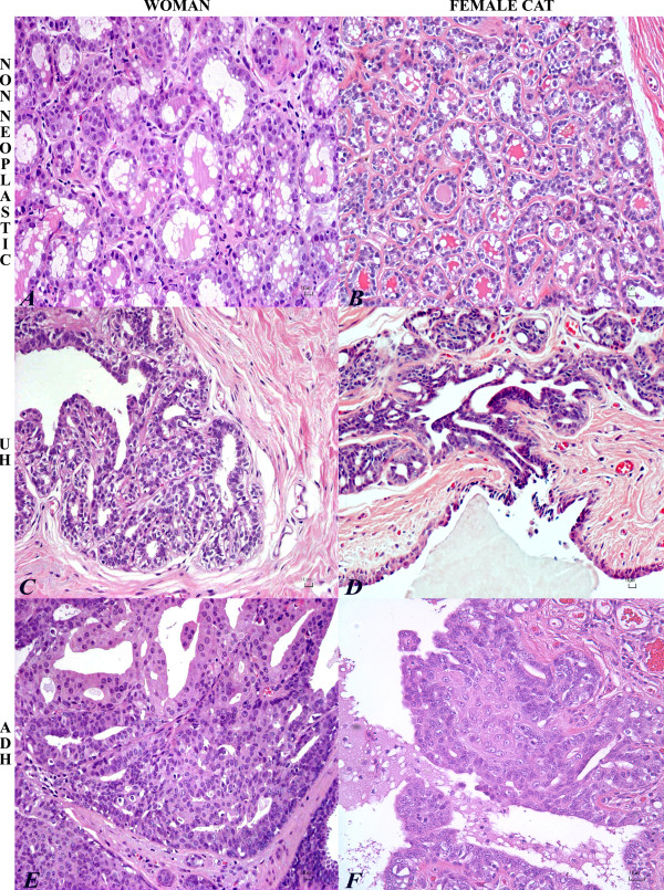
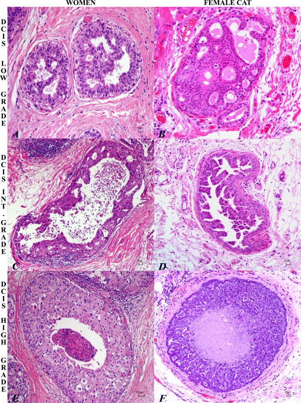
Background: Breast cancer is the most frequently diagnosed cancer in women. Intraepithelial lesions (IELs), such as usual ductal hyperplasia (UH), atypical ductal hyperplasia (ADH), and ductal carcinoma in situ (DCIS) are risk factors that predict a woman's chance of developing invasive breast cancer. Therefore, a comparative study that establishes an animal model of pre-invasive lesions is needed for the development of preventative measures and effective treatment for both mammary IELs and tumors. The purpose of this study was to characterize the histologic and molecular features of feline mammary IELs and compare them with those in women.

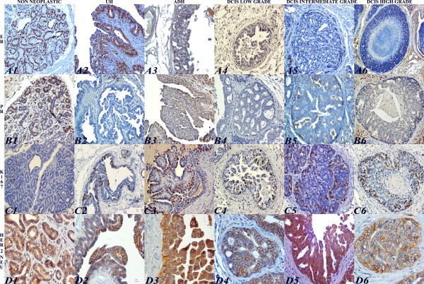
Methods: Formalin-fixed, paraffin-embedded specimens (n = 205) from 203 female cats with clinical mammary disease were retrieved from the archives of the Purdue University Animal Disease Diagnostic Laboratory and Veterinary Teaching Hospital (West Lafayette, IN), and the Department of Pathology and Veterinary Clinic, School of Veterinary Medicine (Sassari, Italy). Histologic sections, stained with hematoxylin and eosin (HE), were evaluated for the presence of IELs in tissue adjacent to excised mammary tumors. Lesions were compared to those of humans. Immunohistochemistry for estrogen receptor (ER-alpha), progesterone receptor (PR), human epidermal growth factor receptor 2 (HER-2/neu) and Ki-67 was performed in IELs and adjacent tumor tissues.

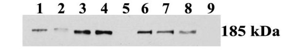
Results: Intraepithelial lesions were found in 57 of 203 (28%) feline mammary specimens and were categorized as UH (27%), ADH (29%), and DCIS (44%). Most IELs with atypia (ADH and DCIS) were associated with mammary cancer (91%), whereas UH was associated with benign lesions in 53% of cases. Feline IELs were remarkably similar to human IELs. No ER or PR immunoreactivity was detected in intermediate-grade or high-grade DCIS or their associated malignant tumors. HER-2 protein overexpression was found in 27% of IELs.

Conclusion: The remarkable similarity of feline mammary IELs to those of humans, with the tendency to lose hormone receptor expression in atypical IELs, supports the cat as a possible model to study ER- and PR-negative breast lesions.


Figures

Figure 1
Figure 1
Histopathology of normal mammary gland, UH, ADH in woman and cat. Normal secretory mammary gland in woman (A) and cat (B): the epithelial cells are vacuolated with cytoplasmic accumulation of fat droplets. HE. Bar = 10 μm. Usual ductal hyperplasia (UH) in woman (C) and cat (D) with typical fenestrated growth pattern. The ducts are lined by monotonous luminal epithelial cells that are cuboidal to columnar, with hyperchromatic nuclei and rarely prominent nucleoli. Few myoepithelial cells are admixed in the hyperplastic epithelium. HE. Bar = 10 μm. Atypical ductal hyperplasia (ADH) in woman (E) and cat (F) with micropapillary projection of disorganized epithelial cells and spindle-shaped (myoepithelial) cells. Epithelial cells are enlarged with round nuclei with coarse chromatin and prominent single nucleoli. HE. Bar = 10 μm
Figure 2
Figure 2
Histopathology of low, intermediate, high-grade DCIS in woman and cat. Low-grade ductal carcinoma in situ (DCIS) in woman (A) and cat (B) with proliferation of monomorphous, small and relatively uniform epithelial cells with moderate and eosinophilic cytoplasm, oval nuclei, arranged to form regular cribriform spaces. HE. Bar = 10 μm. Intermediate-grade ductal carcinoma in situ (DCIS) in woman (C) and cat (D) with proliferation of pleomorphic cuboidal cells with moderate eosinophilic cytoplasm, oval to elongate nuclei, and single prominent nucleoli; micropapillary pattern. Central secretion, necrotic cell debris and periductal inflammatory reaction is depicted in the human intermediate-grade DCIS. HE. Bar = 10 μm. High-grade comedo ductal carcinoma in situ in woman (E) and cat (F) with solid proliferation of epithelial cells in a distended duct with central necrosis. Highly pleomorphic cuboidal to oval cells with abundant eosinophilic granular cytoplasm, round vescicular nuclei and prominent nucleoli. Lymphocytes and plasma cells infiltrate the periductal stroma. Bar = 10 μm.
Figure 3
Figure 3
Immunohistochemical evaluation of ER-α, PR, Ki-67, HER-2/neu in feline normal mammary gland, UH, ADH and DCIS. Strong and diffuse nuclear expression of ER-α in non neoplastic feline mammary gland (A1) and in UH (A2). ADH with patchy ER-α expression in epithelial cells (A3). Low (A4) intermediate (A5) and high grade (A6) DCIS with no ER-α expression. Immunoperoxidase-DAB. Bar = 10 μm. Strong and multifocal nuclear immunoreactivity of PR in non neoplastic feline mammary gland (B1). Lack of PR immunoreactivity in UH (B2), ADH (B3), low (B4), intermediate (B5), and high grade (B6) DCIS. Immunoperoxidase-DAB. Bar = 10 μm. Few and strong Ki-67 nuclear immunoreactivity in non neoplastic feline mammary gland (C1). UH displaying strong and scattered Ki-67 immunolabeling (C2). ADH with widespread and strong Ki-67 expression (C3). DCIS low (C4), intermediate (C5), and high grade (C6) with strong and multifocal Ki-67 positive cells. Immunoperoxidase-DAB. Bar = 10 μm. Non neoplastic feline mammary gland with strong complete cell-membrane staining for HER-2/neu. IHC score: 3+ (D1). UH displaying moderate complete membrane HER-2/neu expression in few cells. IHC score: 2+ (D2). ADH showing strong complete membrane HER-2/neu expression. IHC score: 3+ (D3). Low-grade DCIS depicting strong complete membrane for HER-2/neu expression. IHC score: 3+ (D4). Intermediate-grade DCIS showing moderate complete membrane HER-2/neu immunoreactivity. IHC score: 2+ (D5). High-grade DCIS showing strong and complete membrane HER-2/neu staining. IHC score: 3+ (D6). Immunoperoxidase-DAB. Bar = 10 μm
Figure 4
Figure 4
Reactivity observed by western immunoblot with the Dako Cytomation anti-HER-2/neu antibody in 4 feline mammary tumors. Lanes 1-4: Feline mammary tumors; Lanes 5-8: non-neoplastic feline mammary tissues; Lane 9: feline liver. The molecular weight of the reactive protein band is indicated on the right.

References

    1. Jemal A, Siegel R, Ward E, Hao Y, Xu J, Thun MJ. Cancer statistics, 2009. CA Cancer J Clin. 2009;58:71–96. doi: 10.3322/CA.2007.0010. - DOI - PubMed
    1. O'Shaughnessy JA, Kelloff GJ, Gordon GB, Dannenberg AJ, Hong WK, Fabian CJ, Sigman CC, Bertagnolli MM, Stratton SP, Lam S, Nelson WG, Meyskens FL, Alberts DS, Follen M, Rustgi AK, Papadimitrakopoulou V, Scardino PT, Gazdar AF, Wattenberg LW, Sporn MB, Sakr WA, Lippman SM, Von Hoff DD. Treatment and prevention of intraepithelial neoplasia: an important target for accelerated new agent development. Clin Cancer Res. 2002;8:314–46. - PubMed
    1. Dupont WD, Page DL. Risk factors for breast cancer in women with proliferative breast disease. N Engl J Med. 1985;312(3):146–51. - PubMed
    1. Page DL, Jensen RA, Simpson JF, Dupont WD. Historical and epidemiologic background of human premalignant breast disease. J Mammary Gland Biol Neoplasia. 2000;5:341–349. doi: 10.1023/A:1009521726605. - DOI - PubMed
    1. Arpino G, Laucirica R, Elledge RM. Premalignant and in situ breast disease: biology and clinical implications. Ann Intern Med. 2005;143:446–457. - PubMed

Publication types

MeSH terms