Review
. 2010 Apr 20;18(4):510-25.
doi: 10.1016/j.devcel.2010.03.010.

MicroRNA regulatory networks in cardiovascular development

Affiliations
Review

MicroRNA regulatory networks in cardiovascular development

Ning Liu et al. Dev Cell. .

Abstract

The heart, more than any other organ, requires precise functionality on a second-to-second basis throughout the lifespan of the organism. Even subtle perturbations in cardiac structure or function have catastrophic consequences, resulting in lethal forms of congenital and adult heart disease. Such intolerance of the heart to variability necessitates especially robust regulatory mechanisms to govern cardiac gene expression. Recent studies have revealed central roles for microRNAs (miRNAs) as governors of gene expression during cardiovascular development and disease. The integration of miRNAs into the genetic circuitry of the heart provides a rich and robust array of regulatory interactions to control cardiac gene expression. miRNA regulatory networks also offer opportunities for therapeutically modulating cardiac function through the manipulation of pathogenic and protective miRNAs. We discuss the roles of miRNAs as regulators of cardiac form and function, unresolved questions in the field, and issues for the future.


Figures

Figure 1
Figure 1. MicroRNA biogenesis
miRNAs are transcribed as long precursors (pri-miRNAs), which are processed by Drosha and DGCR8 into hairpins called pre-miRs, which are processed by Dicer and TRBP to form mature miRNAs as a heteroduplex with miRNA*. miRNAs regulate numerous processes as shown.
Figure 2
Figure 2. miRNA feedback loops
In feed-forward regulation (A), miRNAs repress a repressor, leading to activation of transcription factors that activate miRNAs expression. The regulatory loop formed between miR-1, HDAC4 and MEF2 exemplifies this form of regulation. In negative feedback loop (B), miRNAs repress transcription factors that are required for miRNA expression, leading to decreased expression of miRNAs. The regulatory loop formed between miR-133 and SRF exemplifies this form of regulation.
Figure 3
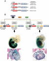
Figure 3. Genomic organization and transcriptional regulation of the miR-1/miR-133 cluster
(A) Bicistronic miR-1/miR-133 clusters and the muscle tissues in which they are expressed are shown. (B) Transcriptional regulation of miR-1/miR-133a genes in heart and skeletal muscle. The upstream and intragenic enhancers of each locus, and the transcription factors that act on these cis-regulatory elements are shown. The expression patterns of lacZ transgenes controlled by the intragenic enhancers of each locus are shown in mouse embryos at E11.5. The bottom panel shows expression patterns of these enhancers in transverse sections of the hearts. a, atrium; lv, left ventricle; rv, right venticle. Embryo images are from Liu et al 2007.
Figure 4
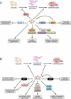
Figure 4. Functions of miR-1 and 133 in heart and skeletal muscle
(A) Targets of miR-1 and processes they regulate are shown. (B) Targets of miR-133 and processes they regulate are shown.
Figure 5
Figure 5. Functions of MyomiRs in cardiac and skeletal muscle
MyomiRs are encoded by myosin heavy chain (MHC) genes. Targets of MyomiRs and processes they regulate are shown.
Figure 6
Figure 6. Model for the regulation of smooth muscle phenotypes and actin dynamics by miR-143 and 145
miR-143 and 145 are cotranscribed as a bicistronic unit. The targets of these miRNAs and the processes they regulate are shown.
Figure 7
Figure 7. Model for miR-126 function in endothelial cells
miR-126 is encoded by an intron of the Egfl7 gene. Targets of miR-126 and the processes they regulate are shown.

References

    1. Ambros V. The functions of animal microRNAs. Nature. 2004;431:350–355. - PubMed
    1. Bartel DP. MicroRNAs: genomics, biogenesis, mechanism, and function. Cell. 2004;116:281–297. - PubMed
    1. Bartel DP. MicroRNAs: target recognition and regulatory functions. Cell. 2009;136:215–233. - PMC - PubMed
    1. Bhattacharyya SN, Habermacher R, Martine U, Closs EI, Filipowicz W. Relief of microRNA-mediated translational repression in human cells subjected to stress. Cell. 2006;125:1111–1124. - PubMed
    1. Boettger T, Beetz N, Kostin S, Schneider J, Kruger M, Hein L, Braun T. Acquisition of the contractile phenotype by murine arterial smooth muscle cells depends on the Mir143/145 gene cluster. J Clin Invest. 2009;119:2634–2647. - PMC - PubMed

Publication types

LinkOut - more resources