. 2010 Apr 20;18(4):592-604.
doi: 10.1016/j.devcel.2010.03.008.

FoxOs inhibit mTORC1 and activate Akt by inducing the expression of Sestrin3 and Rictor

Affiliations

FoxOs inhibit mTORC1 and activate Akt by inducing the expression of Sestrin3 and Rictor

Chia-Chen Chen et al. Dev Cell. .

Abstract

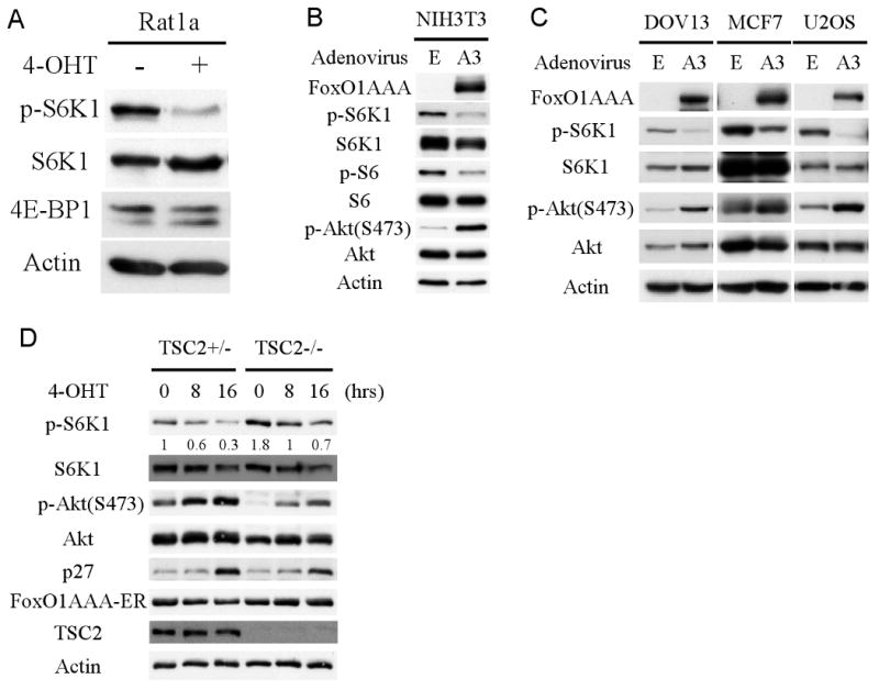
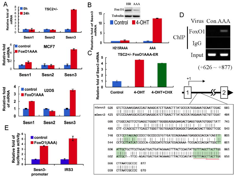
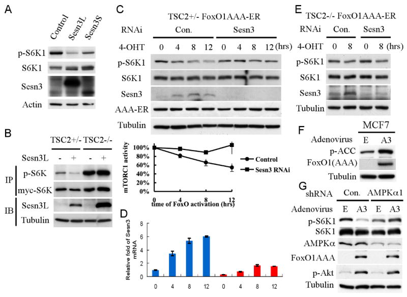
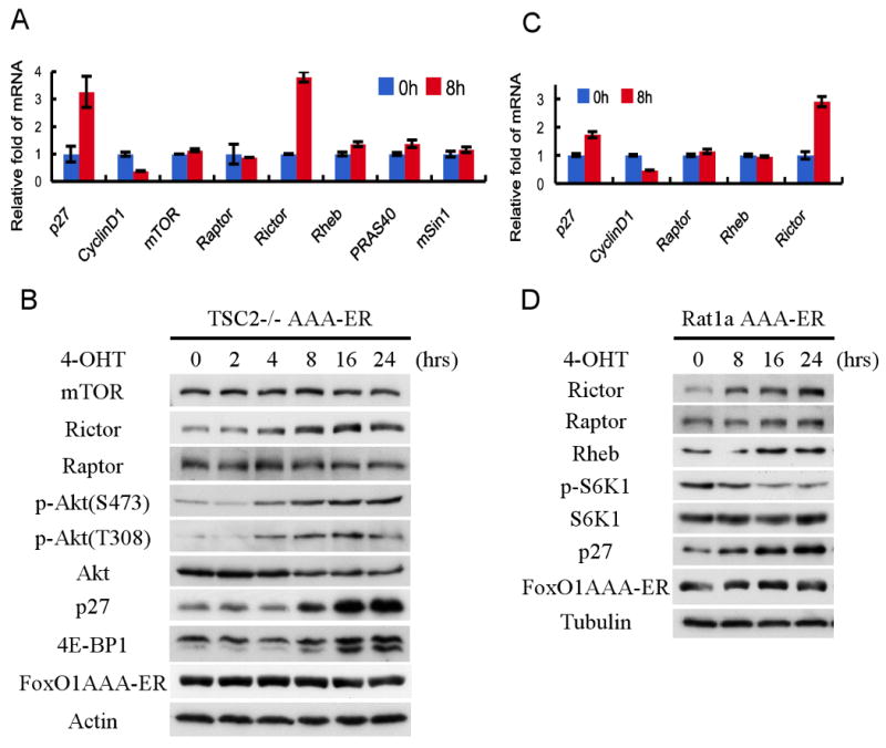
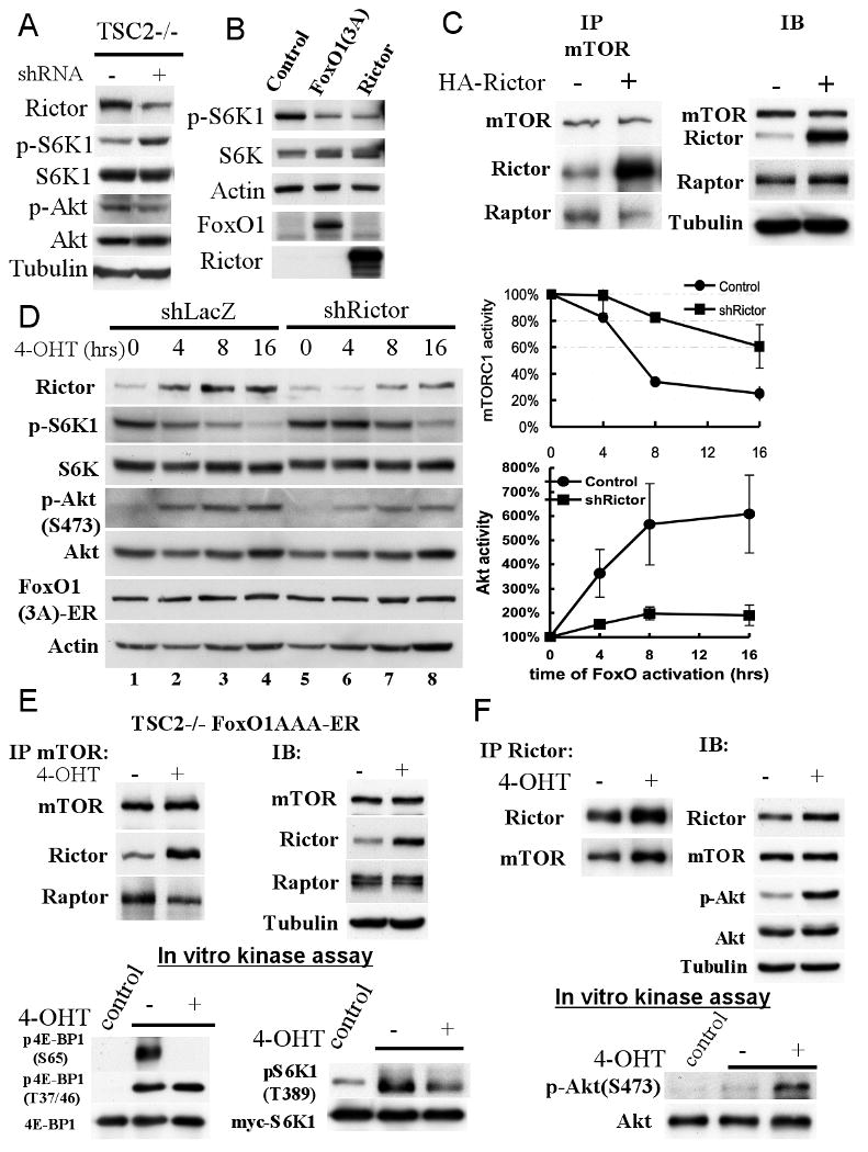
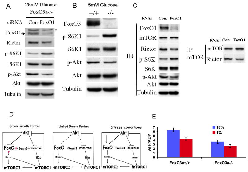
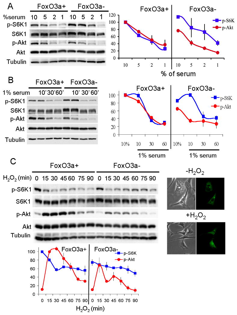
FoxO transcription factors and TORC1 are conserved downstream effectors of Akt. Here, we unraveled regulatory circuits underlying the interplay between Akt, FoxO, and mTOR. Activated FoxO1 inhibits mTORC1 by TSC2-dependent and TSC2-independent mechanisms. First, FoxO1 induces Sestrin3 (Sesn3) gene expression. Sesn3, in turn, inhibits mTORC1 activity in Tsc2-proficient cells. Second, FoxO1 elevates the expression of Rictor, leading to increased mTORC2 activity that consequently activates Akt. In Tsc2-deficient cells, the elevation of Rictor by FoxO increases mTORC2 assembly and activity at the expense of mTORC1, thereby activating Akt while inhibiting mTORC1. FoxO may act as a rheostat that maintains homeostatic balance between Akt and mTOR complexes' activities. In response to physiological stresses, FoxO maintains high Akt activity and low mTORC1 activity. Thus, under stress conditions, FoxO inhibits the anabolic activity of mTORC1, a major consumer of cellular energy, while activating Akt, which increases cellular energy metabolism, thereby maintaining cellular energy homeostasis.


Figures

Figure 1
Figure 1. Activation of FoxO1 down-regulates mTORC1 but elevates Akt activity
A. Activated FoxO1 represses mTORC1 activity. Rat1a cells stably expressing FoxO1(AAA)-ER were treated with 300 nM 4-hydroxy tamoxifen (4-OHT) to induce FoxO1(AAA) activity. Total proteins were extracted, adjusted for similar amounts of total S6K1, and subjected to immunoblotting with the indicated antibodies. B, C. FoxO1 down-regulates mTORC1 activity but elevates Akt activity. NIH3T3, DOV13, MCF7, and U2OS cells were infected with either FoxO1(AAA) adenovirus or control virus. Forty-eight hours after adenovirus infection, total protein was extracted and subjected to immunoblotting with the indicated antibodies. D. Tsc2+/- and Tsc2-/- MEFs stably expressing FoxO1(AAA)-ER were treated with 4-OHT to activate FoxO1. Sixteen hours after total proteins were extracted and subjected to immunoblotting.
Figure 2
Figure 2. FoxO1 binds the promoter region of Sestrin3 gene and directly induces its mRNA levels
A. FoxO1 increases Sesn3 mRNA level. Total RNA was isolated from Tsc2+/- MEFs stably expressing FoxO1(AAA)-ER in the absence or presence of 4-OHT or from FoxO1(AAA) adenovirus or control adenovirus infected MCF7 and U2OS cells. mRNA levels were quantified by qRT-PCR as described in Experimental Procedures. Fold change in mRNA levels was calculated by normalizing to actin mRNA. B. DNA binding deficient FoxO1 cannot induce Sesn3 mRNA. Total RNAs were extracted from Tsc2+/-cells stably expressing FoxO1(AAA)-ER or FoxO1H215R(AAA)-ER in the absence or presence of 4-OHT. Relative mRNA levels were determined by qRT-PCR. Insert shows the levels of FoxO1AAA and FoxO1H215R(AAA). C. The induction of Sesn3 mRNA by FoxO1 does not require de novo protein synthesis. Tsc2+/- FoxO1(AAA)-ER cells were treated with cycloheximide (CHX) for one hour prior to the addition of 4-OHT to prevent de-novo protein synthesis. After addition of 4-OHT for four hours, relative levels of mRNA were determined by qRT-PCR. D. FoxO1 directly binds to the promoter region of the Sesn3 gene. U2OS cells were infected with FoxO1(AAA) adenovirus and then subjected to a ChIP assay using FoxO1 antibodies, as described in Experimental Procedures. The localization of the binding region within the first intron of Sesn3 gene is indicated schematically. Sequences show four predicted consensus FoxO binding sites highlighted within the binding region. The high homology of these sequences between the mouse and human Sesn3 genes is shown. E. FoxO1 regulates transcription from the Sesn3 promoter in a luciferase reporter assay. FoxO1(AAA) MEFs were transfected with pGL3-Sesn3 promoter or conventional pGL3-IRS3 (three copies of insulin response element of IGFBP1 promoter) construct as a positive control. Cells were treated with or without 4-OHT and subjected to Dual-Luciferase reporter assay (Promega). Luciferase activities were normalized to a co-expressed Renilla luminescent signal, and is shown as relative folds against control samples.
Figure 3
Figure 3. FoxO1 inhibits mTORC1 activity by elevating Sesn3 expression
A. Overexpressing of Sesn3 downregulates mTORC1 activity. HEK293 cells were transiently transfected with Sesn3L, Sesn3S, or control plasmid. Forty-eight hour after transfection, total proteins were extracted and subjected to immunobloting with the specified antibodies. B. Sesn3 represses mTORC1 activity in a TSC2-dependent manner. Tsc2+/- and Tsc2-/- cells were transiently co-transfected with Sesn3 and myc-S6K1 plasmids. Twenty-four hour after transfection, total protein extracts were subjected to immunoprecipitation using 9E-10 myc-tag antibody followed by immunobloting with anti-p-S6K1, 9B-11 myc-tag, and anti-Sesn3 antibodies. C, D. Knockdown of Sesn3 attenuates the inhibition of mTORC1 activity by FoxO. Tsc2+/- FoxO1(AAA)-ER cells were transfected with Sesn3 or control RNAi. Forty-eight hour after transfection, cells were treated with 4-OHT and harvested at the indicated time points for protein (C) and RNA analyses (D). mTORC1 activity was deduced from three independent experiments and was quantified by the ratio of pS6K1/S6K1. E. The knockdown of Sesn3 has no effect in Tsc2-/- cells. Tsc2-/- FoxO1(AAA)-ER cells were transfected with Sesn3 or control RNAi. Forty-eight hour after transfection, cells were treated with 4-OHT and harvested at the indicated time points for protein and RNA analyses. F. Activated FoxO1 elevates AMPK activity as measured by pACC. MCF7 cells were infected with either FoxO1(AAA) (A3) adenovirus or control virus (E) for twenty-four hours. Cell lysates were subjected to immunobloting with indicated antibodies. G. The knockdown of AMPKα hindered FoxO1-mediated inhibition of mTORC1 activity without an effect on the increased Akt activity by FoxO1. MCF7 cells stably expressing AMPKα or control shRNA were infected with either FoxO1(AAA) (A3) adenovirus or control virus (E) for twenty-four hours. Whole cell lysates were subjected to immunoblotting with the indicated antibodies.
Figure 4
Figure 4. FoxO1 upregulates Rictor mRNA and protein levels, which coincide with mTORC1 inhibition and Akt activation in Tsc2-/- cells
A, B. FoxO1 elevates Rictor mRNA and protein levels, but not other components of mTOR complexes in Tsc2-/- MEFs. Tsc2-/- FoxO1(AAA)-ER cells were untreated or treated with 4-OHT, and were harvested at the indicated time points for RNA and protein analyses. Total RNA was analyzed by qRT-PCR as described in Experimental Procedures. Total protein was subjected to immunoblotting with the specified antibodies. C, D. FoxO1 elevates Rictor mRNA and protein levels, but not other components of mTOR complexes in Rat1a cells. Experiments were performed as described above for Tsc2-/- MEFs.
Figure 5
Figure 5. Rictor regulates mTORC1 and mTORC2 activities downstream of FoxO
A. Knockdown of Rictor elevates mTORC1 activity. Tsc2-/- MEFs were infected with Lentivirus expressing shRNA targeting Rictor. Seventy-two hour after infection, cells were harvested for immunoblotting with the indicated antibodies. B. Overexpressing of Rictor represses mTORC1 activity to a similar extent as activated FoxO1. HEK293 cells were transiently transfected with FoxO1(AAA), Rictor, or control vectors. Twenty-four hour after transfection, total protein were extracted and subjected to immunoblotting. C. Overexpression of Rictor increases mTOR-Rictor interaction and decreases Raptor-mTOR interaction. HEK293 cells were transiently transfected with HA-Rictor or control DNA. Forty-eight hour after transfection, total protein were harvested and subjected to co-immunoprecipitation with mTOR antibody and immunoblotting with specific antibodies. D. The knockdown of Rictor in Tsc2-/- MEFs impairs the ability of FoxO1 to regulate mTORC1 and Akt activities. Tsc2-/- FoxO1(AAA)-ER cells were infected with lentivirus carrying shRNA targeting Rictor. Forty-eight hour after infection, cells were treated with 4-OHT. At the indicated time points cell lysates were prepared and subjected to immunobloting with the indicated antibodies. A representative immunoblot is shown. Right panels show quantification of mTORC1 and Akt activities after FoxO1 activation in the absence or presence of Rictor shRNA. Relative mTORC1 activity, as quantified by the ratio of pS6K1/S6K1, and relative Akt activity was quantified by the ratio pAkt/Akt in three independent experiments. F. Activated FoxO1 decreases Raptor-mTOR interaction, increases Rictor-mTOR interaction, and inhibits mTORC1 kinase activity in vitro. Tsc2-/- FoxO1(AAA)-ER cells were treated with or without 4-OHT and harvested for co-immunoprecipitation. Samples were immunoprecipitated by mTOR antibody and subjected to immunoblotting and in vitro kinase assays (bottom panels). In vitro kinase assays for mTORC1 activity was done using unphosphorylated S6K1 and recombinant 4E-BP1 (see Experimental Procedures). E. Activated FoxO1 increases mTORC2 kinase activity in vitro. Tsc2-/- FoxO1(AAA)-ER cells were treated with or without 4-OHT and harvested for co-immunoprecipitation. Samples were immunoprecipitated by Rictor antibody and subjected to immunoblotting and in vitro kinase assays (bottom panel). In vitro kinase assay for mTORC2 activity was done using unphosphorylated Akt1 (see Experimental Procedures).
Figure 6
Figure 6. FoxOs-deficiency affects mTORC1 and mTORC2 activities
A. The knockdown of FoxO1 in FoxO3a-/- MEFs, under standard culture conditions, increases mTORC1 activity and decreases mTORC2 activity. FoxO3a-/- MEFs were transiently transfected with FoxO1 or control RNAi. Forty-eight hour after transfection, total protein lysates were subjected to immunobloting. mTORC1 and mTORC2 activities were deduced from the levels of Ser371 phosphorylation on S6K1 and of Ser473 on Akt respectively. B. mTORC1 activity is higher while Akt activity is lower in FoxOa3-/- cells than in control wild-type cells when cultured at low glucose levels. C. Knocking down of FoxO1 in Tsc2-/- cells decreases Rictor expression and Rictor-mTOR complex, decreasing Akt activity while increasing mTORC1 activity. TSC2-/- MEFs were transiently transfected with FoxO1 or control RNAi. Forty-eight hour after transfection, total proteins were harvested and subjected to co-immunoprecipitation with mTOR antibody and immunoblotting with specific antibodies. D. Schematic illustration depicting interplays between Akt, FoxO, and mTOR. In the excess of growth factors and nutrients, Akt inactivates FoxO and activates mTORC1. mTORC1 then induces a negative feedback loop to inhibit Akt. The inhibition of Akt by mTORC1 would activate FoxO to elevate Sesn3 and Rictor, which in turn inhibit mTORC1 and subsequently activate Akt, thereby maintaining a homeostatic balance between Akt and mTOR activities. In limited growth factors and nutrients or other stress conditions, the ability of Akt to inhibit FoxO is impaired or FoxO is activated regardless of Akt, thereby further elevating Sesn3, and Rictor to suppress mTORC1 activity and reduce energy consumption, while maintaining Akt activity to increase energy production. E. Intracellular ATP/ADP ratio in FoxO3a-proficient and FoxO3a-deficeint cells.
Figure 7
Figure 7. FoxO is required to maintain homeostatic balance between Akt and mTORC1 activities under stress conditions
A. FoxO regulates the relative activities mTORC1 and Akt in response to growth factor limitations. Immortalized isogenic wild-type or FoxO3a-/- MEFs were cultured in DMEM medium contained different percentage of serum for twenty-four hours. Whole cell lysates and mRNA were extracted and subjected to immunoblotting. Representative immunoblot is shown (upper panel). Relative mTORC1 activity (p-S6K/S6K) and Akt activity (p-Akt/Akt) were quantified from two independent experiments (bottom panel). B. Immortalized FoxO3a wild-type and knockout MEFs were cultured in 10% serum containing medium for overnight. Cells were placed in 1% serum containing medium for the indicated time points. Total cell lysates were extracted and subjected to immunoblotting analyses. Representative immunoblot is shown (upper panel). Relative mTORC1 activity (p-S6K/S6K) and Akt activity (p-Akt/Akt) were quantified from two independent experiments (bottom panel). C. FoxO regulates the relative activities mTORC1 and Akt in response to oxidative stress. Immortalized isogenic wild-type or FoxO3a-/- MEFs were treated with 0.5 mM H2O2, and were harvested at the indicated time points at 26°C, and then subjected to immunoblotting analyses. Upper panel- shows the translocation of FoxO to the nucleus, in a response to H2O2 at 26°C. Wild-type MEFs were transiently transfected with FoxO1-GFP plasmid and treated with 0.5 mM H2O2. Subcellular localization of FoxO was observed by following GFP signal with fluorescence microscopy. FoxO's nuclear translocation was observed 15min after exposure to H2O2. Middle panel-representative immunoblot shows mTORC1 and Akt activities following exposure to H2O2. Bottom panel- relative mTORC1 activity (p-S6K/S6K) and Akt activity (p-Akt/Akt) were quantified from three independent experiments.

References

    1. Bhaskar PT, Hay N. The Two TORCs and Akt. Dev Cell. 2007;12:487–502. - PubMed
    1. Brunet A, Sweeney LB, Sturgill JF, Chua KF, Greer PL, Lin Y, Tran H, Ross SE, Mostoslavsky R, Cohen HY, et al. Stress-dependent regulation of FOXO transcription factors by the SIRT1 deacetylase. Science. 2004;303:2011–2015. - PubMed
    1. Budanov AV, Karin M. p53 Target Genes Sestrin1 and Sestrin2 Connect Genotoxic Stress and mTOR Signaling. Cell. 2008;134:451–460. - PMC - PubMed
    1. Costanzo-Garvey DL, Pfluger PT, Dougherty MK, Stock JL, Boehm M, Chaika O, Fernandez MR, Fisher K, Kortum RL, Hong EG, et al. KSR2 is an essential regulator of AMP kinase, energy expenditure, and insulin sensitivity. Cell Metab. 2009;10:366–378. - PMC - PubMed
    1. Du K, Herzig S, Kulkarni RN, Montminy M. TRB3: a tribbles homolog that inhibits Akt/PKB activation by insulin in liver. Science. 2003;300:1574–1577. - PubMed

Publication types

MeSH terms

Substances