. 2010 Apr 20;18(4):633-42.
doi: 10.1016/j.devcel.2010.01.016.

beta-catenin activity in the dermal papilla regulates morphogenesis and regeneration of hair

Affiliations

beta-catenin activity in the dermal papilla regulates morphogenesis and regeneration of hair

David Enshell-Seijffers et al. Dev Cell. .

Abstract

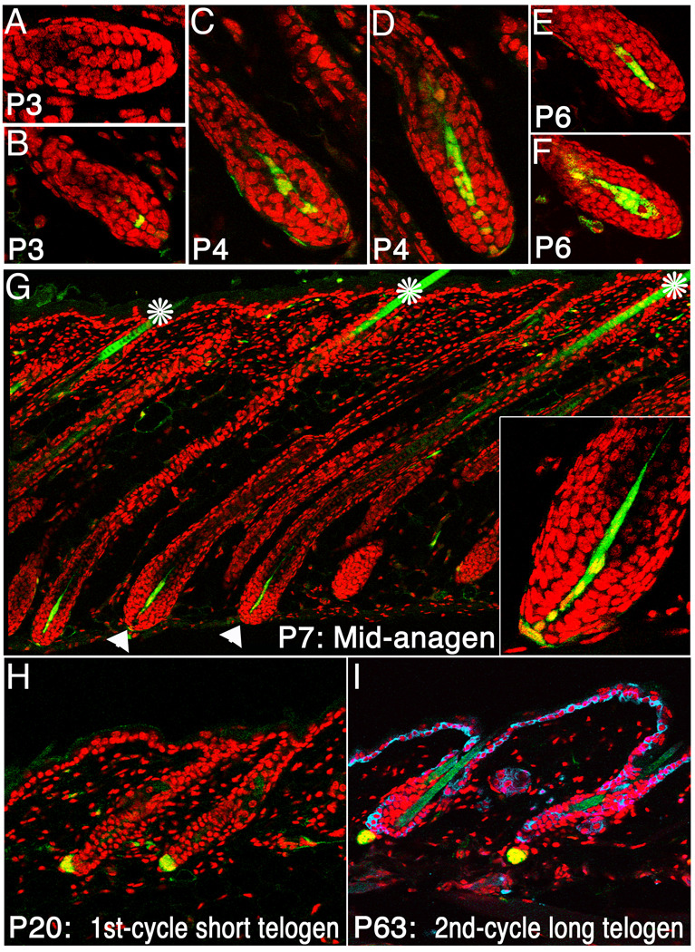
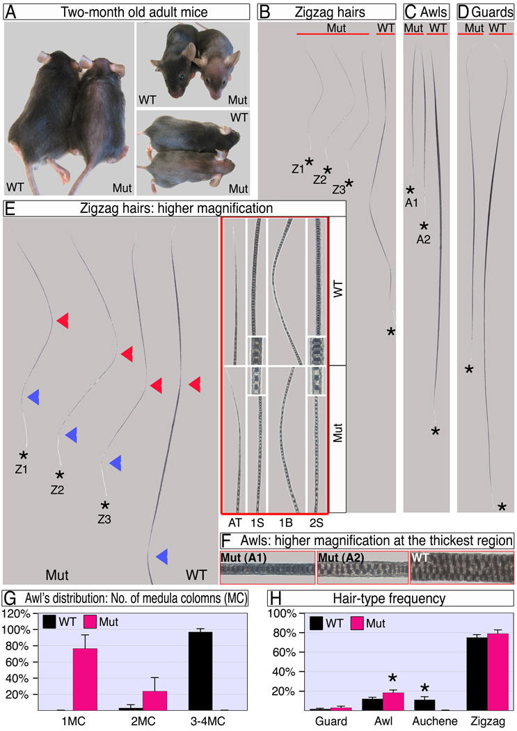
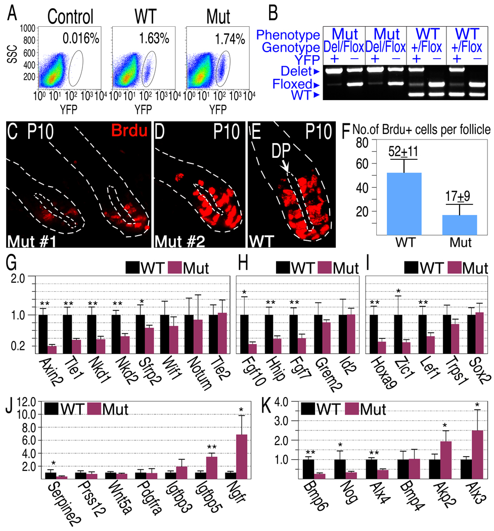
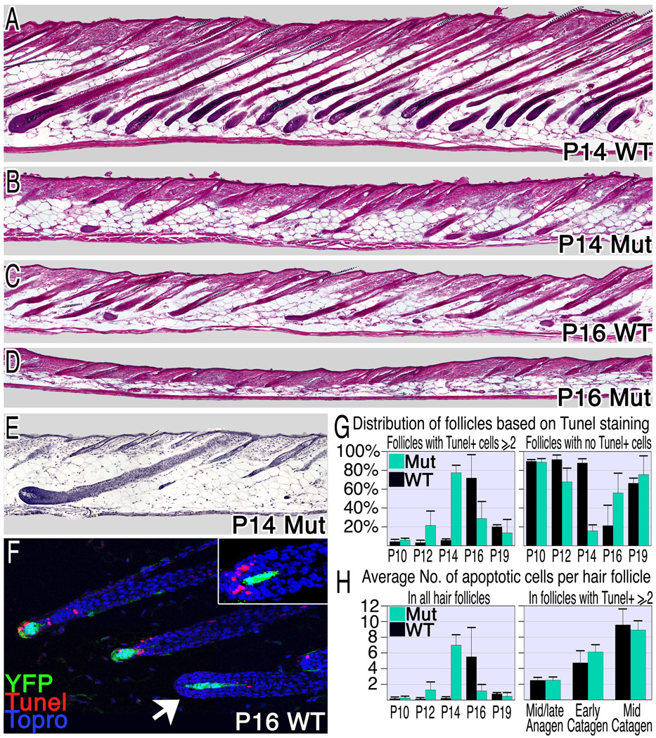
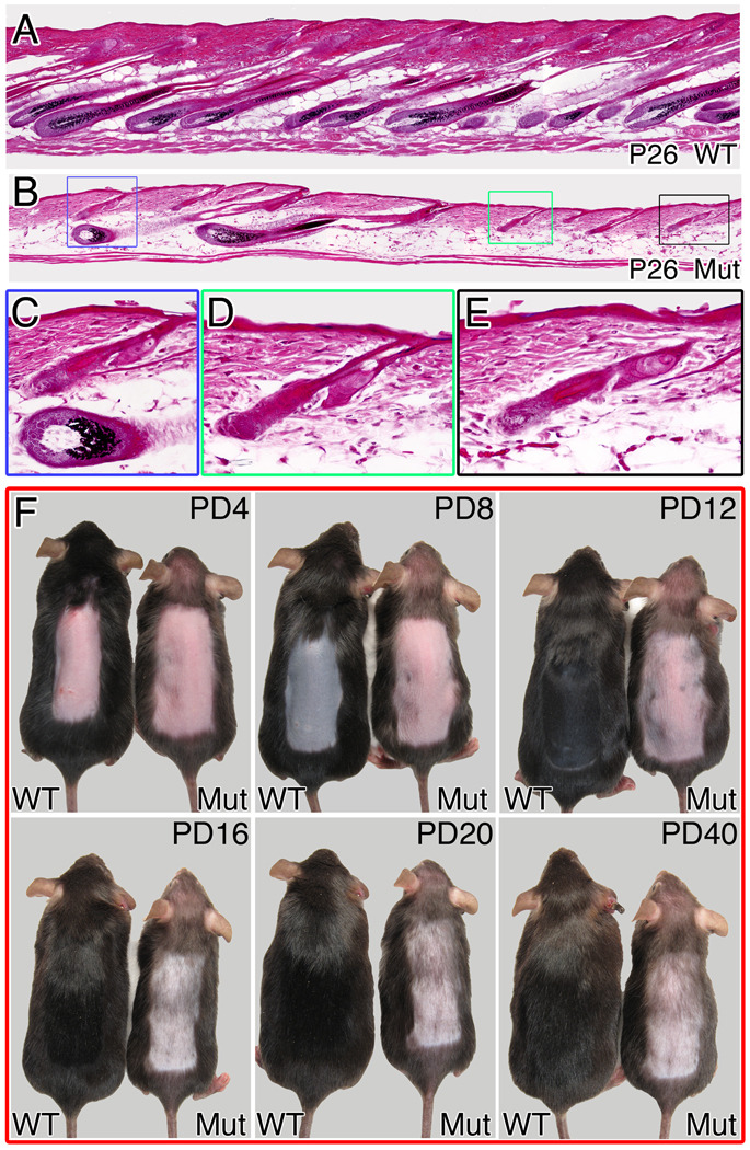
The activity of keratinocytes in the hair follicle is regulated by signals from a specialized mesenchymal niche, the dermal papilla (DP). Here, mice expressing cre recombinase in the DP were developed to probe the interaction between follicular keratinocytes and the DP in vivo. Inactivation of the beta-catenin gene within DP of fully developed hair follicles results in dramatically reduced proliferation of the progenitors and their progeny that generate the hair shaft, and, subsequently, premature induction of the destructive phase of the hair cycle. It also prevents regeneration of the cycling follicle from stem cells. Gene expression analysis reveals that beta-catenin activity in the DP regulates signaling pathways, including FGF and IGF, that can mediate the DP's inductive effects. This study reveals a signaling loop that employs Wnt/beta-catenin signaling in both epithelial progenitor cells and their mesenchymal niche to govern and coordinate the interactions between these compartments to guide hair morphogenesis.


Figures

Figure 1
Figure 1. Cre activity is largely confined to the dermal papilla
Cre-mediated activation of the r26YFP locus reveals the timing and distribution of cre recombinase activity in Cor-cre/+; r26YFP/+ mice. YFP (Green) was first detected at P3 in some but not all follicles (A,B) and increasing numbers of DP cells that express YFP were observed through the mid-anagen phase (C–F). By P7, virtually all DP cells express YFP (G, arrowheads). Inset in G shows higher magnification of a hair bulb. The hair shaft is autofluorescent and its green color is not an indication of cre activity (asterisk). Cre activity remains restricted to the DP as the mice age, as shown during the first (H) and second (I) telogen phases. In I, K14 staining (blue) labels the basal layer of the inter-follicular epidermis and the ORS of the hair follicle. Nuclei-red.
Figure 2
Figure 2. Ablation of β-catenin in the DP results in dramatic hair shortening and thinning
(A) Adult wild type and mutant mice. Note the epidermis is visible through the thin mutant hair coat. (B–D) Zigzag, awl and guard hair-types after the first hair cycle. The apical tips of all hairs are aligned next to the red line and hair-clubs are marked by stars. Mutant hairs within a hair-type population are classified into sub-types based on their different length and/or thickness. Z1–Z3 in B represent a first segment 50% or less, between 50–100%, and similar in length to wild type respectively. A1–A2 in C represent 1 or 2 columns of medulla cells respectively. (E) Mutant zigzag hairs and the apical portion of a wild type zigzag are shown at left. The first and second bends from the apical tip are marked by red and blue arrowheads, respectively. At right, framed with red line, higher magnifications of the apical tip (AT), mid-domain of the first segment (1S), first bend (1B) and mid-region of the second segment (2S) reveal the reduced thickness in mutant hair along the entire hair shaft. Insets show the thickness and organization of the medulla column. (F) Higher magnifications of the thickest region of awl hairs show the number, structure and organization of the medulla columns. (G) Distribution of awl hairs according to the number of medulla columns (MC) at the thickest region. (H) Frequency of hair types in wild type and mutant. Two-tailed unpaired students T-test was employed (*, p<0.0001). See also Figure S1.
Figure 3
Figure 3. Deletion of β-catenin in the DP results in reduced proliferation rates of matrix cells and alterations of gene expression in the DP
(A) FACS analysis of dissociated single cells derived from a back skin of “wild type” (Cor-cre/+; Ctnnb1+/Flox ;r26YFP/+), mutant (Cor-cre/+; Ctnnb1Del/Flox; r26YFP/+) and YFP-negative control (+/+; Ctnnb1+/+;r26YFP/+) P9 mice. (B) Deletion efficiency of the β-catenin gene is 90% at P9. YFP positive (DP) and negative (control) cells were FACS sorted from individual mice and used to genotype the β-catenin gene. Representative examples at P9 are shown. Note that “wild type” mice carry one allele of floxed β-catenin. (C–F) BrdU incorporation in P10 mice. Representative examples from two mutants (C,D) and one wild-type (E) are shown to demonstrate BrdU incorporation (red) in the bulb region. The inner and outer dashed lines demarcate the DP and hair follicle respectively. (F) The average number of BrdU-positive cells in the hair matrix per follicle per section is shown. (G–K) Gene expression analysis of Wnt/β-catenin target and DP signature genes. DP cells purified from individual P9 mice were analyzed for gene expression by real-time PCR (mean±s.d.; n=8 for each genotype). The Y axis represents fold change in expression with wild type levels set to 1. Two-tailed unpaired students T-test was employed (*, p<0.005; **p<0.0001). (G) β-catenin target genes known to be involved in negative feedback loop of Wnt/β-catenin pathway. (H) Other confirmed and inferred β-catenin target genes. (I–J) Transcriptional regulators (I), secreted factors and transmembrane receptors (J) known to be expressed in the DP. (K) Components of the Bmp signaling pathway expressed in the DP and Bmp signaling target genes shown to be modulated in the DP in-vitro. See also Figure S2.
Figure 4
Figure 4. Compromising β-catenin in the DP induces premature catagen
(A–D) Hematoxylin and eosin stained sections from wild type and mutant mice reveal premature and asynchronous induction of the catagen phase in mutant mice. (E) An anagen follicle surrounded by late catagen and telogen follicles in P14 mutant skin. (F–H) TUNEL analysis of apoptosis. Follicles in catagen exhibit 2 or more apoptotic cells, while the absence of apoptotic cells in the hair matrix or bulge was used as a marker for anagen or telogen follicles respectively. (F) TUNEL staining (red) of P16 wild type skin at catagen onset reveals the rapid transition from TUNEL negative follicles (arrowhead) to those with multiple apoptotic cells/follicle. YFP expression marks the DP (green) and nuclei are blue. Inset shows a follicle morphology typical of the transition from late anagen to early catagen where abundant TUNEL staining reveals entry to the catagen phase. Many catagen follicles were observed in mutant skin at P12 (G: left), 4 days earlier than wild type. However, anagen-follicles are still predominant at this stage (G: right). (H) The average number of apoptotic cells per hair follicle per section was calculated. On the left all follicles were included and on the right only those follicles that have entered catagen were scored to eliminate the diluting effects of follicles that remain in anagen. See also Figure S3.
Figure 5
Figure 5. β-catenin activity in the DP is involved in follicular regeneration
Hematoxylin and eosin staining of P26 skin sections from wild type (A) and mutant (B) unperturbed mice. A higher magnification of the colored squares in (B) identifies follicles in advanced anagen (C: lower) and early anagen (C: upper, D, E) in the same region. (F) Anagen-inducing signal was provided by depilation at P70. Pictures of the same wild type and mutant mice were taken every 4 days until hair coat was fully regenerated in the wild type. Note that in the mutant, the depilated region is largely devoid of hairs with only sparse escapers and this remains unaltered even 40 days post depilation. PD-post depilation. See also Figure S5.

References

    1. Andl T, Reddy ST, Gaddapara T, Millar SE. WNT signals are required for the initiation of hair follicle development. Dev Cell. 2002;2:643–653. - PubMed
    1. Barreca A, De Luca M, Del Monte P, Bondanza S, Damonte G, Cariola G, Di Marco E, Giordano G, Cancedda R, Minuto F. In vitro paracrine regulation of human keratinocyte growth by fibroblast-derived insulin-like growth factors. J Cell Physiol. 1992;151:262–268. - PubMed
    1. Botchkarev VA, Botchkareva NV, Albers KM, Chen LH, Welker P, Paus R. A role for p75 neurotrophin receptor in the control of apoptosis-driven hair follicle regression. FASEB J. 2000;14:1931–1942. - PubMed
    1. Brault V, Moore R, Kutsch S, Ishibashi M, Rowitch DH, McMahon AP, Sommer L, Boussadia O, Kemler R. Inactivation of the beta-catenin gene by Wnt1-Cre-mediated deletion results in dramatic brain malformation and failure of craniofacial development. Development. 2001;128:1253–1264. - PubMed
    1. DasGupta R, Fuchs E. Multiple roles for activated LEF/TCF transcription complexes during hair follicle development and differentiation. Development. 1999;126:4557–4568. - PubMed

Publication types