. 2010 Oct;24(7):1078-88.
doi: 10.1016/j.bbi.2010.04.006. Epub 2010 Apr 20.

Thy-1 mRNA destabilization by norepinephrine a 3' UTR cAMP responsive decay element and involves RNA binding proteins

Affiliations

Thy-1 mRNA destabilization by norepinephrine a 3' UTR cAMP responsive decay element and involves RNA binding proteins

Melissa D LaJevic et al. Brain Behav Immun. 2010 Oct.

Abstract

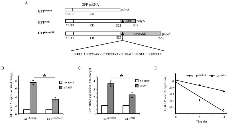
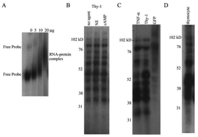
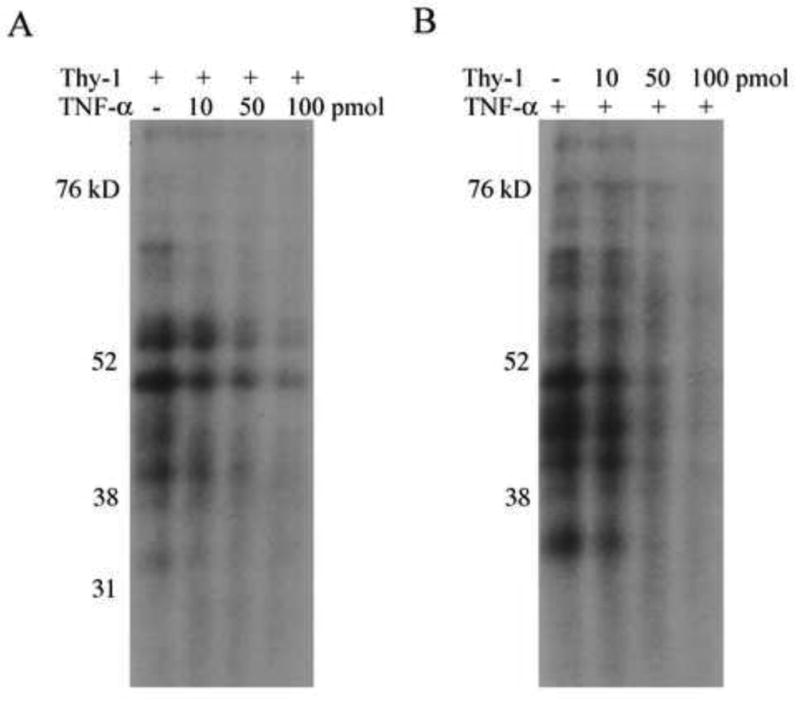
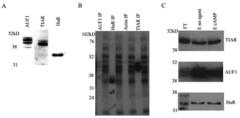
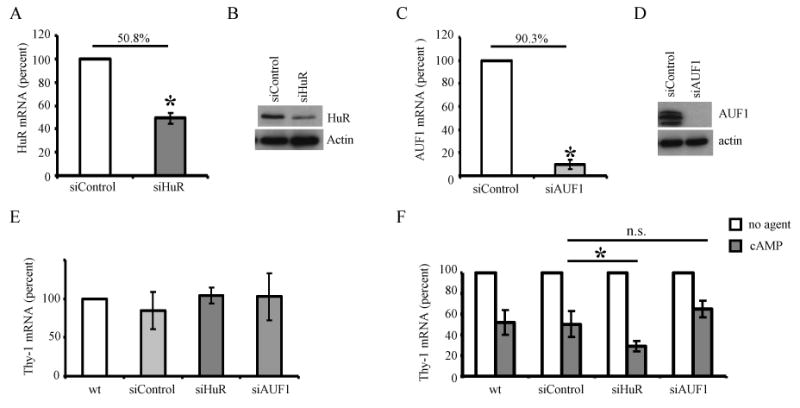
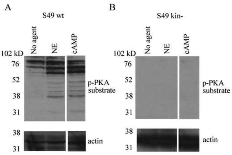
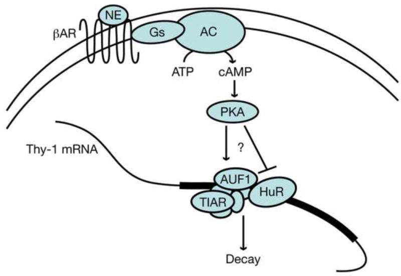
Thy-1 is a cell surface protein important in immunologic and neurologic processes, including T cell activation and proliferation, and neuronal outgrowth. In murine thymocytes, Thy-1 is downregulated in response to norepinephrine (NE) through posttranscriptional destabilization of its mRNA mediated by βAR/AC/cAMP/PKA signaling. In this study we investigated factors involved in NE/cAMP-mediated Thy-1 mRNA destabilization in S49 thymoma cells, and identified a region containing two copies of the AUUUA regulatory element (ARE), a motif commonly associated with mRNA decay, in the Thy-1 mRNA 3' UTR. Insertion of the Thy-1 ARE region into a reporter gene, resulted in cAMP induced destabilization of the reporter gene mRNA. RNA-protein binding studies revealed multiple Thy-1 ARE binding proteins, including AUF1, HuR, and TIAR. RNA silencing of HuR enhanced cAMP-mediated downregulation of Thy-1 mRNA, in contrast, silencing AUF1 had no effect. Immunoblotting revealed multiple proteins phosphorylated by PKA as a result of NE or cAMP signaling. These results reveal that the machinery of NE/cAMP modulation of Thy-1 mRNA decay involves a cAMP responsive ARE in its 3' UTR and multiple site specific ARE binding proteins. These findings add to our knowledge of Thy-1 mRNA regulation and provide insight into the regulation of ARE containing mRNAs, which impacts stress-related immunosuppression.


Conflict of interest statement

Disclosures: The authors have no financial conflict of interest.

Figures

Figure 1
Figure 1. Thy-1 ARE mediates destabilization of GFP mRNA in the presence of 8-bromo cAMP
A. Schematic representation of the constructs used for the reporter gene assay. S49 cells were stably transfected with plasmids expressing GFP mRNA (GFPcontrol), or GFP mRNA with the Thy-1 LongARE sequence (GFPLongARE), or the Thy-1 ARE sequence (GFPARE) inserted in its 3′ UTR. The putative Thy-1 ARE region contains two copies of the AUUUA pentamer (bolded) clustered within a U rich sequence. The transfection efficiency for GFPARE and GFPControl were 50 %, and 10% for GFPLongARE. B. Cells containing GFPcontrol or GFPLongARE were treated in the presence or absence of 8-bromo cAMP (600 μM) for 4 h. Steady state GFP mRNA levels were determined by qPCR and normalized to HPRT1. Data was normalized to the untreated condition and the mean fold change in GFP mRNA levels ± SD for three independent experiments is represented. * p < 0.05. C. Cells containing GFPcontrol or GFPARE were treated in the presence or absence of 8-bromo cAMP (600 μM) for 6 h. Steady state GFP mRNA levels were determined by qPCR and normalized to HPRT1. Data was normalized to the untreated condition and the mean fold change in GFP mRNA levels ± SD for three independent experiments is represented. * p < 0.05. D. Cells containing GFPcontrol or GFPARE were exposed to α-amanitin for four hours followed by treatment with 8-bromo cAMP (600 μM) for 0, 2, and 4 h. GFP mRNA levels were determined by qPCR, normalized to HPRT1, and depicted as the natural log (Ln) of mRNA remaining vs time (h). Data shown is representative of 3 independent experiments.
Figure 2
Figure 2. Detection of Thy-1 ARE-protein complexes
A. Radiolabeled Thy-1 ARE probe was incubated with cytoplasmic protein, and the resulting complexes were separated on nondenaturing gels and visualized by autoradiography. Addition of increasing amounts of cytoplasmic protein (0, 5, 10, and 20 μg) resulted in an upward shift in mobility of the probe indicating RNA-protein complex formation. The results shown represent one of three independent experiments. B-D. Radiolabeled RNA probes for Thy-1 ARE, TNF-α ARE, or control mRNA (obtained from GFP coding region) were incubated with 20 μg of cytoplasmic protein. RNA-protein complexes were fixed with UV irradiation, unbound RNA was digested with RNAse T1/A, and the complexes were subjected to electrophoresis under denaturing conditions and visualized by autoradiography. B. Thy-1 ARE probe crosslinked with protein from untreated, NE (150 μM), or 8-bromo cAMP (600 μM) treated S49 cells for 4 h, resolved on a 12% gel. The results shown represent one independent trial of n > 5. C. Thy-1 ARE, TNF-α ARE, and GFP probes crosslinked to protein from untreated S49 cells, resolved on a 10% gel. The results shown represent one of 3 independent experiments. D. Thy-1 ARE probe crosslinked to protein from untreated thymocytes isolated from BALB/c mice, resolved on a 12% gel. The results shown represent one of 3 independent experiments.
Figure 3
Figure 3. Thy-1 and TNF-α AREs compete for the same binding proteins
A. Radiolabeled Thy-1 ARE probe and increasing amounts of unlabeled TNF-α probe (0, 10, 50, 100 pmol) were incubated with 20 μg cytoplasmic protein. The RNA-protein complexes were fixed by UV irradiation, and the free RNA was digested with RNAse T1/A. Image shows autoradiograph of samples run on denaturing polyacrylamide gels. B. Same conditions as described in A with radiolabeled TNF-α ARE probe competed with unlabeled Thy-1 ARE probe. The results shown are from one representative experiment of three independent trials.
Figure 4
Figure 4. Identification of Thy-1 ARE RNA-binding proteins by immunoprecipitation of RNA-protein complexes
A. S49 cellular extract (20μg) was run on 10% SDS-PAGE and AUF1, HuR, and TIAR were detected by western blot. Images are representative of n > 5 independent experiments. B. Radiolabeled Thy-1 ARE probe was incubated with cytoplasmic protein and the RNA-protein complexes formed were fixed by UV irradiation. Unbound RNA was digested with RNAse T1/A. Antibodies specific for AUF1, HuR, or TIAR were used for immunoprecipitation. The immunoprecipitate was separated by SDS-PAGE and AUF1, HuR, and TIAR bound to labeled probe, were visualized by autoradiography. The results shown represent one experiment of five independent trials. C. RNA binding proteins interacting with a biotinylated Thy-1 ARE probe were isolated by affinity chromatography as described in materials and methods. S49 cytoplasmic protein extract from untreated or 8-bromo cAMP (600 μM, 30 min) treated cells were fractionated by RNA affinity chromatography. The flow through, and eluted fractions were resolved by 10% SDS-PAGE and AUF1, HuR, and TIAR were identified by western blotting. Results shown were from one representative experiment of three independent trials. The gel lanes contain eluted fractions (E) from untreated or treated extract, or flow through (FT) from untreated extract.
Figure 5
Figure 5. Effect of HuR or AUF1 on the regulation of Thy-1 mRNA in S49 cells
S49 cells express a nontargeting shRNA control (siControl) or a targeting shRNA against HuR (siHuR) or AUF1(siAUF1). A. HuR mRNA levels in the control and siHuR cells were analyzed by qPCR (normalized to L32). The data was normalized to the wt condition and the mean percent change ± SD for six independent experiments is represented. The percent decrease is 50.8 +/- 4.8 %. B. HuR and actin immunoblots using whole cell protein from siControl, and siHuR cells (n=6). C. AUF1 mRNA levels in the control and siAUF1 cells as detected by qPCR (normalized to L32). The data was normalized to the wt condition and the mean percent change ± SD for three independent experiments is represented. The percent decrease is 90.3 +/- 3.9 %. D. AUF1 and actin protein levels as detected by immunoblot of whole cell protein (n=3). E. Steady state levels of Thy-1 mRNA in S49 wt, siControl, siAUF1, and siHuR cells as detected by qPCR (normalized to HPRT1). F. S49 wt, siControl, siHuR, or siAUF1 cells were treated with or without 8-bromo cAMP (600 μM) for 4 h. The levels of Thy-1 mRNA were determined by qPCR (normalized to HPRT1). Data from each individual trial was normalized to the untreated condition and the mean percent change in Thy-1 mRNA levels ± SD for five independent experiments is represented. * p < 0.05.
Figure 6
Figure 6. Identification of NE/cAMP mediated phosphorylation of PKA substrates
A. Immunoblot detection of phosphorylated PKA substrates and actin from S49 wild type cells (A) or kin- cells (B) stimulated with NE or 8-bromo cAMP for 30 min. Images are from a representative experiment (impertinent lanes have been removed) of 3 independent trials.
Figure 7
Figure 7. Proposed mechanism of NE mediated Thy-1 mRNA decay

References

    1. Abdelmohsen K, Pullmann R, JR, Lal A, Kim HH, Galban S, Yang X, Blethrow JD, Walker M, Shubert J, Gillespie DA, Furneaux H, Gorospe M. Phosphorylation of HuR by Chk2 regulates SIRT1 expression. Mol Cell. 2007;25:543–557. - PMC - PubMed
    1. Abdelmohsen K, Srikantan S, Kuwano Y, Gorospe M. miR-519 reduces cell proliferation by lowering RNA-binding protein HuR levels. Proc Natl Acad Sci U S A. 2008;105:20297–20302. - PMC - PubMed
    1. Ader R, Cohen N, Felton D. Psychoneuroimmunology: interactions between the nervous system and the immune system. Lancet. 1995;345:99–103. - PubMed
    1. Anderson P. Post-transcriptional control of cytokine production. Nat Immunol. 2008;9:353–359. - PubMed
    1. Barker TH, Hagood JS. Getting a grip on Thy-1 signaling. Biochim Biophys Acta. 2009;1793:921–923. - PMC - PubMed

Publication types

MeSH terms