. 2010 Apr 22;7(4):265-276.
doi: 10.1016/j.chom.2010.03.004.

A pathobiont of the microbiota balances host colonization and intestinal inflammation

Affiliations

A pathobiont of the microbiota balances host colonization and intestinal inflammation

Janet Chow et al. Cell Host Microbe. .

Abstract

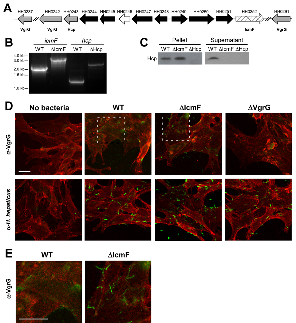
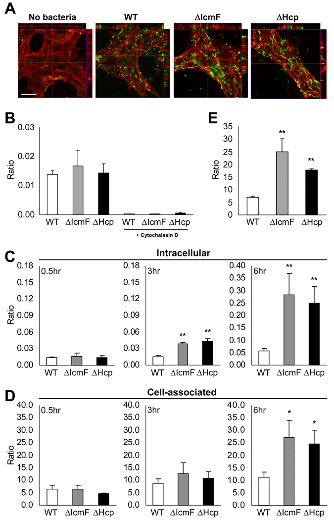
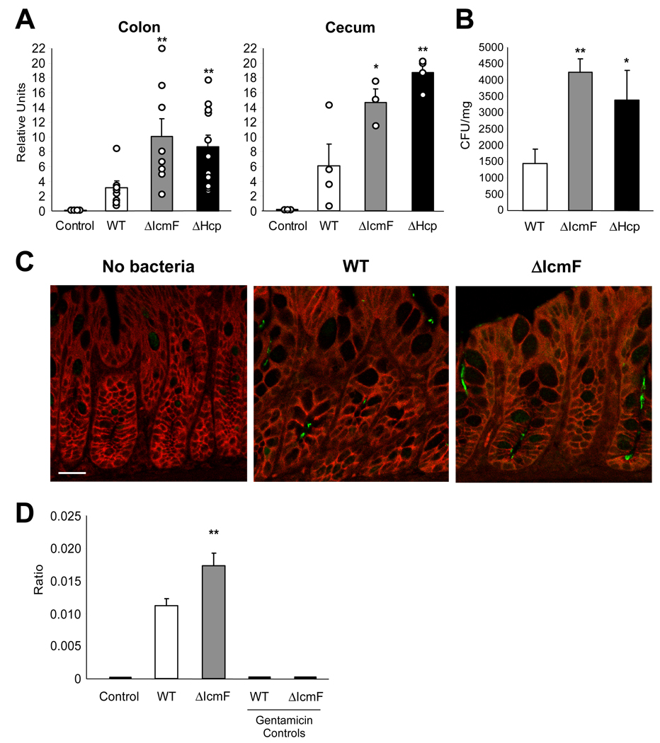
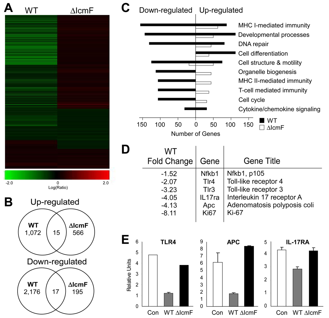
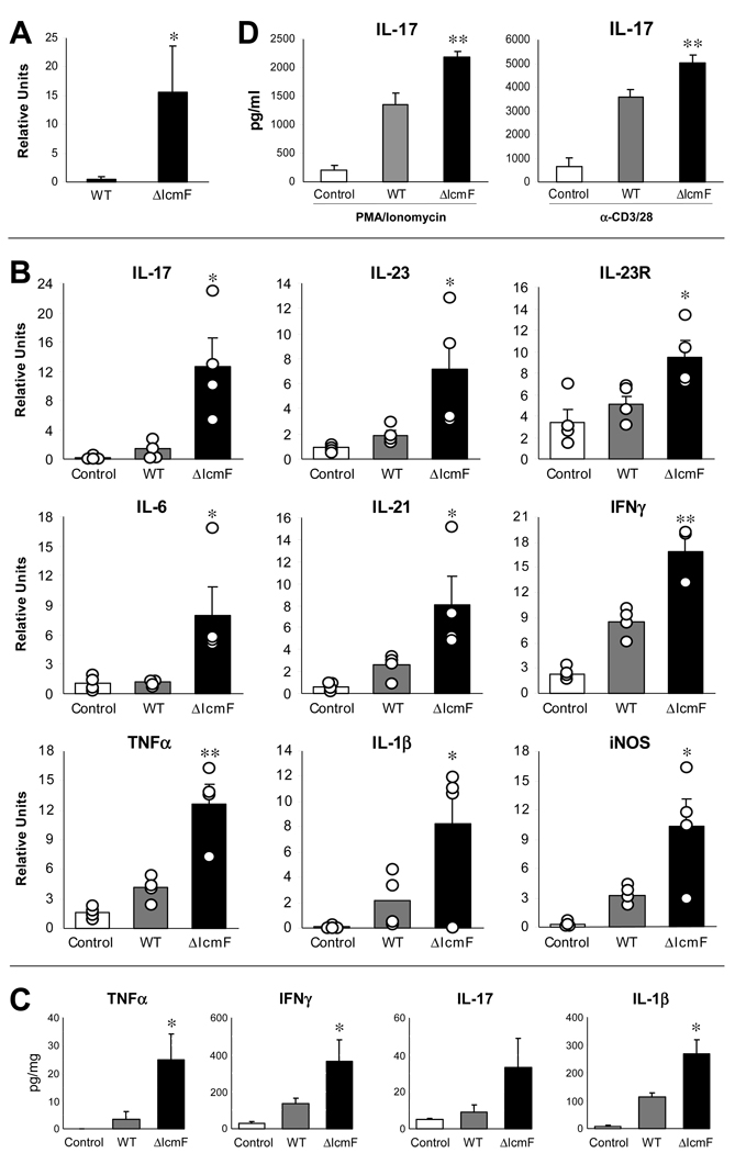
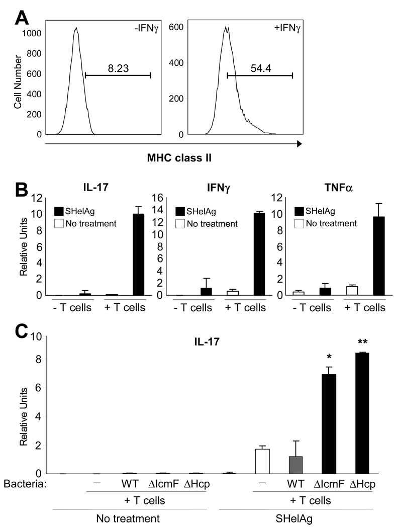
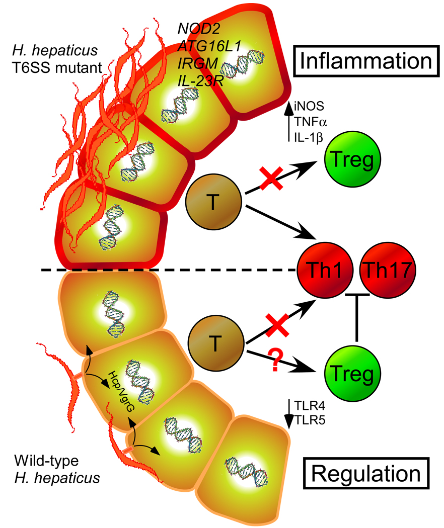
The gastrointestinal tract harbors a diverse microbiota that has coevolved with mammalian hosts. Though most associations are symbiotic or commensal, some resident bacteria (termed pathobionts) have the potential to cause disease. Bacterial type VI secretion systems (T6SSs) are one mechanism for forging host-microbial interactions. Here we reveal a protective role for the T6SS of Helicobacter hepaticus, a Gram-negative bacterium of the intestinal microbiota. H. hepaticus mutants with a defective T6SS display increased numbers within intestinal epithelial cells (IECs) and during intestinal colonization. Remarkably, the T6SS directs an anti-inflammatory gene expression profile in IECs, and CD4+ T cells from mice colonized with T6SS mutants produce increased interleukin-17 in response to IECs presenting H. hepaticus antigens. Thus, the H. hepaticus T6SS limits colonization and intestinal inflammation, promoting a balanced relationship with the host. We propose that disruption of such balances contributes to human disorders such as inflammatory bowel disease and colon cancer.


Figures

Figure 1
Figure 1. H. hepaticus Encodes for a Functional T6SS
(A) Schematic diagram of the genetic organization of H. hepaticus T6SS genes. Gray arrows represent Hcp and VgrG genes, cross-hatched arrow indicates IcmF homologue, black arrows represent T6SS homologues of unknown function, and white arrow indicates a gene non-homologous to other T6SS genes. See also Table S1. (B) Genomic DNA collected from mid-log cultures of WT, ΔIcmF, or ΔHcp H. hepaticus was amplified by PCR using icmF or hcp specific primers. Insertion of the eryR gene was detected by a 1.1kb-increase in the resulting PCR band. (C) Hcp is undetected in supernatants from ΔIcmF and ΔHcp T6SS mutants. Equal amounts of mid-log bacterial cultures of WT, ΔIcmF, or ΔHcp were centrifuged to separate bacterial pellets and supernatant. Supernatants were subsequently filtered to ensure removal of all bacteria. Bacterial pellets and supernatants were analyzed by Western blot. Membranes were blotted with anti-Hcp antibody. (D, E) Confocal images of bacteria incubated with MODE-K cells. WT, ΔIcmF, or ΔVgrG (ΔHH0242) H. hepaticus were incubated with MODE-K cells for 5hr. MODE-K cells were subsequently rinsed with PBS, fixed in 4% PFA, and stained for the eukaryotic cell membrane marker wheat germ agglutinin (red) and either H. hepaticus or VgrG (green). Outlined regions for WT and ΔIcmF in (D) are shown at higher magnification (E). Scale bar represents 20µm.
Figure 2
Figure 2. T6SS Mutants Display Higher Intracellular and Cell-associated Accumulation in MODE-K cells
(A) Confocal image of bacteria inside MODE-K cells. WT, ΔIcmF, or ΔHcp was incubated with MODE-K for 6hr. MODE-K cells were rinsed with PBS, fixed in 4% PFA, and stained for H. hepaticus (green) and the eukaryotic cell membrane marker wheat germ agglutinin (red). Scale bar represents 30µm. See also Movie S1. (B) Cytochalasin D inhibits uptake of H. hepaticus. Prior to incubation with bacteria, MODE-K cells were treated with 10µM cytochalasin D for 1hr. Bacteria were added at an MOI of 100. After 0.5hr incubation at 37°C under mi croaerophilic conditions, cells were treated with 100µg/ml gentamicin, and intracellular bacteria plated for enumeration. Results are expressed as colony-forming units (CFUs) of intracellular bacteria divided by number of MODE-K cells. Error bars indicate SEM from 3 experiments. (C, D) Gentamicin protection assay in which MODE-K cells were incubated with bacteria at an MOI of 100. After 0.5, 3, or 6hr incubation, media was replaced with 100µg/ml gentamicin for enumeration of intracellular bacteria (C) or without gentamicin for cell-associated bacteria (D). Cells were washed and bacteria plated for quantification. Ratios are expressed as CFUs of bacteria divided by number of MODE-K cells. Error bars indicate SEM from 3–5 experiments. *p<0.05, **p<0.01 vs WT. (E) Increased adherence of T6SS mutants is not dependent on bacterial internalization. Prior to co-culture, MODE-K cells were treated with 10µM cytochalasin D. Bacteria were added at an MOI of 100 for 6hr at 37°C under microa erophilic conditions. Bacteria were plated for enumeration. Results are expressed as CFUs of bacteria divided by number of MODE-K cells. Error bars indicate SD from 2 experiments. **p<0.01 vs WT. See also Figure S1.
Figure 3
Figure 3. T6SS Mutants Have Increased Colonization Levels within the Colon
(A) ΔIcmF- and ΔHcp-mono-colonized animals have greater amounts of H. hepaticus 16S rRNA in the colon and cecum compared to WT-mono-colonized animals. RNA was collected from colon and cecum. Levels of 16S were quantified by qRT-PCR using H. hepaticus-specific 16S primers. Error bars show SEM, n=4–11 animals per group. *p<0.05, **p<0.01 vs WT. See also Figure S2A. (B) ΔIcmF- and ΔHcp-mono-colonized animals have higher levels of viable H. hepaticus in the colon compared to WT-mono-colonized animals. Colon tissues were homogenized in BHI and plated for quantification. Bacterial numbers were normalized to colon tissue weights. Units are expressed as CFUs per mg of tissue. Error bars show SEM from n=4 animals per group. *p<0.05, **p<0.01 vs WT. (C) H. hepaticus is found in colonic intestinal crypts. Colons from WT- and ΔIcmF-mono-colonized mice were paraffin-embedded and sectioned. Colon sections were stained for H. hepaticus (green) and E-cadherin (red). Animals were colonized for 8 weeks. Scale bar represents 20µm. See also Figures S2B–D. (D) ΔIcmF-colonized animals have increased levels of intracellular H. hepaticus compared to WT-colonized animals. Purified IECs were treated with gentamicin either prior to or following lysis with saponin (which selectively permeablizes eukaryotic membranes). Bacteria were enumerated by plating on TVP (trimethoprim, vancomycin, polymyxin B) plates which are known to select for H. hepaticus, as confirmed by control mice which received no H. hepaticus. In ‘Gentamicin Controls’ IECs were lysed and then treated with gentamicin. Ratio is expressed as CFUs of bacteria divided by number of IECs. Error bars indicate SEM from n=12 animals per group. **p<0.01 vs WT. See also Figure S2E.
Figure 4
Figure 4. Wild-type H. hepaticus Induces a Wide Repertoire of Responses in MODE-K cells
RNA was harvested from MODE-K cells incubated for 6hr with either wild-type H. hepaticus, ΔIcmF mutant, or no bacteria and analyzed by an Agilent Whole Mouse Genome Microarray. Gene expression of MODE-K cells incubated with bacteria was compared to transcript levels from RNA of untreated MODE-K cells. Only genes with a p-value <0.5 and fold change >1.5 were used for subsequent analysis. (A) Heat-map analysis of MODE-K gene expression in the presence of WT or ΔIcmF shows massive down-regulation by wild-type H. hepaticus. (B) Venn diagram showing up- and down- regulation of gene expression in the presence of WT or ΔIcmF. (C) Gene ontogeny analysis of changes in MODE-K transcript levels for various functional groups. Wild-type bacteria down-regulate numerous cellular pathways. (D) Fold change of select innate and adaptive immune genes in the presence of WT H. hepaticus. (E) qRT-PCR analysis of RNA from MODE-K cells for genes associated withinflammation and colon cancer. Error bars show SEM.
Figure 5
Figure 5. T6SS Mutant Leads to Greater Inflammation in the Colon in the Rag Adoptive Transfer Model of Colitis
(A) ΔIcmF-colonized animals have greater amounts of H. hepaticus 16S rRNA in the colon compared to WT-colonized animals. RNA was collected from colons. Levels of 16S were quantified by qRT-PCR. Data are representative of 3 experiments, with n=4 per group. Error bars show SEM. *p<0.05 vs WT. (B) RNA from the colon was analyzed by qRT-PCR for inflammatory cytokines. Experimental values were normalized against L32. Results are representative of 5 experiments, with n=4 per group. Each circle represents an individual animal. Error bars show SEM. *p<0.05, **p<0.01 vs WT. See also Figure S3. (C) Colons were cultured ex vivo for 24hrs. Supernatants were assayed for cytokine by ELISA. Samples were normalized to total protein. Data are representative of 2 experiments, with n=4 per group. Each circle represents an individual animal. Error bars show SEM. *p<0.05 vs WT. (D) Mesenteric lymph nodes pooled from each experimental group were restimulated with either PMA and ionomycin, or α-CD3 and α-CD28 for 24hrs. Supernatants were assayed by ELISA. Results are from 2 experiments, with n=4 mice per group. Error bars show SEM. **p<0.01 vs WT.
Figure 6
Figure 6. MODE-K Cells are Capable of Stimulating CD4+ T cell Responses with H. hepaticus Antigens
(A) MODE-K cells were treated with 100U/ml of IFNγ for 7 days or left untreated. Cells were removed from the plate with trypsin, stained for MHC class II antigens, and analyzed by flow cytometry. See also Figure S4. (B) MODE-K cells pre-treated with 100U/ml of IFNγ for 7 days were pulsed with either 20 µg/ml of soluble H. hepaticus antigen (SHelAg) from wild-type bacteria or no SHelAg for 24hrs. Media was changed prior to addition of CD4+ T cells harvested from syngeneic C3H/HeJ Helicobacter-free animals or no T cells. The ratio of MODE-K:T cells was 1:10. Cells were co-cultured for 3d. RNA was collected from total cells and analyzed by qRT-PCR. Error bars indicate SD from 2 independent experiments. (C) MODE-K cells pre-treated with IFNγ were pulsed with either 20 µg/ml of SHelAg from wild-type bacteria or no SHelAg for 24hrs. Media was changed prior to addition of CD4+ T cells collected from animals colonized with wild-type, ΔIcmF, ΔHcp, or no H. hepaticus. The ratio of MODE-K:CD4+ T cells was 1:10. Cells were co-cultured for 3d. RNA was collected from total cells and analyzed by qRT-PCR. Error bars indicate SD from 2 independent experiments. *p<0.05, **p<0.01 vs WT treated with SHelAg.
Figure 7
Figure 7. Proposed Interactions between H. hepaticus T6SS and the Intestinal Immune Response during Colonization
During prolonged intestinal colonization of animals, H. hepaticus intimately contacts the epithelium and uses its T6SS to create a tolerogenic immune environment (possibly through down-regulating TLR expression and/or promoting Treg development). Crosstalk between host and bacteria maintains a balanced symbiotic interaction. This balance can be disturbed by genetic mutations associated with IBD (NOD2, ATG16L1, IRGM, IL-23R) and/or dysbiosis caused by external disturbances (e.g. antibiotics, enteric infections, diet, etc.), which may result in elevated immune responses (increased Th17) in genetically susceptible hosts. Based on our and previous studies, it appears that the combination of host genotype and microbial status contributes to intestinal disease.

References

    1. Bingle LE, Bailey CM, Pallen MJ. Type VI secretion: a beginner's guide. Curr Opin Microbiol. 2008;11:3–8. - PubMed
    1. Bladergroen MR, Badelt K, Spaink HP. Infection-blocking genes of a symbiotic Rhizobium leguminosarum strain that are involved in temperature-dependent protein secretion. Mol Plant Microbe Interact. 2003;16:53–64. - PubMed
    1. Bland PW, Warren LG. Antigen presentation by epithelial cells of the rat small intestine. I. Kinetics, antigen specificity and blocking by anti-Ia antisera. Immunology. 1986;58:1–7. - PMC - PubMed
    1. Bohr UR, Glasbrenner B, Primus A, Zagoura A, Wex T, Malfertheiner P. Identification of enterohepatic Helicobacter species in patients suffering from inflammatory bowel disease. J Clin Microbiol. 2004;42:2766–2768. - PMC - PubMed
    1. Das S, Chakrabortty A, Banerjee R, Chaudhuri K. Involvement of in vivo induced icmF gene of Vibrio cholerae in motility, adherence to epithelial cells, and conjugation frequency. Biochem Biophys Res Commun. 2002;295:922–928. - PubMed

Publication types

MeSH terms

Associated data