. 2010 Apr 22;7(4):302-313.
doi: 10.1016/j.chom.2010.03.006.

Virus inhibition of RIP3-dependent necrosis

Affiliations

Virus inhibition of RIP3-dependent necrosis

Jason W Upton et al. Cell Host Microbe. .

Abstract

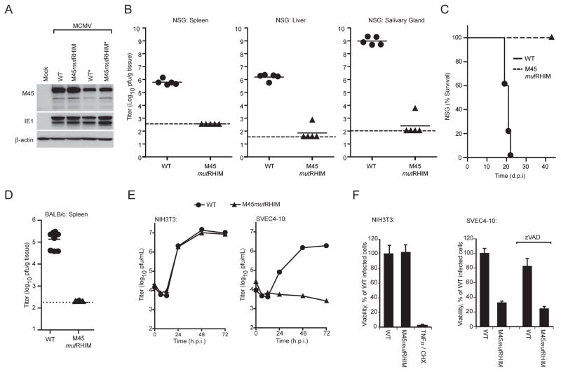
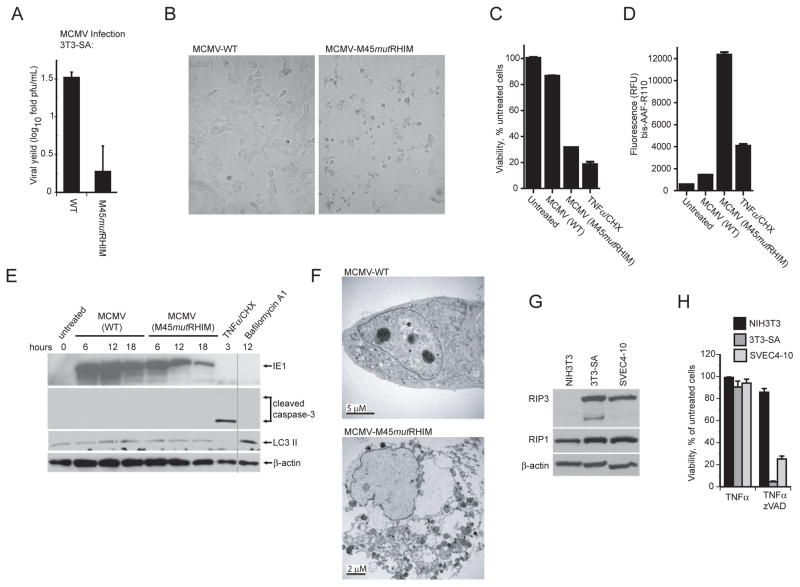
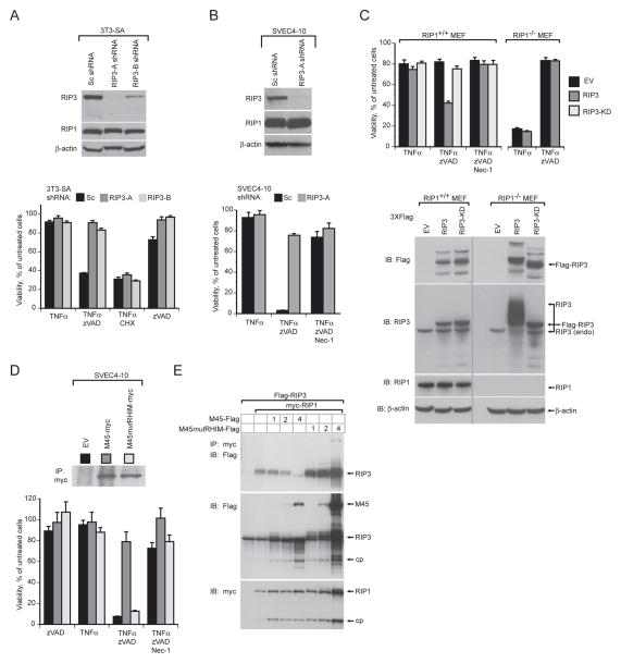
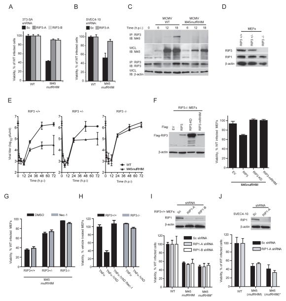
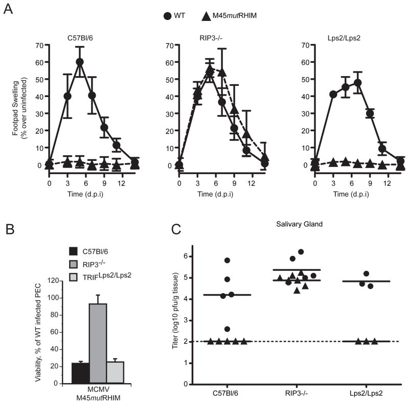
Viral infection activates cytokine expression and triggers cell death, the modulation of which is important for successful pathogenesis. Necroptosis is a form of programmed necrosis dependent on two related RIP homotypic interaction motif (RHIM)-containing signaling adaptors, receptor-interacting protein kinases (RIP) 1 and 3. We find that murine cytomegalovirus infection induces RIP3-dependent necrosis. Whereas RIP3 kinase activity and RHIM-dependent interactions control virus-associated necrosis, virus-induced death proceeds independently of RIP1 and is therefore distinct from TNFalpha-dependent necroptosis. Viral M45-encoded inhibitor of RIP activation (vIRA) targets RIP3 during infection and disrupts RIP3-RIP1 interactions characteristic of TNFalpha-induced necroptosis, thereby suppressing both death pathways. Importantly, attenuation of vIRA mutant virus in wild-type mice is normalized in RIP3-deficient mice. Thus, vIRA function validates necrosis as central to host defense against viral infections and highlights the benefit of multiple virus-encoded cell-death suppressors that inhibit not only apoptotic, but also necrotic mechanisms of virus clearance.


Figures

Figure 1
Figure 1. Role of MCMV M45 RHIM interactions in suppression of virus-induced death
(A) IB analysis of vIRA expression from recombinant viruses to detect vIRA (M45), IE1, and βactin. NIH3T3 fibroblasts were either mock- or virus-infected at a multiplicity of infection (MOI) of 10 with bacmid-derived K181 (WT) or vIRA mutant virus (M45mutRHIM). An asterisk (*) designates a second independent WT or mutant virus isolate. (B) Replication levels and pathogenesis of WT and M45mutRHIM virus in severely immunodeficient NSG mice. At 16 d post inoculation (106 pfu into footpads), spleen (left panel), liver (middle panel), and salivary glands (right panel) were harvested from euthanized mice and viral titers determined by plaque assay. Each symbol represents one animal, solid horizontal lines represent the mean for each group, and dashed lines indicate the limit of detection in each assay. (C) Kaplan-Meier survival plot of NSG mice monitored for 42 days post infection with WT and M45mutRHIM virus (106 pfu into footpads). (D) Replication levels of WT and M45mutRHIM virus in spleen at 3 d post inoculation (106 pfu into peritoneal cavity) of BALB/c mice. (E) Replication levels of WT and M45mutRHIM virus in infected NIH3T3 fibroblasts (left panel) and SVEC4-10 endothelial cells (right panel) (MOI=10). Viral titers were determined by plaque assay at the indicated times post infection. (F) Viability of NIH3T3 fibroblasts (left panel) or SVEC4-10 cells (right panel) following infection with WT and M45mutRHIM virus. Where noted, zVAD-fmk (50 μM) was included throughout infection. Cell viability was determined by measuring intracellular ATP levels with a Cell Titer-Glo Luminescent Cell Viability Assay kit. Treatment with TNFα (25 ng/mL) and cycloheximide (CHX; 5 μg/mL) was used to induce apoptosis in uninfected NIH3T3 cells as a control. Error bars indicate standard deviation (SD) of the mean. See also the related Figure S1.
Figure 2
Figure 2. Cells sensitive to MCMV-associated programmed necrosis express high levels of RIP3
(A) Viral yields determined by plaque assay at 72 hpi in 3T3-SA cells following infection with WT or M45mutRHIM virus (MOI=10). (B) Bright field micrographs of 3T3-SA cells infected for 18 h with either WT (left panel) or M45mutRHIM (right panel) virus. Magnification, 10X. (C) Viability of 3T3-SA cells assessed by intracellular ATP levels following infection with WT or M45mutRHIM virus. (D) Release of protease to the medium assessed in 3T3-SA cells using a CytoTox-Fluor kit. (E) IB of 3T3-SA cells infected with WT or M45mutRHIM virus (MOI=10) for the indicated times followed by detection of IE1, caspase-3, LC3 II and β-actin. Vertical line shows where lanes from the original gel were brought adjacent. In C - E, uninfected cells were treated to induce apoptosis (TNFα/CHX) as described in Figure 1F or with Bafilomycin A1 (250 nM) to induce LC3 II, or left untreated. (F) Transmission electron micrographs of 3T3-SA cells at 18 hpi with WT (top panel) or M45mutRHIM (bottom panel) virus. Size bars are indicated. (G) IB analysis of NIH3T3, 3T3-SA, and SVEC4-10 cells to detect RIP1, RIP3, and β-actin. (H) Viability of NIH3T3, 3T3-SA, and SVEC4-10 cells treated for 18 h with TNFα (25 ng/mL) in the absence or presence of zVAD-fmk (25 μM). See also the related Figure S2.
Figure 3
Figure 3. MCMV RHIM-dependent suppression of RIP3-mediated necrosis
(A) IB analysis (top panel) to detect RIP3, RIP1, and β-actin, and viability (lower panel) of 3T3-SA fibroblasts expressing a scramble control (Sc) or one of two RIP3-specific shRNAs (RIP3-A and RIP3-B) and treated to induce necroptosis as described in Figure 2H or to induce apoptosis as described in Figure 1F. (B) IB analysis (top panel) to detect RIP1, RIP3, and β-actin and viability (bottom panel) of SVEC4-10 cells expressing Sc or RIP3-A shRNA. Cells were treated to induce necroptosis as described in Figure 2H in the absence or presence of Nec-1 (30 μM). (C) Viability (top panel) and IB analysis to detect RIP1, RIP3, β–actin and Flag-epitope tagged proteins (lower panel) in RIP1+/+ and RIP1−/− MEFs transduced with empty vector (EV), Flag-tagged RIP3 (RIP3), or Flag-tagged kinase-deficient RIP3 (RIP3-KD) retroviral constructs. Transduced cells were treated as described in (B). The vertical line shows where lanes from the original gel were brought adjacent. (D) IB of myc-tagged proteins following IP with anti-myc conjugated agarose (top panel) of SVEC4-10 cells transduced with EV, M45-myc, or M45mutRHIM-myc retroviral constructs and viability (bottom panel) of these cells after treatment as described in (B). (E) IB of 293T cells transfected with Flag-tagged RIP3, myc-tagged RIP1 and 1, 2, or 4 μg of either M45-Flag or M45mutRHIM-Flag expression plasmids after IP with anti-myc agarose beads. In all samples, the amount of transfected DNA was held constant with addition of control plasmid DNA. IB detection was with anti-Flag or anti-c-Myc (9E10) antibody. Major protein species are identified, with “cp” indicating a processed form of RIP1 or RIP3. Error bars indicate SD of the mean.
Figure 4
Figure 4. RIP3 is required, and RIP1 is dispensable for MCMV-associated programmed necrosis
(A) Viability of 3T3-SA cells expressing Sc, RIP3-A or RIP3-B shRNAs determined 18 hpi with WT or M45mutRHIM virus (MOI of 10). (B) Viability of SVEC4-10 cells using a subset of conditions described in (A). (C) IB of 3T3-SA cells infected with WT or M45mutRHIM virus (MOI of 5), harvested at indicated times for IP of RIP3 followed by detection of vIRA (M45) and RIP3. IB using ~5% of cell lysate to detect vIRA (M45) and β–actin. (D) IB of RIP3+/+, RIP3+/−, and RIP3−/− MEFs to detect RIP3, RIP1, and β–actin. (E) Replication of WT and M45mutRHIM viruses (MOI of 5) on RIP3+/+ (left panel), RIP3+/− (middle panel), and RIP3−/− (right panel) MEFs over a 72 h time course. Viral titers were determined by plaque assay with the first (0 h) time point representing the amount of virus in the inoculum. (F) IB analysis for FLAG-tagged proteins as well as β-actin (left panel) in RIP3−/− MEFs expressing FLAG-tagged RIP3, RIP3-KD, or RIP3-mRHIM and viability of reconstituted cells (right panel) infected with M45mutRHIM and WT virus. (G) Viability of RIP3+/+, RIP3+/−, and RIP3−/− MEFs infected with WT or M45mutRHIM virus in the presence or absence of Nec-1 (30 μM). (H) Viability of RIP3+/+ and RIP3−/− MEFs treated to induce necroptosis as described in Figure 2H in the presence or absence of Nec-1 (30 μM). (I) IB analysis for RIP1 as well as β–actin (top panel) and viability (bottom panel) of WT (RIP3+/+) MEFs stably expressing Sc, RIP1-A or RIP1-B shRNAs. Cell viability was determined for cells infected with WT or either of two independent isolates of M45mutRHIM virus. (J) IB analysis (top panel) and viability (bottom panel) of SVEC4-10 cells using a subset of conditions applied in (I). Error bars indicate SD of the mean. See also the related Figure S3.
Figure 5
Figure 5. M45mutRHIM attenuation in vivo is specifically normalized in RIP3-deficient mice
(A) Swelling induced by WT or M45mutRHIM virus infection of C57BL/6 (RIP3+/+), RIP3−/−, and TRIF-deficient (Lps2/Lps2) mice. Groups of five (C57BL/6 and RIP3−/−) or three (Lps2/Lps2) mice were inoculated (106 pfu) in footpads, and thickness was measured with a digital caliper (Saederup et al., 2001), and mean values were plotted at the indicated times over a 14 day time course. Error bars indicate standard error of the mean. (B) Viability of explanted, cultured RIP3−/−, Lps2/Lps2, or C57BL/6 (WT) PECs at 18 hpi with either WT or M45mutRHIM. Error bars indicate SD of the mean. (C) Salivary glands were harvested from euthanized mice (described in A) and titers determined by plaque assay. Each symbol represents one mouse, and solid horizontal lines represent the mean for each group. Dotted line is the limit of detection in this assay.

References

    1. Alexopoulou L, Holt AC, Medzhitov R, Flavell RA. Recognition of double-stranded RNA and activation of NF-kappaB by Toll-like receptor 3. Nature. 2001;413:732–738. - PubMed
    1. Benedict CA. Viruses and the TNF-related cytokines, an evolving battle. Cytokine Growth Factor Rev. 2003;14:349–357. - PubMed
    1. Benedict CA, Banks TA, Ware CF. Death and survival: viral regulation of TNF signaling pathways. Curr Opin Immunol. 2003;15:59–65. - PubMed
    1. Brune W, Menard C, Heesemann J, Koszinowski UH. A ribonucleotide reductase homolog of cytomegalovirus and endothelial cell tropism. Science. 2001;291:303–305. - PubMed
    1. Bubic I, Wagner M, Krmpotic A, Saulig T, Kim S, Yokoyama WM, Jonjic S, Koszinowski UH. Gain of virulence caused by loss of a gene in murine cytomegalovirus. J Virol. 2004;78:7536–7544. - PMC - PubMed

Publication types

MeSH terms

Substances