. 2010 Aug 15;182(4):489-500.
doi: 10.1164/rccm.200906-0925OC. Epub 2010 Apr 22.

Activating transcription factor 3 confers protection against ventilator-induced lung injury

Affiliations

Activating transcription factor 3 confers protection against ventilator-induced lung injury

Ali Akram et al. Am J Respir Crit Care Med. .

Abstract

Rationale: Ventilator-induced lung injury (VILI) significantly contributes to mortality in patients with acute respiratory distress syndrome, the most severe form of acute lung injury. Understanding the molecular basis for response to cyclic stretch (CS) and its derangement during high-volume ventilation is of high priority.

Objectives: To identify specific molecular regulators involved in the development of VILI.

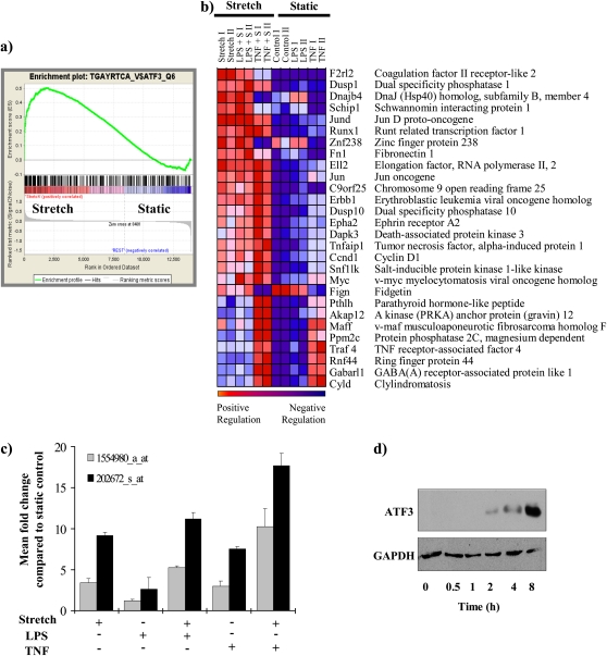
Methods: We undertook a comparative examination of cis-regulatory sequences involved in the coordinated expression of CS-responsive genes using microarray analysis. Analysis of stretched versus nonstretched cells identified significant enrichment for genes containing putative binding sites for the transcription factor activating transcription factor 3 (ATF3). To determine the role of ATF3 in vivo, we compared the response of ATF3 gene-deficient mice to wild-type mice in an in vivo model of VILI.

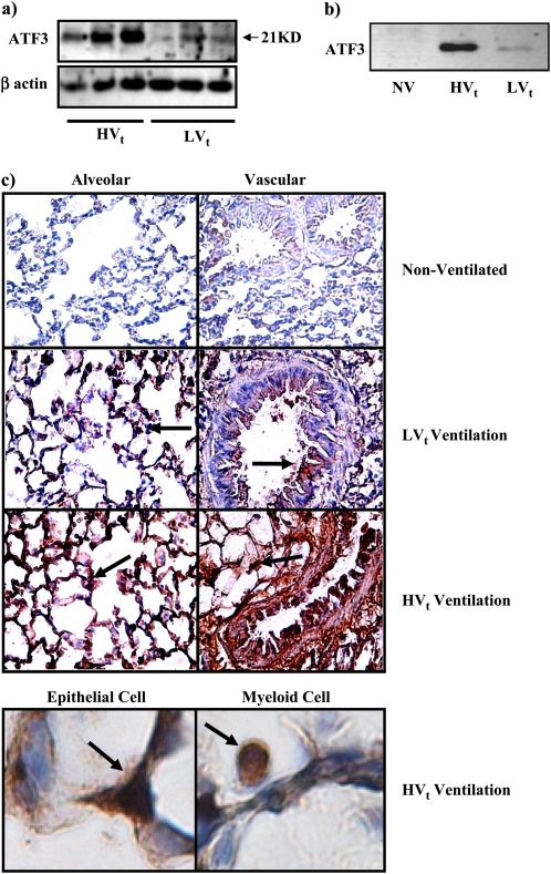
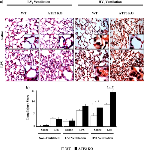
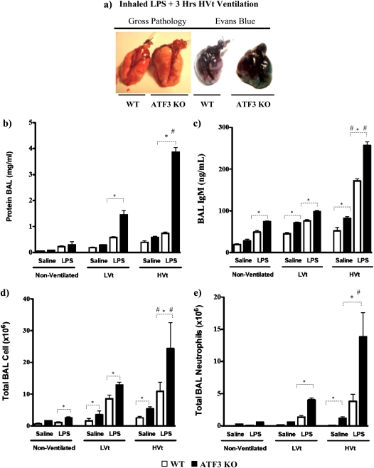
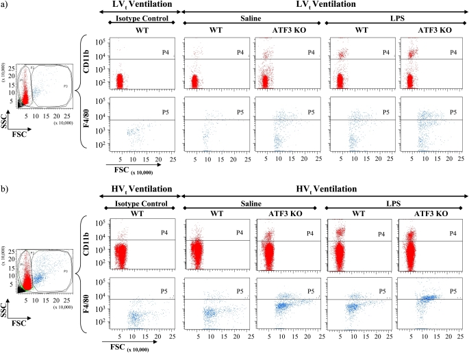
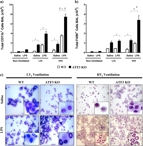
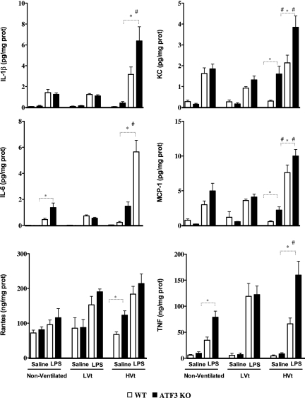
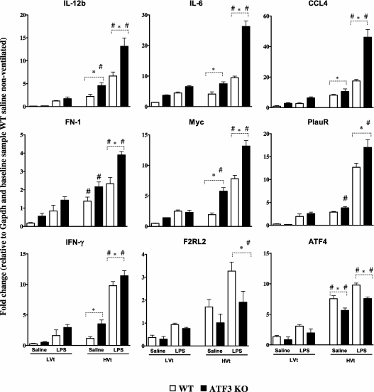
Measurements and main results: ATF3 protein expression and nuclear translocation is increased in the lung after mechanical ventilation in wild-type mice. ATF3-deficient mice have greater sensitivity to mechanical ventilation alone or in conjunction with inhaled endotoxin, as demonstrated by increased cell infiltration and proinflammatory cytokines in the lung and bronchoalveolar lavage, and increased pulmonary edema and indices of tissue injury. The expression of stretch-responsive genes containing putative ATF3 cis-regulatory regions was significantly altered in ATF3-deficient mice.

Conclusions: ATF3 deficiency confers increased sensitivity to mechanical ventilation alone or in combination with inhaled endotoxin. We propose ATF3 acts to counterbalance CS and high volume-induced inflammation, dampening its ability to cause injury and consequently protecting animals from injurious CS.


Figures

Figure 1.
Figure 1.
Enrichment for genes containing putative activating transcription factor 3 (ATF3) cis-regulatory binding sequences. (a) Individual plots for the progression of the running enrichment score (ES) and the maximum peak therein for the top gene set containing genes enriched for ATF3 cis-regulatory regions. The middle part shows the genes in each gene set as “hits” (black vertical lines) against the ranked list of genes. The bottom portion of the plot shows the value of the ranking metric as you move down the list of ranked genes. The ranking metric measures a gene's correlation with the phenotype. A positive value indicates correlation with the phenotype profile (stretch) and a negative value indicates no correlation or inverse correlation with the profile (no-stretch). (b) Corresponding heat map for the expression values for genes containing putative ATF3 cis-regulatory sequence TGACGTCA in their 5′ untranslated region (n = 12 chips) that are significantly differentially expressed between stretch and static. By convention, expression values are represented as a color spectrum going from red (up-regulated) to blue (down-regulated) depending on the correlation with each specific phenotype (stretched versus nonstretched). (c) Mean fold change in expression of ATF3 message in each group compared with static control. Two separate probes for ATF3 are spotted in the array and fold change for each probe is plotted separately. (d) Representative Western blot for ATF3 protein expression in bronchial epithelial airway (Beas-2B) cells exposed to CS for the time periods indicated (n = 3). GAPDH = glyceraldehyde-3-phosphate dehydrogenase; TNF = tumor necrosis factor.
Figure 2.
Figure 2.
High or low tidal volume ventilation (HVt or LVt) for 4 hours increased activating transcription factor 3 (ATF3) expression in rats. Western blot analysis showing ATF3 protein expression was increased both in (a) whole lung tissue cell lysate and (b) nuclear protein extracts from lung tissue of animals breathing spontaneously (nonventilated, NV) or ventilated with either HVt or LVt ventilation strategy. (c) ATF3 protein expression shown by immunohistochemistry (×60) in alveolar and vascular structures. Brown staining (arrows) indicates elevated ATF3 expression. Bottom panel shows detail of both epithelial and myeloid cells demonstrating ubiquitously increased ATF3 protein expression in lung tissues.
Figure 3.
Figure 3.
Activating transcription factor 3 (ATF3) deficiency results in increased lung injury. (a) ATK3 knockout (KO) and wild-type (WT) mice subjected to intratracheal instillation of LPS or equal volume saline. Animals were randomized to either nonventilation or ventilation with low tidal volume (LVt) or injurious high volume ventilation (HVt) for 3 hours. Representative photomicrographs with hematoxylin and eosin staining (×60). ATF3 KO mice had enhanced hyaline membrane formation, severe lung interstitial edema, and inflammatory cell infiltration compared with WT mice. Both LPS and HVt increased lung injury (n = 4 per group). (b) Lung injury scores were determined based on leukocyte infiltration, exudative edema, hemorrhage, and alveolar wall thickness. Data are presented as means ± SEM (n = 4 per group). *P < 0.05 versus corresponding WT.
Figure 4.
Figure 4.
Activating transcription factor 3 (ATF3) deficiency results in increased lung permeability and cellular infiltration. (a) Representative images of gross pathology and Evans blue stain 3 hours after exposure to inhaled LPS and high tidal volume (HVt) ventilation in wild-type (WT) and ATF3 knockout mice (ATF3 KO). Note increased pulmonary edema in ATF3 KO mice compared with WT. (b) The effect of saline (single hit) and LPS (double hit) and mechanical ventilation with either low tidal volume (LVt), HVt, or nonventilated (NV) on protein influx in the bronchoalveolar lavage (BAL) fluid. (c) Extravasation of IgM in BAL. (d) Total cell count. (e) Total neutrophil count. Data are presented as means ± SEM (n = 6). *P < 0.05 between WT and ATF3 mice of similar ventilation strategy (NV, LVt, or HVt) and pretreatment (saline or LPS). #P < 0.05 versus LVt-ventilated mice of similar phenotype (WT or ATF3 KO) and pretreatment (saline or LPS).
Figure 5.
Figure 5.
Fluorescence-activated cell sorting (FACS) analysis of bronchoalveolar lavage (BAL) collected from activating transcription factor 3 (ATF3) knockout (KO) animals treated with LPS along with (a) low tidal volume (LVt) and (b) high tidal volume (HVt) ventilation showing increased infiltration of CD11b+ and F4/80+ cells in BAL. Aliquot of BAL was stained with fluorescein isothiocyanate (FITC)-conjugated anti-CD11b and anti-F4/80 antibodies for FACS analysis as described in detailed Methods. As controls, other aliquots of BAL samples were stained with FITC-conjugated anti-CD11b and anti-F4/80 isotype antibodies. Analyzed cells were divided into two populations based on cell size as exemplified in forward and sideward scatter dot blots (FSC and SSC). Using isotype controls, appropriate gates (P4 and P5) were defined to differentiate between CD11b+ and CD11b cells in the subpopulation of smaller, and between F4/80+ and F4/80 cells in the subpopulation of larger BAL cells. CD11b+ cells likely reflect monocytes, neutrophils, and NK cells but not lymphocytes or nonmyeloid cells, whereas F4/80+ cells are considered to reflect macrophages. (a) Representative staining of BAL collected from mice treated with LVt ventilation strategy stained with anti-CD11b and anti-F4/80 antibodies and with their corresponding isotype control antibodies. Compared with (b) HVt, modest increases of CD11b+ and F4/80+ cell numbers were detected after 3 hours of LVt ventilation and LPS treatment. (b) Representative staining of BAL collected from mice treated with HVt ventilation strategy stained with anti-CD11b and anti-F4/80 antibodies and with their corresponding isotype control antibodies. Higher number of CD11b+ and F4/80+ cells was detected after 3 hours of HVt ventilation as compared with LVt-ventilated mice and after LPS treatment as compared with HVt ventilation in saline-treated mice. This effect was more pronounced in ATF3 KO mice as compared with respective wild-type (WT) control mice. *P < 0.05 as indicated. Data are presented as means ± SEM (n = 6 per group).
Figure 6.
Figure 6.
Activating transcription factor 3 (ATF3) deficiency results in increased cellular infiltrates and lung injury. Pulmonary sequestration of (a) CD11b+ and (b) F4/80+ cells was analyzed by flow cytometry and is expressed as total positive cells (106) per lung. In ATF3 knockout (KO) mice, the combination of mechanical ventilation and LPS inhalation led to increased influx of CD11b+ and F4/80+ cells into the alveolar space compared with wild-type (WT) mice. Injurious ventilation significantly increased cellular influx compared with the lung-protective low tidal volume strategy. Data are presented as means ± SEMs (n = 6). *P < 0.05 between WT and ATF3 KO mice of similar ventilation strategy (nonventilated [NV], low tidal volume [LVt] or high tidal volume [HVt]) and pretreatment (saline or LPS). #P < 0.05 versus LVt-ventilated mice of similar phenotype (WT or ATF3 KO) and pretreatment (saline or LPS). (c) Representative hematoxylin and eosin staining photomicrographs of cytospin of BAL (×40) from WT and ATF3 KO mice ventilated with either LVt or HVt after exposure to either saline or LPS.
Figure 7.
Figure 7.
Levels of inflammatory mediators in lung tissue homogenates of ATK3 knockout (KO) and wild-type (WT) mice. Activating transcription factor 3 (ATF3) KO and WT mice subjected to saline or LPS followed by low tidal volume (LVt) ventilation, high tidal volume (HVt) ventilation, or nonventilation (NV) as detailed in Figure 4. All of the mediator profiles altered by ventilator-induced lung injury that differed between ATF3 KO and WT mice are shown. Data are presented as means ± SEM (n = 8). *P < 0.05 between WT and ATF3 mice of similar ventilation strategy (NV, LVt or HVt) and pretreatment (saline or LPS). #P < 0.05 versus LVt-ventilated mice of similar phenotype (WT or ATF3) and pretreatment (saline or LPS).
Figure 8.
Figure 8.
Quantitative real-time polymerase chain reaction of selected genes containing activating transcription factor 3 (ATF3) cis-regulatory binding sequences. Lung tissues of ATK3 knockout (KO) and wild-type (WT) mice subjected to high tidal volume (HVt) ventilation or low tidal volume (LVt) ventilation as detailed in Figure 4. The gene expression was normalized against glyceraldehyde-3-phosphate dehydrogenase. The changes in gene expression are expressed as fold change relative to the baseline sample from nonventilation (NV) WT mice exposed to saline. ATF3 deficiency led to increased mRNA expression of IL-6, IL-12b, chemokine C-C motif ligand 4 (CCL-4), INF-γ, urokinase receptor (PlauR), coagulation factor II receptor-like 2 (F2rl2), fibronectin (FN1), Myc oncogene (Myc), and activating transcription factor 4 (ATF4). Data are presented as means ± SEMs (n = 6). *P < 0.05 between WT and ATF3 mice of similar ventilation strategy (NV, LVt or HVt) and pretreatment (saline or LPS). #P < 0.05 versus LVt-ventilated mice of similar phenotype (WT or ATF3) and pretreatment (saline or LPS).

References

    1. Ware LB, Matthay MA. The acute respiratory distress syndrome. N Engl J Med 2000;342:1334–1349. - PubMed
    1. ARDSnetwork. Ventilation with lower tidal volumes as compared with traditional tidal volumes for acute lung injury and the acute respiratory distress syndrome. N Engl J Med 2000;342:1301–1308. - PubMed
    1. dos Santos CC, Okutani D, Hu P, Han B, Crimi E, He X, Keshavjee S, Greenwood C, Slutsky AS, Zhang H, et al. Differential gene profiling in acute lung injury identifies injury-specific gene expression. Crit Care Med 2008;36:855–865. - PubMed
    1. dos Santos CC, Han B, Andrade CF, Bai X, Uhlig S, Hubmayr R, Tsang M, Lodyga M, Keshavjee S, Slutsky AS, et al. DNA microarray analysis of gene expression in alveolar epithelial cells in response to TNFalpha, LPS, and cyclic stretch. Physiol Genomics 2004;19:331–342. - PubMed
    1. Han B, Mura M, Andrade CF, Okutani D, Lodyga M, dos Santos CC, Keshavjee S, Matthay M, Liu M. TNFalpha-induced long pentraxin PTX3 expression in human lung epithelial cells via JNK. J Immunol 2005;175:8303–8311. - PubMed

Publication types

MeSH terms