

Imaging in radiation oncology: a perspective
- PMID: 20413639
- PMCID: PMC3227970
- DOI: 10.1634/theoncologist.2009-S106
Imaging in radiation oncology: a perspective
Abstract
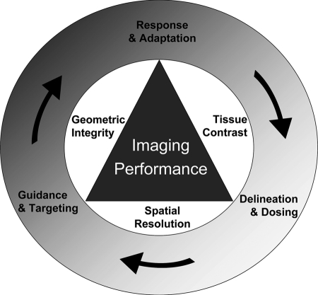
An inherent goal of radiation therapy is to deliver enough dose to the tumor to eradicate all cancer cells or to palliate symptoms, while avoiding normal tissue injury. Imaging for cancer diagnosis, staging, treatment planning, and radiation targeting has been integrated in various ways to improve the chance of this occurring. A large spectrum of imaging strategies and technologies has evolved in parallel to advances in radiation delivery. The types of imaging can be categorized into offline imaging (outside the treatment room) and online imaging (inside the treatment room, conventionally termed image-guided radiation therapy). The direct integration of images in the radiotherapy planning process (physically or computationally) often entails trade-offs in imaging performance. Although such compromises may be acceptable given specific clinical objectives, general requirements for imaging performance are expected to increase as paradigms for radiation delivery evolve to address underlying biology and adapt to radiation responses. This paper reviews the integration of imaging and radiation oncology, and discusses challenges and opportunities for improving the practice of radiation oncology with imaging.
Conflict of interest statement
The content of this article has been reviewed by independent peer reviewers to ensure that it is balanced, objective, and free from commercial bias. No financial relationships relevant to the content of this article have been disclosed by the independent peer reviewers.
Figures
Similar articles
-
How rapid advances in imaging are defining the future of precision radiation oncology.Br J Cancer. 2019 Apr;120(8):779-790. doi: 10.1038/s41416-019-0412-y. Epub 2019 Mar 26. Br J Cancer. 2019. PMID: 30911090 Free PMC article. Review.
-
[Image-guided radiotherapy].Cancer Radiother. 2007 Nov;11(6-7):296-304. doi: 10.1016/j.canrad.2007.08.002. Epub 2007 Sep 21. Cancer Radiother. 2007. PMID: 17889585 Review. French.
-
Functional anatomic imaging in radiation therapy planning.Cancer J. 2004 Jul-Aug;10(4):214-20. doi: 10.1097/00130404-200407000-00002. Cancer J. 2004. PMID: 15383202 Review.
-
Imaging-Based Treatment Adaptation in Radiation Oncology.J Nucl Med. 2015 Dec;56(12):1922-9. doi: 10.2967/jnumed.115.162529. Epub 2015 Oct 1. J Nucl Med. 2015. PMID: 26429959 Review.
-
If you can't see it, you can miss it: the role of biomedical imaging in radiation oncology.Radiat Prot Dosimetry. 2010 Apr-May;139(1-3):321-6. doi: 10.1093/rpd/ncq022. Epub 2010 Mar 11. Radiat Prot Dosimetry. 2010. PMID: 20223847
Cited by
-
Stopping Criteria for Log-Domain Diffeomorphic Demons Registration: An Experimental Survey for Radiotherapy Application.Technol Cancer Res Treat. 2016 Feb;15(1):77-90. doi: 10.7785/tcrtexpress.2013.600269. Epub 2014 Dec 2. Technol Cancer Res Treat. 2016. PMID: 24000996 Free PMC article.
-
Clinical validation of a commercially available deep learning software for synthetic CT generation for brain.Radiat Oncol. 2021 Apr 7;16(1):66. doi: 10.1186/s13014-021-01794-6. Radiat Oncol. 2021. PMID: 33827619 Free PMC article.
-
Emerging Magnetic Resonance Imaging Technologies for Radiation Therapy Planning and Response Assessment.Int J Radiat Oncol Biol Phys. 2018 Aug 1;101(5):1046-1056. doi: 10.1016/j.ijrobp.2018.03.028. Epub 2018 Mar 30. Int J Radiat Oncol Biol Phys. 2018. PMID: 30012524 Free PMC article. Review.
-
Quantitative evaluation of internal marks made using MRgFUS as seen on MRI, CT, US, and digital color images - a pilot study.Phys Med. 2014 Dec;30(8):941-6. doi: 10.1016/j.ejmp.2014.04.007. Epub 2014 May 17. Phys Med. 2014. PMID: 24842080 Free PMC article.
-
Ultrasmall Silica-Based Bismuth Gadolinium Nanoparticles for Dual Magnetic Resonance-Computed Tomography Image Guided Radiation Therapy.Nano Lett. 2017 Mar 8;17(3):1733-1740. doi: 10.1021/acs.nanolett.6b05055. Epub 2017 Feb 2. Nano Lett. 2017. PMID: 28145723 Free PMC article.
References
-
- Eisbruch A, Dawson LA, Kim HM, et al. Conformal and intensity modulated irradiation of head and neck cancer: The potential for improved target irradiation, salivary gland function, and quality of life. Acta Otorhinolaryngol Belg. 1999;53:271–275. - PubMed
-
- van Tinteren H, Hoekstra OS, Smit EF, et al. Effectiveness of positron emission tomography in the preoperative assessment of patients with suspected non-small-cell lung cancer: The PLUS multicentre randomised trial. Lancet. 2002;359:1388–1393. - PubMed
-
- Khoo VS, Joon DL. New developments in MRI for target volume delineation in radiotherapy. Br J Radiol. 2006;79:S2–S15. - PubMed
-
- Haie-Meder C, Pötter R, Van Limbergen E, et al. Recommendations from Gynaecological (GYN) GEC-ESTRO Working Group (I): Concepts and terms in 3D image based 3D treatment planning in cervix cancer brachytherapy with emphasis on MRI assessment of GTV and CTV. Radiother Oncol. 2005;74:235–245. - PubMed
-
- Krempien RC, Daeuber S, Hensley FW, et al. Image fusion of CT and MRI data enables improved target volume definition in 3D-brachytherapy treatment planning. Brachytherapy. 2003;2:164–171. - PubMed
Publication types
MeSH terms
LinkOut - more resources
Full Text Sources
Other Literature Sources
Medical
