. 2010 May 11;107(19):8730-5.
doi: 10.1073/pnas.1002769107. Epub 2010 Apr 22.

Prolonged antigen survival and cytosolic export in cross-presenting human gammadelta T cells

Affiliations

Prolonged antigen survival and cytosolic export in cross-presenting human gammadelta T cells

Simone Meuter et al. Proc Natl Acad Sci U S A. .

Abstract

Human blood Vgamma9Vdelta2 T cells respond to signals from microbes and tumors and subsequently differentiate into professional antigen-presenting cells (gammadelta T-APCs) for induction of CD4(+) and CD8(+) T cell responses. gammadelta T-APCs readily take up and degrade exogenous soluble protein for peptide loading on MHC I, in a process termed antigen cross-presentation. The mechanisms underlying antigen cross-presentation are ill-defined, most notably in human dendritic cells (DCs), and no study has addressed this process in gammadelta T-APCs. Here we show that intracellular protein degradation and endosomal acidification were significantly delayed in gammadelta T-APCs compared with human monocyte-derived DCs (moDCs). Such conditions are known to favor antigen cross-presentation. In both gammadelta T-APCs and moDCs, internalized antigen was transported across insulin-regulated aminopeptidase (IRAP)-positive early and late endosomes; however, and in contrast to various human DC subsets, gammadelta T-APCs efficiently translocated soluble antigen into the cytosol for processing via the cytosolic proteasome-dependent cross-presentation pathway. Of note, gammadelta T-APCs cross-presented influenza antigen derived from virus-infected cells and from free virus particles. The robust cross-presentation capability appears to be a hallmark of gammadelta T-APCs and underscores their potential application in cellular immunotherapy.


Conflict of interest statement

The authors declare no conflict of interest.

Figures

Fig. 1.
Fig. 1.
γδ T-APCs delay endosomal acidification and antigen proteolysis compared with moDCs. (A) Residual fluorescence of FITC-BSA–pulsed γδ T-APCs and moDCs over a 6 h chase period. Values were calculated as percentage of maximal fluorescence after the pulse and expressed as mean ± SEM (n = 8–10). (B) Kinetics of endosomal pH in γδ T-APCs and moDCs, expressed as mean ± SEM of four independent experiments. (C) Western blot analysis of protein degradation in FITC-BSA–pulsed γδ T-APCs and moDCs (top), and quantification of residual protein expressed as percentage of maximal intensity after the pulse (mean ± SEM, n = 6–8) (bottom). *P < 0.05; **P < 0.01; ***P < 0.005.
Fig. 2.
Fig. 2.
Endocytosed BSA is transferred from early to late endosomes/lysosomes in γδ T-APCs and moDCs. Cells were pulsed for 1 h with BSA (green) and analyzed by confocal microscopy using the markers EEA-1, IRAP, Lamp-1, and rab11 (red) to identify early endosomes, late endosomes and lysosomes, and recycling endosomes, respectively. Nuclei are shown in blue. Examples of colocalizing vesicles are marked with arrows. Original magnification, 600×. Confocal images of selected single z-planes are shown, representing the analysis of 12–31 cells (30–40 z-planes/each) from two or three independent experiments.
Fig. 3.
Fig. 3.
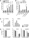
γδ T-APCs, but not moDCs, export internalized protein into the cytosol for degradation by the proteasome. (A) γδ T-APCs were cultured for 6 h with cyt c with or without the caspase inhibitor Z-VAD-FMK. Results are expressed as percentage of apoptotic annexin V+ cells (mean ± SEM; n = 4–6). (B) Apoptosis induction on incubation with 5 mg/mL of cyt c in human γδ T-APCs, moDCs, plasmacytoid DCs (pDCs), conventional DCs (cDCs), and murine (mu) spleen CD8+ and CD8- DCs, expressed as percentage of annexin V+ cells (mean ± SEM; n = 3–6). *P < 0.05; **P < 0.01; ***P < 0.005. (C) Mean fluorescence intensity (MFI) of cells pulsed for 30 min with DQ-BSA and chased for up to 3 h, presented as mean ± SEM (n = 9). (D) Inhibition of protein degradation in DQ-BSA–pulsed cells by lactacystin, NH4Cl, and leupeptin over a 3 h chase period. Results are expressed as mean ± SEM from three to five independent experiments.
Fig. 4.
Fig. 4.
γδ T-APCs take up and cross-present cellular debris. (A) γδ T-APCs (red) were incubated with debris (green), stained with the nuclear dye DRAQ5 (blue), and analyzed by confocal microscopy. A single section in the middle of the cell is shown. Original magnification, 600×. Pictures are representative of two independent experiments. (B) γδ T-APCs were incubated with debris from influenza-infected cells (flu-debris) and cocultured with M1p58-66–specific CD8+ T cells. Results are expressed as percentage of IFN-γ–producing cells, gated on M1p58-66 Tet+ cells (mean ± SEM, n = 4). **P = 0.0054. (C) Blood CD8+ T cells were cocultured with debris-loaded γδ T-APCs, and M1p58-66 Tet+ cells were quantified after 11 days. One of two independent experiments is shown. Negative controls included debris from noninfected cells [debris (no flu)] and infected debris without γδ T-APCs.
Fig. 5.
Fig. 5.
γδ T-APCs cross-present live and inactivated virus. (A) Percentage of IFN-γ–producing M1p58-66 Tet+ T cells in response to γδ T-APCs incubated with increasing concentrations of live influenza virus. Results are expressed as mean ± SEM from three to seven independent experiments. (B) Blood CD8+ T cells were cocultured with virus-treated γδ T-APCs, and M1p58-66 Tet+ cells were quantified by FACS after 11 days. Controls included medium and M1 protein–loaded γδ T-APCs. One of three independent experiments is shown. (C) Residual efficiency of γδ T-APCs to cross-present virus (MOI 1) in the IFN-γ responder cell assay after pretreatment of γδ T-APCs with lactacystin (Left) or after processing of UV or heat-inactivated virus (Right). Data are presented as percentage of activity (mean ± SEM; n = 4–5). (D) Influenza-infected cells (flu-cells) were incubated with γδ T-APCs for 18 h, and cross-presentation was assessed as IFN-γ production by M1p58-66–specific CD8+ T cells (mean ± SEM; n = 4–7). Negative controls included noninfected cells (no flu), HLA-A2 γδ T-APCs, and virus-infected cells in the absence of γδ T-APCs. *P = 0.015; ***P = 0.0018.

References

    1. Cresswell P, Ackerman AL, Giodini A, Peaper DR, Wearsch PA. Mechanisms of MHC class I–restricted antigen processing and cross-presentation. Immunol Rev. 2005;207:145–157. - PubMed
    1. Lin ML, Zhan Y, Villadangos JA, Lew AM. The cell biology of cross-presentation and the role of dendritic cell subsets. Immunol Cell Biol. 2008;86:353–362. - PubMed
    1. Villadangos JA, Schnorrer P. Intrinsic and cooperative antigen-presenting functions of dendritic-cell subsets in vivo. Nat Rev Immunol. 2007;7:543–555. - PubMed
    1. Vyas JM, Van der Veen AG, Ploegh HL. The known unknowns of antigen processing and presentation. Nat Rev Immunol. 2008;8:607–618. - PMC - PubMed
    1. Di Pucchio T, et al. Direct proteasome-independent cross-presentation of viral antigen by plasmacytoid dendritic cells on major histocompatibility complex class I. Nat Immunol. 2008;9:551–557. - PMC - PubMed

Publication types

MeSH terms