Review
. 2010 May;28(3):180-203.
doi: 10.1055/s-0030-1251476. Epub 2010 Apr 22.

Proinflammatory and profibrotic mediators: principal effectors of leiomyoma development as a fibrotic disorder

Affiliations
Review

Proinflammatory and profibrotic mediators: principal effectors of leiomyoma development as a fibrotic disorder

Nasser Chegini. Semin Reprod Med. 2010 May.

Erratum in

  • Semin Reprod Med. 2010 Jul;28(4):345-6

Abstract

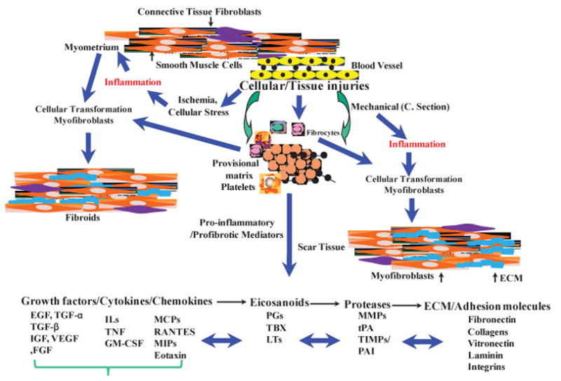
Leiomyomas are believed to derive from the transformation of myometrial smooth muscle cells/connective tissue fibroblasts. Although the identity of the molecule(s) that initiate such cellular transformation and orchestrate subsequent growth is still unknown, conventional evidence indicates that ovarian steroids are essential for leiomyoma growth. Ovarian steroid action in their target cell/tissue is mediated in part through local expression of various growth factors, cytokines, and chemokines. These autocrine/paracrine molecules with proinflammatory and profibrotic activities serve as major contributing factors in regulating cellular transformation, cell growth and apoptosis, angiogenesis, cellular hypertrophy, and excess tissue turnover, events central to leiomyoma growth. This review addresses the key regulatory functions of proinflammatory and profibrotic mediators and their molecular mechanisms, downstream signaling that regulates cellular events that result in transformation, and commitments of specific cells into forming a cellular environment with a possible role in development and subsequent growth of leiomyomas.


Figures

Figure 1
Figure 1
Schematic diagram illustrating an overview of cellular injury and inflammatory-mediated steps in development of leiomyoma as compared with uterine scar formation caused by mechanical injury (i.e., myomectomy and cesarean delivery). Cellular/tissue injuries and generation of an inflammatory response, either at a small scale (microenvironment) or an extended area (myomectomy/cesarean delivery), results in individual and combined regulatory interactions among several proinflammatory and profibrotic mediators. These mediators, including cytokines, chemokines, growth factors, eicosanoids, proteases, and extracellular matrix (ECM), activate and cause myoblasts and resident fibroblasts differentiation into myofibroblastic phenotype. In addition, possible participation of fibrocytes, which are derived from bone marrow and through circulation reside at the site of inflamed/injury, as well as transformation of vascular endothelial cell into mesenchymal cells, also exist to transform into myofibroblastic phenotype. Collectively, myofibroblasts are highly responsive to the action of various mediators, including cytokines and chemokines, and they produce and deposit various components of ECM that are essential for tissue repair process. Continuous inflammation, excess myofibroblastic transformation, and production of large quantity of ECM with concurrent reduced degradation represent a pathway that leads to development of either leiomyoma and/or scar tissue formation. EGF, endothelial growth factor; FGF, fibroblast growth factor; GM-CSF, granulocyte-macrophage colony-stimulating factor; IGF, insulinlike growth factor; IL, interleukin; LT, leukotriene; MCP, monocyte chemoattractant protein; MIP, macrophage inflammatory protein; MMP, metalloproteinase; PAI, plasminogen activator inhibitor; PG, prostaglandin; RANTES, regulated upon activation, normal T-cell expressed and secreted; TBX, thromboxane; TGF, transforming growth factor; TNF, tumor necrosis factor; TIMP, tissue inhibitor of metalloproteinase; tPA, tissue plasminogen activator; VEGF, vascular endothelial growth factor.
Figure 2
Figure 2
Schematic diagram of pathways involving cellular injury and inflammatory-mediated regulation of fibrinolysis through the individual and combined actions of several cytokines and chemokines. Because fibrinolysis is an essential component of provisional matrix establishment and resolution, and angiogenesis and a key part of cellular/tissue repair involving the activation of transforming growth factor (TGF-β), any alteration in this process can often lead to defective fibrinogenic activity and tumorigenesis, including leiomyomas. IFN, interferon; IL, interleukin; MMP, metalloproteinase; PAI, plasminogen activator inhibitor; TIMP, tissue inhibitor of metalloproteinase; TNF, tumor necrosis factor.
Figure 3
Figure 3
Schematic diagram of pathways involving cellular injury and inflammatory-mediated regulation of proteolytic activities involving matrix metalloproteinases (MMPs) and their regulation by several cytokines and chemokines. MMPs and their tissue inhibitors play a central role in provisional matrix degradation, cellular migration, angiogenesis, as well as tissue turnover, processes required for normal cellular/tissue regeneration and repair. Aberrant expression and activation of MMPs has been associated with various fibrinogenic and tumorigenic disorders, including leiomyomas. ECM, extracellular matrix; tPA, tissue plasminogen activator; uPA, urokinase plasminogen activator.
Figure 4
Figure 4
Schematic diagram of pathways involving cellular injury and inflammatory-mediated regulation through the action of individual or combined interaction of several cytokines and chemokines, including ELR- and ELR+chemokines. Because angiogenesis is a key step in neovacuolization and the reparative process of cellular/tissue injury, any alteration in this process can often lead to increased fibrinogenesis and tumorigenesis, including leiomyomas. IFN, interferon; IL, interleukin; TNF, tumor necrosis factor.

References

    1. Holly J. Physiology of the IGF system. Novartis Found Symp. 2004;262:19–26. 26–35, 265–268. - PubMed
    1. Levy B, Mukherjee T, Hirschhorn K. Molecular cytogenetic analysis of uterine leiomyoma and leiomyosarcoma by comparative genomic hybridization. Cancer Genet Cytogenet. 2000;121(1):1–8. - PubMed
    1. Ligon AH, Morton CC. Leiomyomata: heritability and cytogenetic studies. Hum Reprod Update. 2001;7(1):8–14. - PubMed
    1. Lobel MK, Somasundaram P, Morton CC. The genetic heterogeneity of uterine leiomyomata. Obstet Gynecol Clin North Am. 2006;33(1):13–39. - PubMed
    1. Parker WH. Uterine myomas: management. Fertil Steril. 2007;88(2):255–271. - PubMed

Publication types