Review
. 2010 Apr;15(3):433-50.
doi: 10.1111/j.1440-1843.2010.01739.x.

The immunology of tuberculosis: from bench to bedside

Affiliations
Review

The immunology of tuberculosis: from bench to bedside

Keertan Dheda et al. Respirology. 2010 Apr.

Abstract

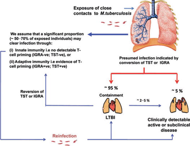
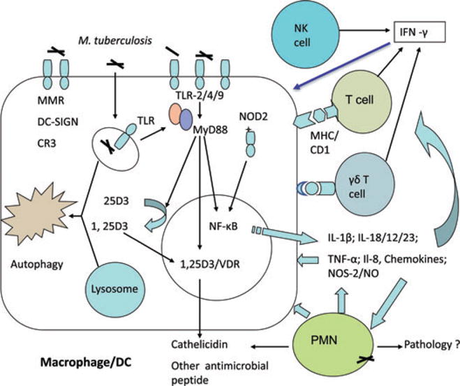
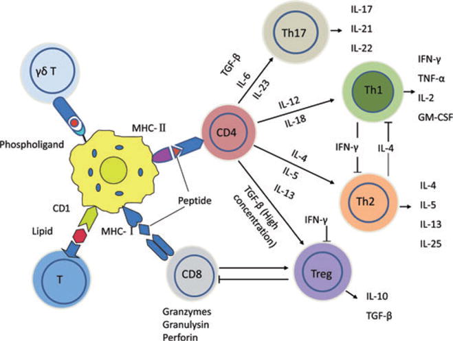
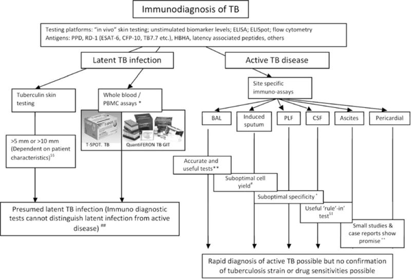
Tuberculosis (TB) is an international public health priority and kills almost two million people annually. TB is out of control in Africa due to increasing poverty and HIV coinfection, and drug-resistant TB threatens to destabilize TB control efforts in several regions of the world. Existing diagnostic tools and therapeutic interventions for TB are suboptimal. Thus, new vaccines, immunotherapeutic interventions and diagnostic tools are urgently required to facilitate TB control efforts. An improved understanding of the immunopathogenesis of TB can facilitate the identification of correlates of immune protection, the design of effective vaccines, the rational selection of immunotherapeutic agents, the evaluation of new drug candidates, and drive the development of new immunodiagnostic tools. Here we review the immunology of TB with a focus on aspects that are clinically and therapeutically relevant. An immunologically orientated approach to tackling TB can only succeed with concurrent efforts to alleviate poverty and reduce the global burden of HIV.


Figures

Figure 1
Figure 1
The spectrum of Mycobacterium tuberculosis infection and the life cycle of M. tuberculosis. Many exposed individuals (~30–50%) have no immunodiagnostic evidence of M. tuberculosis infection or T-cell priming despite heavy exposure to M. tuberculosis. These individuals, although unproven, may be innately resistant to M. tuberculosis infection. Of those who have immunodiagnostic evidence of T-cell priming (+ve TST or IGRA) it is likely that a substantial proportion have LTBI (clinically asymptomatic M. tuberculosis infection during which organisms are in a state of non-replicating persistence). Some individuals have transiently positive responses, and may subsequently ‘revert’ their reactions—they may have ‘acute resolving infection’ or clear their infection. These individuals may become reinfected and progress to active tuberculosis. A proportion of patients with LTBI may also progress to active disease. Given the lack of a gold diagnostic standard for LTBI some of these postulates are based on circumstantial evidence and remain unproven. A multitude of clinical conditions (HIV-1 infection, diabetes mellitus, malnutrition, tobacco smoking, TNF-α inhibitor therapy, helminth coinfection) may permit tipping of the immunological balance and promote transition from latent infection to active disease. IGRA, interferon-γ release assay; LTBI, latent tuberculosis infection; TNF-α, tumour necrosis factor-α; TST, tuberculin skin test.
Figure 2
Figure 2
Innate immunity to tuberculosis infection. Mycobacterium tuberculosis is phagocytosed by macrophages and dendritic cells through membrane-bound receptors such as CR3, scavenger receptor, MMR, TLR, NOD2 and DC-SIGN. These lead to activation of macrophage signalling pathways (NF-κB), causing secretion of pro-inflammatory cytokines, chemokines, and antimicrobial molecules, and activation of VDR, which induces the expression of the antimicrobial peptides cathelicidin and β-defensin. In addition, induction of autophagy mediates antimicrobial activity. PMN cells recognize and engulf M. tuberculosis and secrete antimicrobial peptides to kill bacteria. NK cells, γδ T cells and CD1-restricted T cells are also be activated by specific ligands and cytokines, release cytotoxic factors and secrete IFN-γ, which activates macrophages. CR3, complement receptor 3; DC-SIGN, dendritic cell-specific intercellular-adhesion-molecule-3-grabbing non-integrin; INF, interferon; MMR, macrophage mannose receptor; NK, natural killer; PMN, polymorphonuclear neutrophils; TLR, toll-like receptors; TNF, tumour necrosis factor; VDR, vitamin D receptor.
Figure 3
Figure 3
Adaptive immunity to tuberculosis infection. The infected macrophages and dendritic cells secrete cytokines that include IL-12, IL-23, IL-7, IL-15 and TNF-α, and present antigens to several T-cell populations including CD4+ T cells (MHC class II), CD8+ T cells (MHC class I), CD1-restricted T cells (glycolipid antigens) and γδ T cells (phospholigands). These T cells produce the effector cytokine IFN-γ, which activates macrophages in conjunction with TNF-α to effect killing of intracellular mycobacteria through reactive oxygen and nitrogen intermediates. In addition, CD8+ cytotoxic T cells can kill intracellular mycobacteria through granulysin and perforin-mediated pathways. However, CD4+ Th2 cells produce immunosuppressive cytokines such as IL-4, and CD4+CD25+FoxP3+ regulatory T (Treg) cells produce IL-10 and TGF-β that may suppress mycobactericidal effector mechanisms. A new subset of T helper cells called Th17 cells that are produced in the presence of IL-23, and are characterized by production of IL-17, is important modulator of inflammation and recall memory responses. Th17 cells can recruit neutrophils and monocytes, and IFN-γ-producing CD4+ T cells, and stimulate chemokine expression. However, IFN-γ in turn can suppress the IL-17 producing Th17 cells. Thus, there appears to be a more complex cross-regulation of Th1, Th2, Th17 and Treg cell responses, than previously recognized and the precise role of individual responses in protective immunity remain controversial. GM-CSF, granulocyte-macrophage colony-stimulating factor; IFN, interferon; TGF, transforming growth factor; TNF, tumour necrosis factor.
Figure 4
Figure 4
Rapid immunodiagnosis of TB. An outline of the different antigens, testing platforms, biological markers and cells from different compartments used for the rapid quantitative immunodiagnosis of active TB and LTBI. *Flow cytometric platforms have been evaluated (Cosmi et al.). $$Cut-points are defined by international guidelines and vary according to geographical location, BCG vaccination and HIV/immune status. ##Preliminary evidence that HBHA may help distinguish latent from active disease. **Jafari et al., Dheda et al. #Cashmore et al.. +Dheda et al. $Thomas et al., Kosters et al., Kim et al.van Zyl-Smit et al. ++Kim et al., Tinelli et al. BCG, Bacille Calmette Guérin; CSF, cerebrospinal fluid; LTBI, latent TB infection; HBHA, heparin-binding haemagglutinin; PBMC, peripheral blood mononuclear cells; PLF, pleural fluid; TB, tuberculosis.

References

    1. World Health Organization. Global Tuberculosis Control 2009: Epidemiology, Strategy, Financing. WHO; Geneva: 2009.
    1. World Health Organization. Anti-Tuberculosis Drug Resistance in the World: The WHO/IUATLD Global Project on Anti-Tuberculosis Drug Resistance Surveillance. WHO; Geneva: 2008.
    1. Cooper AM. Cell-mediated immune responses in tuberculosis. Annu Rev Immunol. 2009;27:393–422. - PMC - PubMed
    1. Hanekom WA, Abel B, Scriba TJ. Immunological protection against tuberculosis. S Afr Med J. 2007;97:973–7. - PubMed
    1. Reece ST, Kaufmann SH. Rational design of vaccines against tuberculosis directed by basic immunology. Int J Med Microbiol. 2008;298:143–50. - PubMed

Publication types

MeSH terms

Substances