

The effect of transforming growth factor-beta1 on nasopharyngeal carcinoma cells: insensitive to cell growth but functional to TGF-beta/Smad pathway
- PMID: 20416076
- PMCID: PMC2865451
- DOI: 10.1186/1756-9966-29-35
The effect of transforming growth factor-beta1 on nasopharyngeal carcinoma cells: insensitive to cell growth but functional to TGF-beta/Smad pathway
Abstract
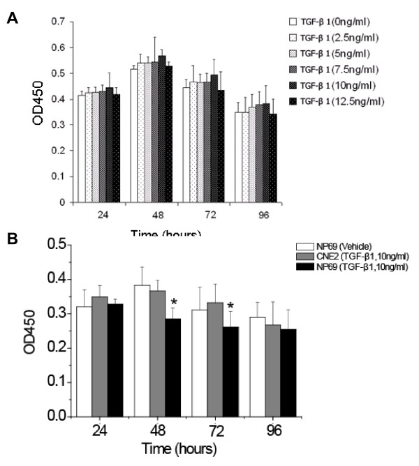
Objectives: This study explored the response of nasopharyngeal carcinoma cells to TGF-beta1-induced growth suppression and investigated the roles of the TGF-beta/Smad signaling pathway in nasopharyngeal carcinoma cells.
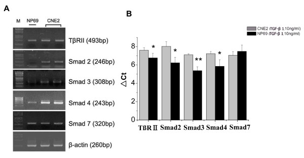
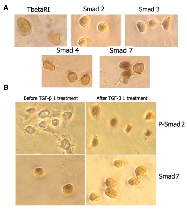
Methods: The cells of nasopharyngeal carcinoma cell line CNE2 were treated with TGF-beta1. The growth responses of CNE2 cells were analyzed by MTT assay. The mRNA expression and protein subcellular localization of the TGF-beta/Smad signaling components in the CNE2 were determined by real time RT-PCR and immunocytochemical analysis.
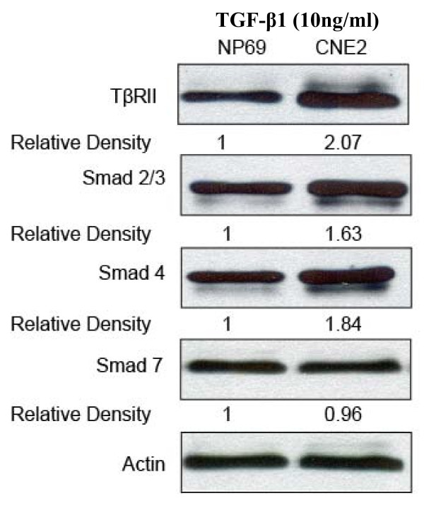
Results: We found that the growth of CNE2 cells was not suppressed by TGF-beta1. The signaling proteins TbetaRII, Smad 7 were expressed normally, while Smad2, Smad3, and Smad4 increased significantly at the mRNA level. TGF-beta type II receptor and Smad7 had no change compared to the normal nasopharyngeal epithelial cells. In addition, Smad2 was phosphorylated to pSmad2, and the activated pSmad2 translocated into the nucleus from the cytoplasm, while the inhibitory Smad-Smad7 translocated from the nucleus to the cytoplasm after TGF-beta1 stimulation.
Conclusion: The results suggested that CNE2 cells are not sensitive to growth suppression by TGF-beta1, but the TGF-beta/Smad signaling transduction is functional. Further work is needed to address a more detailed spectrum of the TGF-beta/Smad signaling pathway in CNE2 cells.
Figures
Similar articles
-
TGF-beta activated Smad signalling leads to a Smad3-mediated down-regulation of DSPP in an odontoblast cell line.Arch Oral Biol. 2004 Nov;49(11):911-8. doi: 10.1016/j.archoralbio.2004.05.005. Arch Oral Biol. 2004. PMID: 15353247
-
Aberrant expression in multiple components of the transforming growth factor-β1-induced Smad signaling pathway during 7,12-dimethylbenz[a]anthracene-induced hamster buccal-pouch squamous-cell carcinogenesis.Oral Oncol. 2011 Apr;47(4):262-7. doi: 10.1016/j.oraloncology.2011.02.003. Epub 2011 Feb 26. Oral Oncol. 2011. PMID: 21356605
-
Inhibition of transforming growth factor-beta1-induced signaling and epithelial-to-mesenchymal transition by the Smad-binding peptide aptamer Trx-SARA.Mol Biol Cell. 2006 Sep;17(9):3819-31. doi: 10.1091/mbc.e05-10-0990. Epub 2006 Jun 14. Mol Biol Cell. 2006. PMID: 16775010 Free PMC article.
-
The transforming growth factor beta 1/SMAD signaling pathway involved in human chronic myeloid leukemia.Tumori. 2010 Sep-Oct;96(5):659-66. doi: 10.1177/030089161009600503. Tumori. 2010. PMID: 21302608 Review.
-
Central role of dysregulation of TGF-β/Smad in CKD progression and potential targets of its treatment.Biomed Pharmacother. 2018 May;101:670-681. doi: 10.1016/j.biopha.2018.02.090. Epub 2018 Mar 22. Biomed Pharmacother. 2018. PMID: 29518614 Review.
Cited by
-
P300 promotes migration, invasion and epithelial-mesenchymal transition in a nasopharyngeal carcinoma cell line.Oncol Lett. 2017 Feb;13(2):763-769. doi: 10.3892/ol.2016.5491. Epub 2016 Dec 13. Oncol Lett. 2017. PMID: 28356956 Free PMC article.
-
ZEB1 Promotes Oxaliplatin Resistance through the Induction of Epithelial - Mesenchymal Transition in Colon Cancer Cells.J Cancer. 2017 Sep 30;8(17):3555-3566. doi: 10.7150/jca.20952. eCollection 2017. J Cancer. 2017. PMID: 29151941 Free PMC article.
-
The Dynamic Roles of TGF-β Signalling in EBV-Associated Cancers.Cancers (Basel). 2018 Jul 27;10(8):247. doi: 10.3390/cancers10080247. Cancers (Basel). 2018. PMID: 30060514 Free PMC article. Review.
-
Limb-bud and Heart (LBH) functions as a tumor suppressor of nasopharyngeal carcinoma by inducing G1/S cell cycle arrest.Sci Rep. 2015 Jan 5;5:7626. doi: 10.1038/srep07626. Sci Rep. 2015. PMID: 25557837 Free PMC article.
-
Epstein-Barr Virus-Encoded Products Promote Circulating Tumor Cell Generation: A Novel Mechanism of Nasopharyngeal Carcinoma Metastasis.Onco Targets Ther. 2019 Dec 31;12:11793-11804. doi: 10.2147/OTT.S235948. eCollection 2019. Onco Targets Ther. 2019. PMID: 32099385 Free PMC article. Review.
References
-
- Shanmugaratnam KSL. WHO. World Health Organization. International Histological Classification of Tumours. 2. Berlin, Springer; 1996. Histological Typing of Tumours of the Upper Respiratory Tract and Ear.
Publication types
MeSH terms
Substances
LinkOut - more resources
Full Text Sources
Miscellaneous
