. 2010 Jul-Aug;41(4):53.
doi: 10.1051/vetres/2010025. Epub 2010 Apr 26.

Essential role of neutrophils but not mammary alveolar macrophages in a murine model of acute Escherichia coli mastitis

Affiliations

Essential role of neutrophils but not mammary alveolar macrophages in a murine model of acute Escherichia coli mastitis

Sharon Elazar et al. Vet Res. 2010 Jul-Aug.

Abstract

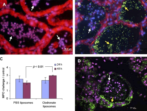
Mastitis, the inflammation of the mammary gland, is an important disease affecting dairy animals worldwide. The disease is caused by mammary pathogenic bacteria and Escherichia coli are frequently implicated. Virulence factors of mammary pathogenic E. coli are only partially known and intramammary challenge with LPS elicits neutrophil recruitment in experimental bovine and murine mastitis models. We have previously shown that neutrophil recruitment in LPS-induced murine mastitis is strictly dependent on mammary alveolar macrophages. However, the relative role of alveolar macrophages and blood neutrophils in E. coli mastitis is not well defined. To this end, we selectively depleted mammary alveolar macrophages or blood neutrophils before intramammary challenge with E. coli strain P4 (ECP4). Mice depleted of alveolar macrophages prior to intramammary challenge recruited neutrophils normally and restricted bacterial growth and interstitial invasion. Importantly however, upon depletion of alveolar macrophages, ECP4 invaded the mammary alveolar epithelial cells and formed intracellular bacterial communities. In contrast, neutrophil depletion prior to intramammary infection with ECP4 was associated with unrestricted bacterial growth, tissue damage, severe sepsis and mortality. This study suggests that neutrophils but not alveolar macrophages provide essential antimicrobial defense against mammary pathogenic E. coli. Furthermore, we show here similar invasion after depletion of alveolar macrophages as in our previous studies showing that LPS/TLR4 signaling on alveolar macrophages abrogates ECP4 invasion of the mammary epithelium. Interestingly, similar ECP4 invasion and formation of intracellular communities were also observed following intramammary infection of either iNOS gene-deficient or IL-1 receptor type 1 gene-deficient mice.


Figures

Figure 1.
Figure 1.
E. coli P4 invades mammary epithelium in alveolar macrophage-depleted mice. Mammary glands of lactating C3H/HeN mice were infused with either PBS-loaded liposomes (A–B) or clodronate-loaded liposomes (D) followed by intramammary infection with E. coli P4. Fluorescence staining of paraffin embedded sections (A and D) or cryosections (B) with DAPI (blue) combined with Sytox orange (red) (A) or phalloidin-rhodamine (red) (B and D) of mammary tissues 48 h after bacterial challenge. GFP expressing bacteria (yellow arrows in B and D) are visible in the alveolar space of PBS-liposome treated mice (B) and inside epithelial cells and in the alveolar space in clodronate-liposome treated mice (D). Neutrophil recruitment into the alveolar space is clearly visible in both treatments (white arrows) which is also reflected by relative (ratio of challenge to control gland) MPO activity (C). Data are mean and S.E., n ≥ 6/group and p value compared PBS-liposomes with clodronate-liposomes infused mice using Student’s t-test. Scale bars 50 μm (A–B), 15 μm (D). All images are representative of the entire sample and the histological morphology and pathology results were very similar for each gland in a given mouse and between mice. (For a color version of this figure please consult www.vetres.org.)
Figure 2.
Figure 2.
Bacterial counts in mammary tissue homogenates after intramammary infection with E. coli P4 in C3H/HeN (A), iNOS −/− (B) and IL1R −/− (C–D) mice. Data represent individual mouse bacterial counts and horizontal bars represent mean bacterial counts (A–B). Total bacterial counts 24 and 48 h after challenge of normal, macrophage depleted and neutrophil-depleted C3H/HeN mice (A) demonstrating the unrestricted overgrowth of bacteria after neutrophil depletion. Differential counts of total, extracellular and intracellular bacteria in mammary tissues of iNOS −/− mice 24 and 48 h after challenged (B) demonstrating epithelial invasion of bacteria 48 h after challenge. Horizontal bars represent the means, n ≥ 6/group and p values were calculated using the non-parametric Mann–Whitney two-independent-samples test for comparison of means. Epithelial invasion in IL1R −/− mice is reflected by differential counts of total (T), extracellular (E) and intracellular (I) bacteria in mammary tissue of IL-1βR −/− mice 24 (C) and 48 h (D) after challenge using drop plating of the dilutions indicated of tissue homogenates (1:10 to 1:107). The challenge dose (C) of the experiment is presented on drop plate C. (For a color version of this figure please consult www.vetres.org.)
Figure 3.
Figure 3.
Similar levels of mammary cytokines and chemokines were produced 24 h after intramammary infusion of E. coli P4 in C3H/HeN and C3H/HeJ mice. Bars represent mean concentrations per mg total protein and S.E. of TNFα, IL-1β, MIP-2 and KC measured by ELISA in tissue homogenates of E. coli P4 and PBS-infused control glands of 6 mice in two different experiments. *p < 0.05 value compared means between challenged and control glands using Student’s t-test.
Figure 4.
Figure 4.
E. coli P4 invades mammary epithelium of iNOS −/− mice 48 h after intramammary infection. Fluorescence staining of cryosections (A and D) or paraffin embedded section (B) with DAPI (blue) combined with phalloidin-rhodamine (red) (A and D) or Sytox orange (red) (B) of mammary tissues 48 h after bacterial challenge. Numerous bacteria are visible in the alveolar space of the wild type (wt) C57BL/6 mouse (yellow arrow in B) while GFP expressing bacteria are visible inside epithelial cells in iNOS −/− mice (yellow arrows in D). Similar neutrophil recruitment (white arrows) into the alveolar space is clearly visible in both C57BL/6 (B) and iNOS −/− (A and D) mice, which is also reflected by relative (ratio of challenge to control gland) MPO activity (C). Data are mean and S.E., n ≥ 6/group and p value compared iNOS −/− with wt mice using Student’s t-test. Scale bars 50 μm (A-B) and 20 μm (D). All images are representative of the entire sample and the histological morphology and pathology results were very similar for each gland in a given mouse and between mice. (For a color version of this figure please consult www.vetres.org.)
Figure 5.
Figure 5.
E. coli P4 invades mammary epithelium of IL1R −/− mice 48 h after intramammary infection. Paraffin embedded sections stained with H&E (A) or Sytox orange (red) combined with DAPI (blue) (B–C). Numerous bacteria are visible in the alveolar space and in the alveolar epithelial cells (yellow arrow). Inflammation is characterized by massive neutrophil recruitment into the alveolar and tubular spaces (black arrows in A) which is better seen in the enlarged alveolus in B (white arrows). Congestion and accumulation of neutrophils in the interstitial spaces is also seen in IL1R −/− mice (white arrows in A and C), which is also reflected by significantly higher relative MPO activity than in wt mice (D). Data are mean and S.E., n ≥ 6/group and p value compared means between IL1R −/− and wt mice using Student t-test. Scale bars 200 μm (A), 20 μm (B) and 50 μm (C). All images are representative of the entire sample and the histological morphology and pathology results were very similar for each gland in a given mouse and between mice. (For a color version of this figure please consult www.vetres.org.)
Figure 6.
Figure 6.
Severe tissue damage and uncontrolled bacterial growth and invasion after E. coli P4 infection in neutrophil-depleted C3H/HeN mice. H&E staining of paraffin sections of mammary tissues 24 (A–B) and 48 h (C–D) after E. coli P4 infection of C3H/HeN mice rendered neutropenic by intraperitoneal injection of anti Gr-1 antibodies 24 h before infection. Normal tissue architecture is maintained and some congestion (blue arrows) is visible 24 h after infection (A–B), and neutrophils are not seen in the alveolar spaces (black arrows in A–B). Fatal septic mastitis 48 h after infection is associated with severe hemorrhages (blue arrows in C–D), massive bacterial growth in the alveolar spaces (black arrows in D) and invasion into alveolar epithelial cells which are undergoing severe swelling, cytoplasmic vacuolization and nuclear condensation (yellow arrow in D). Scale bars 200 μm (A and C), 50 μm (B) and 20 μm (D). All images are representative of the entire sample and the histological morphology and pathology results were very similar for each gland in a given mouse and between mice. (For a color version of this figure please consult www.vetres.org.)

References

    1. Anderson G.G., Palermo J.J., Schilling J.D., Roth R., Heuser J., Hultgren S.J., Intracellular bacterial biofilm-like pods in urinary tract infections, Science (2003) 301:105–107 - PubMed
    1. Berry R.E., Klumpp D.J., Schaeffer A.J., Urothelial cultures support intracellular bacterial community formation by uropathogenic Escherichia coli , Infect. Immun. (2009) 77:2762–2772 - PMC - PubMed
    1. Bradley A.J., Green M.J., Adaptation of Escherichia coli to the bovine mammary gland, J. Clin. Microbiol. (2001) 39:1845–1849 - PMC - PubMed
    1. Burvenich C., Van Merris V., Mehrzad J., Diez-Fraile A., Duchateau L., Severity of E. coli mastitis is mainly determined by cow factors, Vet. Res. (2003) 34:521–564 - PubMed
    1. Burvenich C., Bannerman D.D., Lippolis J.D., Peelman L., Nonnecke B.J., Kehrli M.E. Jr, Paape M.J., Cumulative physiological events influence the inflammatory response of the bovine udder to Escherichia coli infections during the transition period, J. Dairy Sci. (2007) 90:E39–E54 - PubMed

Publication types

MeSH terms