. 2010 Apr 23;17(4):323-32.
doi: 10.1016/j.chembiol.2010.03.009.

Activity-based metabolomic profiling of enzymatic function: identification of Rv1248c as a mycobacterial 2-hydroxy-3-oxoadipate synthase

Affiliations

Activity-based metabolomic profiling of enzymatic function: identification of Rv1248c as a mycobacterial 2-hydroxy-3-oxoadipate synthase

Luiz Pedro S de Carvalho et al. Chem Biol. .

Abstract

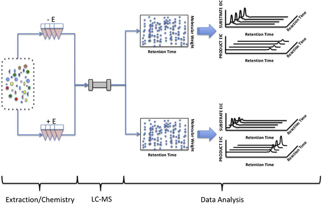
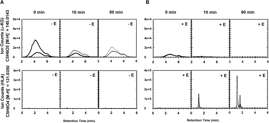
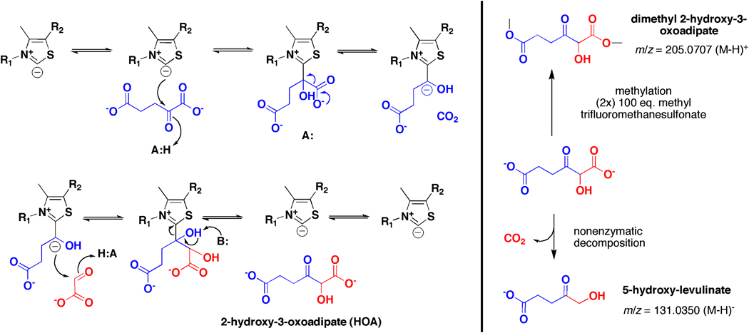
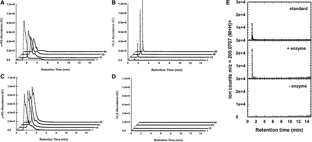
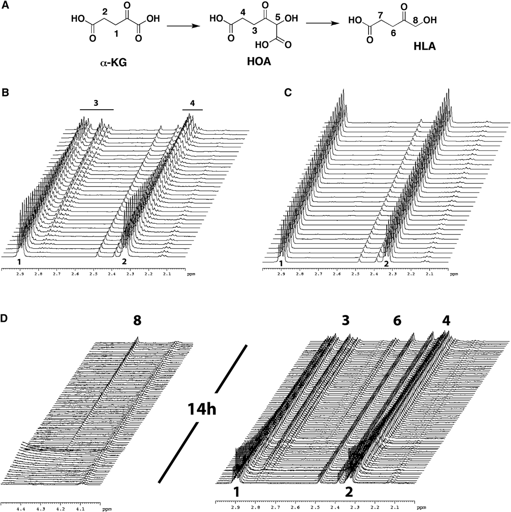
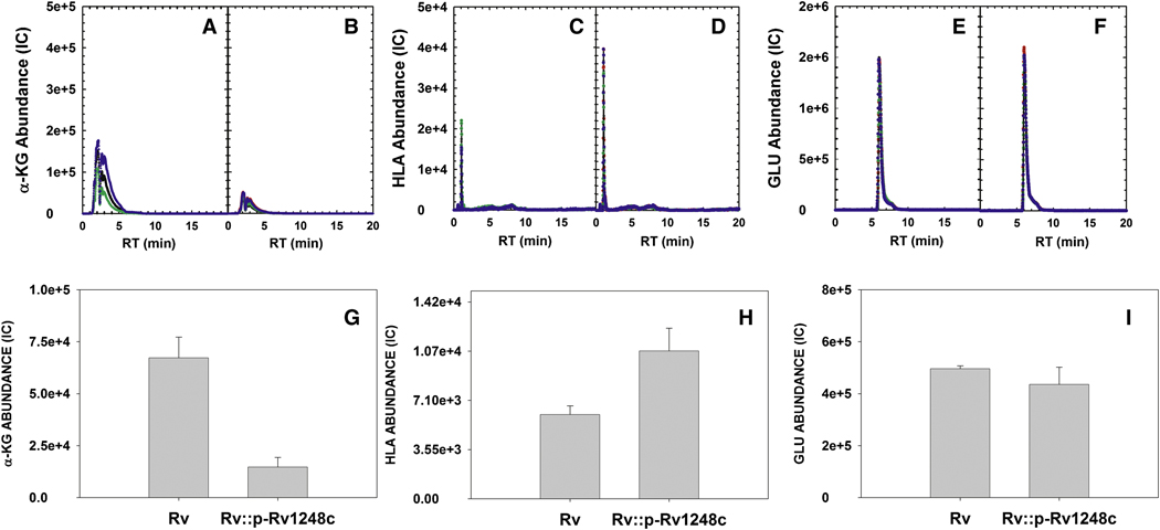
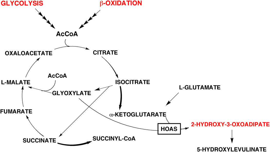
Activity based metabolomic profiling (ABMP) allows unbiased discovery of enzymatic activities encoded by genes of unknown function, and applies liquid-chromatography mass spectrometry (LC-MS) to analyze the impact of a recombinant enzyme on the homologous cellular extract as a physiologic library of potential substrates and products. The Mycobacterium tuberculosis protein Rv1248c was incompletely characterized as a thiamine diphosphate-dependent alpha-ketoglutarate decarboxylase. Here, recombinant Rv1248c catalyzed consumption of alpha-ketoglutarate in a mycobacterial small molecule extract with matched production of 5-hydroxylevulinate (HLA) in a reaction predicted to require glyoxylate. As confirmed using pure substrates by LC-MS, (1)H-NMR, chemical trapping, and intracellular metabolite profiling, Rv1248c catalyzes C-C bond formation between the activated aldehyde of alpha-ketoglutarate and the carbonyl of glyoxylate to yield 2-hydroxy-3-oxoadipate (HOA), which decomposes to HLA. Thus, Rv1248c encodes an HOA synthase.


Figures

Figure 1
Figure 1. Major steps used for activity-based metabolomic profiling
ABMP involves three steps. (1) Incubation of purified recombinant enzyme and potential cofactors with a highly dense, physiologic mixture of potential substrates derived from the native cell type, or closely related source. Metabolite extraction (not depicted) can vary from cell-type to cell-type according to the extraction method and solvent mixture used. Reaction progress is monitored by serial sampling at different time points. (2) Resolution of quenched reaction mixtures using aqueous normal phase liquid chromatography, to achieve chemical class-specific separation, followed by metabolite identification and quantification using accurate mass time-of-flight mass spectrometry. (3) Statistical analysis to identify features that are consumed (putative substrates) and/or produced (putative products) in a time- and enzyme-dependent manner.
Figure 2
Figure 2. ABMP analysis of Rv1248c
Rv1248c-dependent conversion of α-KG and GLX into HLA as followed by LC-MS-based detection in negative mode. (A) Time-course in the absence of enzyme with SME. Extracted ion chromatogram (EIC) for α–KG (m/z (145.0143 [M−H]), top panels, and HLA (m/z 131.0350 [M−H]), bottom panels. (B) Time-course in the presence of enzyme with SME. Extracted ion chromatogram (EIC) for α–KG (m/z (145.0143 [M−H]), top panels, and HLA (m/z 131.0350 [M−H]), bottom panels. Each panel shows the overlay of two replicates. Results are representative of three independent experiments.
Figure 3
Figure 3. Proposed chemical mechanism for Rv1248c-catalyzed formation of HOA from α–KG and GLX followed by non-enzymatic decomposition of HOA to HLA
Unstable HOA was trapped using methyl trifluromethanosulfonate.
Figure 4
Figure 4. Analysis of HOAS activity using authentic substrates and chemical trapping
(A) EIC for α-KG from reaction mix containing Rv1248c. (B) EIC for HLA from reaction mix containing Rv1248c. (C) EIC for α-KG from reaction mix without Rv1248c. (D) EIC for HLA from reaction mix without Rv1248c. Reaction mix contained 50 mM KPi, pH 7.4, 100 µM MgCl2, 100 µM TDP, 100 µM GLX, 100 µM α-KG and 600 nM Rv1248c. Reactions were quenched by addition of cold acidic acetonitrile solution. (E) Trapping of HOA produced by Rv1248c using methyl trifluoromethanesulfonate and comparison with synthetic standard. EIC (m/z = 205.0707 [M+H]+) for synthetic bismethylated HOA (top panel) and the product of trapping reactions performed in reaction mixture containing Rv1248c (middle panel) or not (bottom panel). This experiment was performed in duplicates (black and red symbols), and the results are representative of two independent experiments.
Figure 5
Figure 5. Kinetic analysis of HOAS activity by 1H-NMR
(A) Schematic representation of the reaction and the compounds consumed and generated and designation of the positions followed by NMR. (B) Region of interest from the 1H-NMR spectra of the reaction of Rv1248c with α-KG in the presence of GLX. The spectra were recorded every 4 min for ~ 2 hours. Reaction mix contained 50 mM KPi, pH 7.4, 2 mM MgCl2, 300 µM TDP, 10 mM α-KG, 10 mM GLX and 500 nM Rv1248c. Resonance 5 cannot be seen as it overlaps with the water signal. (C) Region of interest from the 1H-NMR spectra of the reaction of Rv1248c with α-KG. Reaction mix contained 50 mM KPi, pH 7.4, 2 mM MgCl2, 300 µM TDP, 10 mM α-KG and 500 nM Rv1248c. (D) Stack plot of the region of interest from the 1H-NMR spectra of the reaction of Rv1248c with α-KG and GLX. Spectra were recorded every 10 min for ~ 14 hours. Reaction mix contained 50 mM KPi, pH 7.4, 2 mM MgCl2, 100 µM TDP, 2 mM α-KG, 2 mM GLX and 500 nM Rv1248c.
Figure 6
Figure 6. In vivo analysis of α-KG and HLA in M. tuberculosis H37Rv and its Rv1248c merodiploid strain
(A) EIC of α-KG in H37Rv. (B) EIC of α-KG in Rv1248c-merodiploid. (C) EIC of HLA in H37Rv. (D) EIC of HLA in Rv1248c-merodiploid. (E) EIC of GLU in H37Rv. (F) EIC of GLU in Rv1248c-merodiploid. (G) Quantification of the α-KG data in panels A and B. (H) Quantification of the HLA data in panels C and D. (I) Quantification of the GLU data in panels E and F. Data are from a single experiment performed in quadruplicate and are representative of two independent experiments.
Figure 7
Figure 7. Schematic representation of HOAS in Mtb metabolism
Diagram of the glyoxylate shunt and noncanonical Krebs cycle in Mtb, including the new findings on the synthesis of HOA and its conversion to HLA.

Comment in

Similar articles

Cited by

References

    1. Alston TA, Mela L, Bright HJ. 3-Nitropropionate, the toxic substance of Indigofera, is a suicide inactivator of succinate dehydrogenase. Proc Natl Acad Sci U S A. 1977;74:3767–3771. - PMC - PubMed
    1. Argyrou A, Blanchard JS. Mycobacterium tuberculosis lipoamide dehydrogenase is encoded by Rv0462 and not by the lpdA or lpdB genes. Biochemistry. 2001;40:11353–11363. - PubMed
    1. Baughn AD, Garforth SJ, Vilcheze C, W RJ., Jr An anaerobic-type α-Ketoglutarate Ferredoxin Oxidoreductase Completes the Oxidative Tricarboxylic Acid Cycle of Mycobacterium tuberculosis. PLoS Pathog. 2009;5:e1000662. - PMC - PubMed
    1. Brauer MJ, Yuan J, Bennett BD, Lu W, Kimball E, Botstein D, Rabinowitz JD. Conservation of the metabolomic response to starvation across two divergent microbes. Proc Natl Acad Sci U S A. 2006;103:19302–19307. - PMC - PubMed
    1. Brown SC, Kruppa G, Dasseux JL. Metabolomics applications of FT-ICR mass spectrometry. Mass Spectrom Rev. 2005;24:223–231. - PubMed

Publication types

MeSH terms

Substances

LinkOut - more resources