. 2010 Apr 23;17(4):333-41.
doi: 10.1016/j.chembiol.2010.03.005.

Structural characterization of acylimine-containing blue and red chromophores in mTagBFP and TagRFP fluorescent proteins

Affiliations

Structural characterization of acylimine-containing blue and red chromophores in mTagBFP and TagRFP fluorescent proteins

Oksana M Subach et al. Chem Biol. .

Abstract

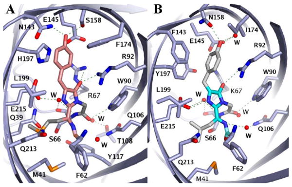
We determined the 2.2 A crystal structures of the red fluorescent protein TagRFP and its derivative, the blue fluorescent protein mTagBFP. The crystallographic analysis is consistent with a model in which TagRFP has the trans coplanar anionic chromophore with the conjugated pi-electron system, similar to that of DsRed-like chromophores. Refined conformation of mTagBFP suggests the presence of an N-acylimine functionality in its chromophore and single C(alpha)-C(beta) bond in the Tyr64 side chain. Mass spectrum of mTagBFP chromophore-bearing peptide indicates a loss of 20 Da upon maturation, whereas tandem mass spectrometry reveals that the C(alpha)-N bond in Leu63 is oxidized. These data indicate that mTagBFP has a new type of the chromophore, N-[(5-hydroxy-1H-imidazole-2-yl)methylidene]acetamide. We propose a chemical mechanism in which the DsRed-like chromophore is formed via the mTagBFP-like blue intermediate.


Figures

Figure 1
Figure 1
The structures of the chromophore-forming tripeptides superimposed onto their (2Fo-Fc) electron density maps. (A) TagRFP, subunit A. (B) mTagBFP, subunit A, two orthogonal views. Oxygen and nitrogen atoms are colored red and blue, respectively. The chromophore backbones for TagRFP and mTagBFP are shown in orange and cyan, respectively. See also Figure S1.
Figure 2
Figure 2
Chromophores and their environments in TagRFP (A) and mTagBFP (B). The chromophore backbones for TagRFP and mTagBFP are shown in orange and cyan, respectively. The hydrogen bonds are indicated with green dashed lines; the atoms are colored by atom type; and the water molecules are shown as red spheres. The Ala59, Thr60 and Ser61 residues are not included for figure clarity. See also description of contacts of the TagRFP and mTagBFP chromophores with the respective β-barrel interiors in the Supplemental Data.
Figure 3
Figure 3
Absorbance spectra of TagRFP (A) and mTagBFP (B) in PBS buffer (solid lines), denatured in 0.7 M NaOH (dashed lines) and denatured in 0.2 M HCl (dotted lines).
Figure 4
Figure 4
Mass-spectral analysis of the chromophore-bearing peptide of mTagBFP. Table representing the distinctive peaks in MS/MS spectra of the chromophore-bearing peptide ions and structure of the mTagBFP chymotrypsin-derived chromopeptide are shown. The most prominent MS/MS fragments are presented in brackets.
Figure 5
Figure 5
Proposed pathways for the formation of the mTagBFP and TagRFP chromophores. The C-form is the chromophore precursor; the B-form is the blue-emitting chromophore form; and the R-form is the red-emitting chromophore form.

References

    1. Andresen M, Wahl MC, Stiel AC, Gräter F, Schäfer LV, Trowitzsch S, Weber G, Eggeling C, Grubmüller H, Hell SW, Jakobs S. Structure and mechanism of the reversible photoswitch of a fluorescent protein. Proc Natl Acad Sci USA. 2005;102:13070–13074. - PMC - PubMed
    1. Barondeau DP, Kassmann CJ, Tainer JA, Getzoff ED. The case of the missing ring: radical cleavage of a carbon-carbon bond and implications for GFP chromophore biosynthesis. J Am Chem Soc. 2007;129:3118–3126. - PubMed
    1. Bogdanov AM, Mishin AS, Yampolsky IV, Belousov VV, Chudakov DM, Subach FV, Verkhusha VV, Lukyanov S, Lukyanov KA. Green fluorescent proteins are light-induced electron donors. Nat Chem Biol. 2009;5:459–461. - PMC - PubMed
    1. Brejc K, Sixma TK, Kitts PA, Kain SR, Tsien RY, Ormö M, Remington SJ. Structural basis for dual excitation and photoisomerization of the Aequorea victoria green fluorescent protein. Proc Natl Acad Sci U S A. 1997;94:2306–2311. - PMC - PubMed
    1. Bulina ME, Verkhusha VV, Staroverov DB, Chudakov DM, Lukyanov KA. Hetero-oligomeric tagging diminishes non-specific aggregation of target proteins fused with Anthozoa fluorescent proteins. Biochem J. 2003;371:109–114. - PMC - PubMed

Publication types

Substances