. 2010 Jun 25;285(26):19738-46.
doi: 10.1074/jbc.M110.101287. Epub 2010 Apr 23.

Functional analysis of the transmembrane domains of presenilin 1: participation of transmembrane domains 2 and 6 in the formation of initial substrate-binding site of gamma-secretase

Affiliations

Functional analysis of the transmembrane domains of presenilin 1: participation of transmembrane domains 2 and 6 in the formation of initial substrate-binding site of gamma-secretase

Naoto Watanabe et al. J Biol Chem. .

Abstract

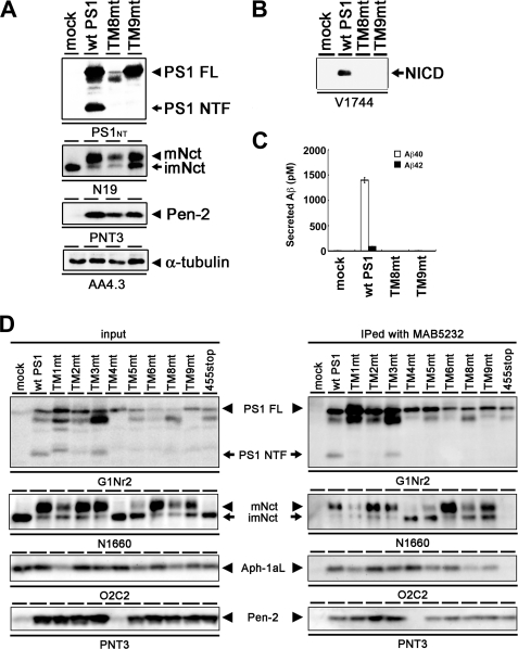
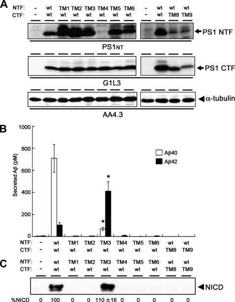
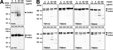
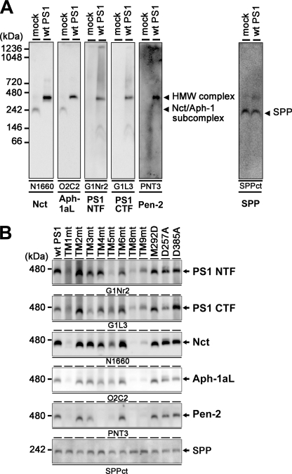
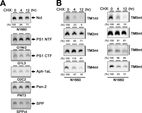
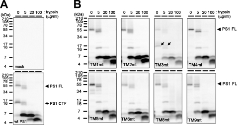
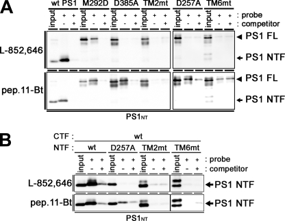
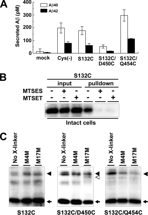
gamma-Secretase is a multimeric membrane protein complex composed of presenilin (PS), nicastrin, Aph-1, and Pen-2, which mediates intramembrane proteolysis of a range of type I transmembrane proteins. We previously analyzed the functional roles of the N-terminal transmembrane domains (TMDs) 1-6 of PS1 in the assembly and proteolytic activity of the gamma-secretase using a series of TMD-swap PS1 mutants. Here we applied the TMD-swap method to all the TMDs of PS1 for the structure-function analysis of the proteolytic mechanism of gamma-secretase. We found that TMD2- or -6-swapped mutant PS1 failed to bind the helical peptide-based, substrate-mimic gamma-secretase inhibitor. Cross-linking experiments revealed that both TMD2 and TMD6 of PS1 locate in proximity to the TMD9, the latter being implicated in the initial substrate binding. Taken together, our data suggest that TMD2 and the luminal side of TMD6 are involved in the formation of the initial substrate-binding site of the gamma-secretase complex.


Figures

FIGURE 1.
FIGURE 1.
Complementation of maturation of Nct and accumulation of Pen-2 by expression of TM8mt and TM9mt in DKO cells. A, immunoblot analysis of DKO cells stably transfected with WT or mutant PS1 (as indicated above the panel). Cell lysates were analyzed by immunoblotting with each antibody (as indicated below the panel). FL, full length; mNct, mature nicastrin; imNct, immature nicastrin. B, immunoblot analysis of the generation of NICD from NotchΔE-stable DKO cells. C, sandwich ELISA analysis of secreted Aβ from APPNL-stable DKO cells transiently expressing TMD-swap PS1 mutants. The levels of Aβ40 and Aβ42 are shown by white and black bars, respectively. Error bars indicate S.E. D, co-immunoprecipitation analysis of 1% CHAPSO-solubilized fractions prepared from DKO cells transiently transfected with WT or mutant PS1. Soluble fractions were precipitated (IPed) by MAB5232 and then analyzed by immunoblotting with each antibody as indicated below the panel.
FIGURE 2.
FIGURE 2.
Intramolecular complementation analysis in DKO cells. A, immunoblot analysis of DKO cells transiently co-expressing NTF and CTF of PS1. B, sandwich ELISA analysis of secreted Aβ from APPNL-stable DKO cells transiently co-expressing mutant NTFs and CTFs. The levels of Aβ40 and Aβ42 are shown by white and black bars, respectively. TM3mt exhibited a significantly decreased Aβ40 activity and increased Aβ42-generating activities (*, statistically significant (n = 3, p < 0.001) when compared with those in cells co-expressing WT NTF/WT CTF by Student's t test). Error bars indicate S.E. C, immunoblot analysis of the generation of NICD from NotchΔE-stable DKO cells using anti-V1744 antibody. Numbers shown below each lane denote the relative band intensities of NICD (%, n = 3) when compared with those in cells co-expressing WT NTF/WT CTF.
FIGURE 3.
FIGURE 3.
Trypsin digestion of Nct polypeptides in DKO cells expressing TMD-swap PS1 mutants. 1% CHAPSO-solubilized lysates prepared from DKO cells transiently transfected with WT PS1 (A) or TMD-swap PS1 mutants (B) were divided into four samples and incubated with trypsin as indicated by concentrations above the panels and analyzed by immunoblotting using anti-Nct antibody (N1660). imNct, immature nicastrin; mNct, mature nicastrin.
FIGURE 4.
FIGURE 4.
HMW complex formation of TMD-swap PS1 mutants. A, BN-PAGE analysis of digitonin-solubilized lysate prepared from DKO cells expressing mock or WT PS1. Antibodies used as well as target proteins are indicated below the panel. Note that the overexpression of PS1 in DKO cells restored the formation of the HMW complex that migrated at 440 kDa. B, BN-PAGE analysis of mutant PS1. SPP was used as loading control as SPP was detected at 200 kDa in this analysis irrespective of PS1 overexpression.
FIGURE 5.
FIGURE 5.
Stability of the HMW complex harboring TMD-swap PS1 mutants. A, analysis of stability of the HMW complex containing WT PS1 in DKO cells incubated in culture medium containing cycloheximide (CHX) (30 mg/ml). DKO cells after various incubation periods (0–12 h) were solubilized in BN-PAGE lysis buffer and analyzed by BN-PAGE analysis with each antibody (as indicated below the panel). In immunoblot using anti-Nct antibody, numbers shown above or below each lane denote the incubation period and the relative band intensity, respectively. Note that all components were stable over 12 h. B, analysis of stability of the HMW complex harboring TMD-swap PS1 mutant probed by anti-Nct antibody.
FIGURE 6.
FIGURE 6.
Trypsin digestion of TMD-swap PS1 polypeptides in DKO cells. 1% CHAPSO-solubilized lysates prepared from DKO cells transiently transfected with WT PS1 (A) or TMD-swap PS1 mutants (B) were divided into four samples and incubated with trypsin at the indicated concentrations above the panels and analyzed by immunoblotting using the anti-PS1 C terminus antibody (anti-PS-C3). Full-length PS1 (PS1 FL) and CTF are shown by black arrowheads and arrows, respectively. Note that the digestion patterns of mutant PS1 are almost similar, whereas the stability of HMW complex is different (see Fig. 5B).
FIGURE 7.
FIGURE 7.
Photoaffinity labeling experiments using TMD-swap PS1 mutants. A, CHAPSO-solubilized DKO cell membranes (DKO cells expressing PS1 holoproteins) were photoactivated in the absence (−) or presence (+) of the parent compound as a competitor and analyzed by immunoblotting with anti-PS1 NTF antibody (PS1NT). PS1 FL, full-length PS1. B, CHAPSO-solubilized DKO cell membranes (DKO cells co-expressing PS1 NTF and PS1 CTF individually from separate vectors) were photoactivated in the absence (−) or presence (+) of the parent compound as a competitor and analyzed by immunoblotting with anti-PS1NT antibody. Probes used are indicated at left of the panel.
FIGURE 8.
FIGURE 8.
Cross-linking experiment using MTS cross-linkers. A, sandwich ELISA analysis of secreted Aβ from APPNL-stable DKO cells transiently expressing cysteine PS1 mutants. Error bars indicate S.E. B, SCAM analysis of S132C mutant PS1 using intact cells. MTSES, 2-sulfonatoethyl methanethiosulfonate; MTSET, 2-(trimethylammonium)-ethyl methanethiosulfonate. C, cross-linking experiments of single- (S132C) or double-Cys (S132C/D450C or S132C/Q454C) mutant PS1 in microsomes and immunoblot analysis using anti-PS1NT antibody. Locations of cysteine mutations are shown below the panel. PS1 FL, NTF, and cross-linked product (NTF-CTF heterodimer) are shown by black arrowheads, black arrows, and white arrowhead, respectively. No X-linker, no cross-linker; M4M, MTS-4-MTS; M17M, MTS-17-MTS.
FIGURE 9.
FIGURE 9.
Malfunction of TMD-swap PS1 mutants in the maturation process of the γ-secretase complex. The maturation process of the γ-secretase complex is indicated as boxes with arrows. PS1 mutants are indicated at right of the process that is defective in the mutant.

References

    1. Tomita T. (2009) Expert Rev. Neurother. 9, 661–679 - PubMed
    1. Wolfe M. S. (2009) Semin. Cell Dev. Biol. 20, 219–224 - PubMed
    1. Takasugi N., Tomita T., Hayashi I., Tsuruoka M., Niimura M., Takahashi Y., Thinakaran G., Iwatsubo T. (2003) Nature 422, 438–441 - PubMed
    1. Esler W. P., Kimberly W. T., Ostaszewski B. L., Ye W., Diehl T. S., Selkoe D. J., Wolfe M. S. (2002) Proc. Natl. Acad. Sci. U.S.A. 99, 2720–2725 - PMC - PubMed
    1. Kornilova A. Y., Bihel F., Das C., Wolfe M. S. (2005) Proc. Natl. Acad. Sci. U.S.A. 102, 3230–3235 - PMC - PubMed

Publication types

MeSH terms

LinkOut - more resources