. 2010 Jul;299(1):H114-24.
doi: 10.1152/ajpheart.00028.2010. Epub 2010 Apr 23.

Alterations in membrane type-1 matrix metalloproteinase abundance after the induction of thoracic aortic aneurysm in a murine model

Affiliations

Alterations in membrane type-1 matrix metalloproteinase abundance after the induction of thoracic aortic aneurysm in a murine model

Jeffrey A Jones et al. Am J Physiol Heart Circ Physiol. 2010 Jul.

Abstract

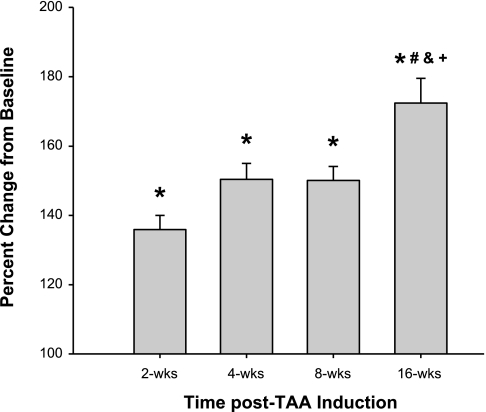
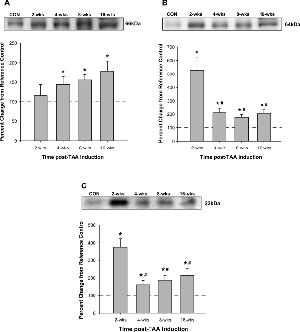
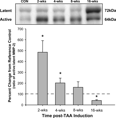
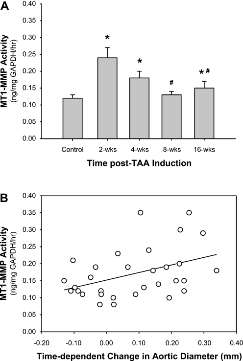
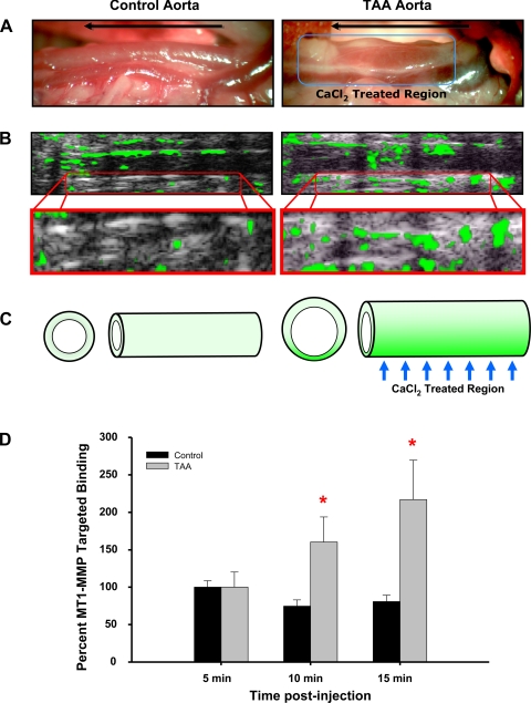
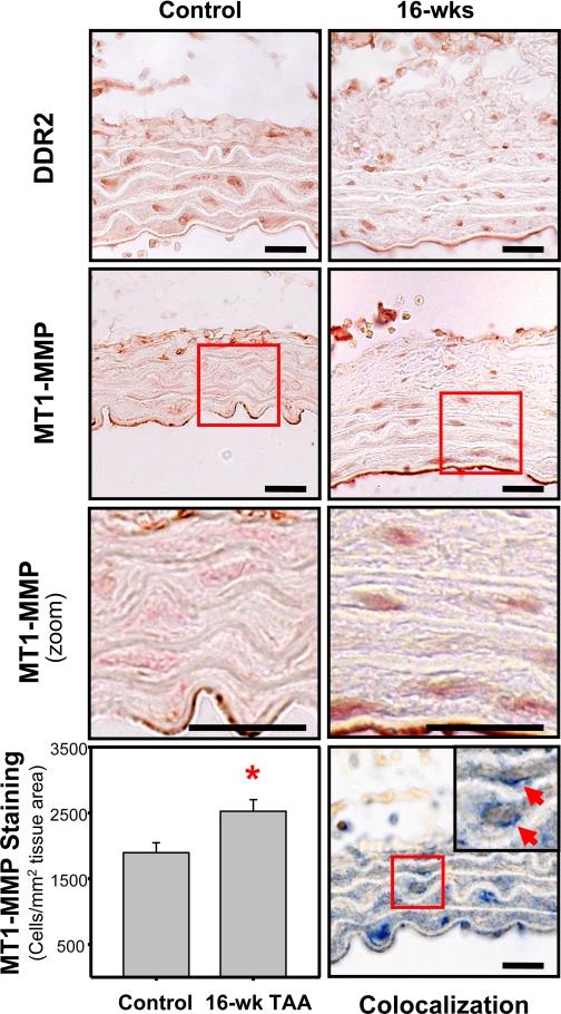
Thoracic aortic aneurysms (TAAs) develop as a result of dysregulated extracellular matrix remodeling mediated by several matrix metalloproteinases (MMPs). Membrane type-1 MMP (MT1-MMP) is the prototypical member of a unique family of membrane-bound MMPs, possessing multiple substrates and functions. The present study tested the hypothesis that MT1-MMP expression, abundance, and activity would be elevated during TAA development and that this protease is produced primarily by mesenchymal cells within the thoracic aorta. Descending thoracic aortas were harvested from C57BL/6J mice at multiple time points (2, 4, 8, and 16 wk, n = 15 each) post-TAA induction (0.5M CaCl(2), 15 min) and compared with reference controls (n = 15). The expression and abundance of MT1-MMP, MMP-2, and tissue inhibitor of metalloproteinase (TIMP)-2 were assessed by quantitative PCR and immunoblot analysis. MT1-MMP activity was determined by fluorescent peptide assay. MT1-MMP was localized within the aortic wall by immunohistochemistry. MT1-MMP abundance and localization in live animals (8 wk post-TAA induction vs. control) was determined by micro-ultrasound imaging with an MT1-MMP-targeted microbubble contrast agent. Aortic diameter was increased 172 +/- 7% at 16 wk post-TAA induction (P < 0.05). MT1-MMP and MMP-2 mRNA levels were elevated at 2 wk post-TAA induction (P < 0.05). MT1-MMP protein abundance increased progressively to a maximum of 178 +/- 26% at 16 wk post-TAA induction, whereas MMP-2 and TIMP-2 peaked at 2 wk post-TAA induction (526 +/- 93% and 376 +/- 48%, respectively, P < 0.05). MT1-MMP colocalized with fibroblasts, and MT1-MMP-targeted contrast binding was elevated in 8-wk TAA-induced mice versus control mice (217 +/- 53% vs. 81 +/- 8%, P < 0.05). In conclusion, these novel results suggest that MT1-MMP plays a dynamic multifunctional role in TAA development and, therefore, may provide a significant target for therapeutic strategies.


Figures

Fig. 1.
Fig. 1.
Change in aortic diameter over time after thoracic aortic aneurysm (TAA) induction. Aortic diameter was determined at baseline and terminal surgery in each animal using digital micrometry. Aortic diameter values were expressed as percent changes from baseline (100%) in each animal. After TAA induction, aortic diameter was elevated at all time points (*P < 0.001 vs. reference control), and 16-wk values were significantly greater than those at the other time points (#P < 0.05 vs. 2 wk, &P < 0.05 vs. 4 wk, and +P < 0.05 vs. 8 wk).
Fig. 2.
Fig. 2.
Quantitative immunoblot analysis was used to measure membrane type-1 (MT1) matrix metalloproteinase (MMP), MMP-2, and tissue inhibitor of metalloproteinase (TIMP)-2 protein abundance. Values are expressed as percent changes from reference control (CON) animals. A: MT1-MMP was elevated at 4 wk post-TAA induction and progressively increased through the 16-wk time course (*P < 0.05 vs. reference control animals). B and C: MMP-2 (B) and TIMP-2 (C) protein levels were increased at all time points after TAA induction (*P < 0.05 vs. reference control animals). Values of the 4-, 8-, and 16-wk time points were significantly different from peak protein abundance observed at 2 wk (#P < 0.05 vs. 2 wk).
Fig. 3.
Fig. 3.
Ratio of active to latent MMP-2 as determined by quantitative immunoblot analysis. The percent change in the ratio of the active (64 kDa) to latent (72 kDa) MMP-2-immunoreactive bands was elevated at 2 and 4 wk post-TAA induction. No change was observed at 8 wk post-TAA induction, and a decrease in the ratio was observed at 16 wk post-TAA induction (*P < 0.05 vs. reference control animals).
Fig. 4.
Fig. 4.
MT1-MMP activity and functional significance. A: activity of MT1-MMP was measured in aortic homogenates from normal and TAA-induced mice using an MT1-MMP-specific quenched fluorogenic peptide activity assay. The results demonstrated elevated MT1-MMP activity (in ng·mg GAPDH−1·h−1) at 2, 4, and 16 wk post-TAA induction (*P < 0.05 vs. reference control animals; #P < 0.05 vs. 2 wk). B: pairwise regression analysis was performed between MT1-MMP activity (in ng·mg GAPDH−1·h−1) and the time-dependent change in aortic diameter (in mm). The results demonstrated a significant correlation (r = 0.4142, P = 0.0205), suggesting that MT1-MMP activity is required for aortic dilatation during TAA development. Linear mixed model analysis demonstrated that significant predictors of MT1-MMP activity included aortic diameter (F = 16.85, P = 0.0004), time (F = 6.09, P = 0.0205), and the interaction between aortic diameter and time (F = 7.62, P = 0.0104).
Fig. 5.
Fig. 5.
MT1-MMP-targeted microbubble contrast imaging. MT1-MMP antibody-conjugated microbubble contrast agent was injected into the tail vein and used to identify elevated MT1-MMP protein levels in the descending thoracic aorta of control and TAA-induced (8 wk) mice. A: intraoperative images of the descending thoracic aorta at terminal surgery showing increased aortic diameter at 8 wk post-TAA induction (right) versus a control animal (left). The direction of blood flow is shown by the arrow in each image, and the CaCl2 treatment region is shown on the right. B: high-resolution ultrasound imaging (equivalent region as shown in A) after the injection of MT1-MMP antibody-conjugated microbubble contrast agent (top). The green overlay indicates areas of enhanced MT1-MMP-targeted contrast binding. Specific binding was quantitated within a region of interest equivalent to the CaCl2-treated region in both control and TAA-induced animals. The bottom images show enlargement of the quantitated region of interest at 15 min after the contrast injection. C: pictogram summarizing the study results showing enhanced aortic dilatation accompanied by increased MT1-MMP-targeted microbubble contrast binding within the CaCl2-treated region. D: quantitative results of MT1-MMP-targeted microbubble contrast imaging demonstrating elevated binding at 10 and 15 min after the contrast injection. *P < 0.05 vs. 5 min. Representative images are shown.
Fig. 6.
Fig. 6.
Immunohistochemical localization of discoidin domain receptor 2 (DDR2) and MT1-MMP. Immunostaining of 3-μm aortic tissue sections from control and TAA-induced mice (16 wk post-TAA induction) demonstrated the presence of DDR2-positive cells (top) and MT1-MMP-positive cells (middle top and middle bottom) within the aortic wall. Bottom left: quantitation of MT1-MMP immunostaining revealed an increased number MT1-MMP-positive cells in the 16-wk TAA-induced sections (33.3% increase vs. control, *P < 0.05). Bottom right: dual sequential immunostaining demonstrated the colocalization of MT1-MMP (blue) with DDR2 (brown) in aortic fibroblasts. Representative images are shown. Bars = 20 μm.

References

    1. Annabi B, Shedid D, Ghosn P, Kenigsberg RL, Desrosiers RR, Bojanowski MW, Beaulieu E, Nassif E, Moumdjian R, Beliveau R. Differential regulation of matrix metalloproteinase activities in abdominal aortic aneurysms. J Vasc Surg 35: 539–546, 2002 - PubMed
    1. Ashworth JL, Murphy G, Rock MJ, Sherratt MJ, Shapiro SD, Shuttleworth CA, Kielty CM. Fibrillin degradation by matrix metalloproteinases: implications for connective tissue remodelling. Biochem J 340: 171–181, 1999 - PMC - PubMed
    1. Barbour JR, Spinale FG, Ikonomidis JS. Proteinase systems and thoracic aortic aneurysm progression. J Surg Res 139: 292–307, 2007 - PubMed
    1. Basalyga DM, Simionescu DT, Xiong W, Baxter BT, Starcher BC, Vyavahare NR. Elastin degradation and calcification in an abdominal aorta injury model: role of matrix metalloproteinases. Circulation 110: 3480–3487, 2004 - PMC - PubMed
    1. Bickerstaff LK, Pairolero PC, Hollier LH, Melton LJ, Van Peenen HJ, Cherry KJ, Joyce JW, Lie JT. Thoracic aortic aneurysms: a population-based study. Surgery 92: 1103–1108, 1982 - PubMed

Publication types

MeSH terms