. 2010 May 18;107(20):9035-42.
doi: 10.1073/pnas.0914013107. Epub 2010 Apr 23.

Dual autonomous mitochondrial cell death pathways are activated by Nix/BNip3L and induce cardiomyopathy

Affiliations

Dual autonomous mitochondrial cell death pathways are activated by Nix/BNip3L and induce cardiomyopathy

Yun Chen et al. Proc Natl Acad Sci U S A. .

Abstract

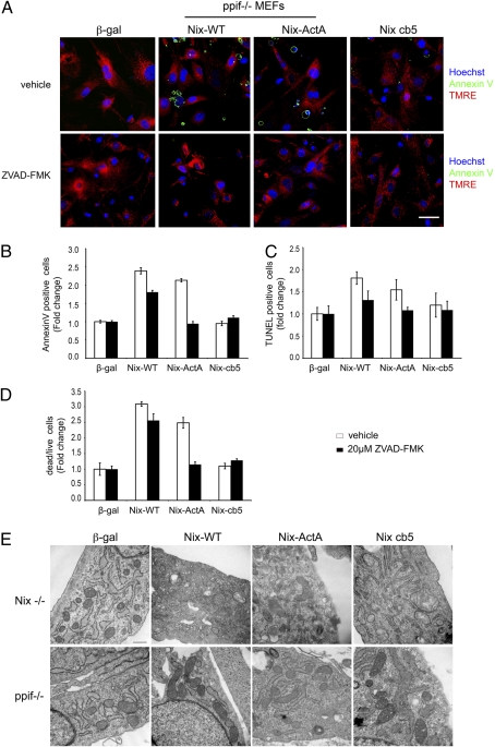
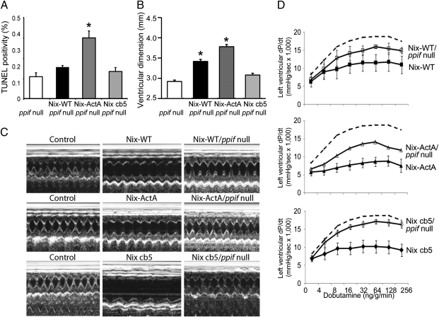
Dysregulation of programmed cell death due to abnormal expression of Bcl-2 proteins is implicated in cancer, neurodegenerative diseases, and heart failure. Among Bcl-2 family members, BNip proteins uniquely stimulate cell death with features of both apoptosis and necrosis. Localization of these factors to mitochondria and endoplasmic reticulum (ER) provides additional complexity. Previously, we observed regulation of intracellular calcium stores by reticular Nix. Here, we report effects of Nix targeting to mitochondria or ER on cell death pathways and heart failure progression. Nix-deficient fibroblasts expressing mitochondrial-directed or ER-directed Nix mutants exhibited similar cytochrome c release, caspase activation, annexin V and TUNEL labeling, and cell death. ER-Nix cells, but not mitochondrial-Nix cells, showed dissipation of mitochondrial inner membrane potential, Deltapsi(m), and were protected from cell death by cyclosporine A or ppif ablation, implicating the mitochondrial permeability transition pore (MPTP). ER-Nix cells were not protected from death by caspase inhibition or combined ablation of Bax and Bak. Combined inhibition of caspases and the MPTP fully protected against Nix-mediated cell death. To determine the role of the dual pathways in heart failure, mice conditionally overexpressing Nix or Nix mutants in hearts were created. Cardiomyocte death caused by mitochondrial- and ER-directed Nix was equivalent, but ppif ablation fully protected only ER-Nix. Thus, Nix stimulates dual autonomous death pathways, determined by its subcellular localization. Mitochondrial Nix activates Bax/Bak- and caspase-dependent apoptosis, whereas ER-Nix activates Bax/Bak-independent, MPTP-dependent necrosis. Complete protection against programmed cell death mediated by Nix and related factors can be achieved by simultaneous inhibition of both pathways.


Conflict of interest statement

The authors declare no conflict of interest.

Figures

Fig. 1.
Fig. 1.
Mitochondrial and reticular Nix are equally effective in causing in vitro cell death. (A) Immunoblot analysis of subcellular fractions: 10,000 × g pellet (10p), 100,000 × g pellet (100p), and 100,000 × g supernatant (S). (B) Confocal colocalization of Nix (green) with MitoFluor Red 589 (mito; Upper) or ER calnexin (ER; Lower) (both red). (Original magnification: ×600.) (C) Quantitative analysis of cell live/dead assay (n = 6 per group). (D) Representative TUNEL (green, with blue DAPI nuclear counterstain) and live (green) dead (red) images. (E) Quantitative TUNEL analysis (n = 6 per group). (Scale bars: 50 μm.) *, P < 0.05 vs β-gal control.
Fig. 2.
Fig. 2.
Mitochondrial and reticular Nix are equally effective in causing in vivo cardiomyopathy. (A) Nix immunoblot analysis of heart homogenates. (B) Mutant Nix localization to cardiac subcellular fractions. (C) TUNEL assays (n = 5 hearts per group). (D) Echocardiographic left ventricular end-diastolic dimension. (E) Echocardiographic left ventricular fractional shortening (n = 26 tet off controls, n = 6 Nix-WT, n = 12 Nix-ActA, n = 10 Nix cb5). (F) Left ventricular peak positive dP/dt as a function of dobutamine infusion dose (n = 9 tet off controls, n = 5 Nix-WT, n = 5 Nix-ActA and n = 4 Nix cb5). *, P < 0.05 compared with tet off control.
Fig. 3.
Fig. 3.
Mitochondrial and reticular Nix cause cell death by different mechanisms. (A) Confocal imaging of: cytochrome c (green) and mitochondrial (red) colocalization (Top), caspase activity (Middle) and annexin V labeling (Bottom). (BD) Group analysis of Annexin V labeling (B), TUNEL staining (C), and cell death (D) as a function of ZVAD-FMK (black bars) or cyclosporin A (gray bars) treatment (n = 6 per group). (E) Confocal imaging of annexin V (green) and TMRE (red) staining (Top), and effects of ZVAD-FMK (Middle) or cyclosporin A (Bottom). (F) Group analysis of live/dead, Annexin V, and TUNEL labeling in Nix-WT expressing MEFS and effects of combined caspase and MPTP inhibition. (Scale bars: 50 μm.)
Fig. 4.
Fig. 4.
Combined bax/bak ablation protects mitochondrial-, but not reticular-, Nix-expressing cells from death. (A) Confocal imaging of cytochrome c (green) and mitochondrial (red) colocalization in bax-bak double null MEFs expressing Nix and Nix mutants. (Top) Vehicle. (Bottom) Cyclosporin A-treated. (B) Confocal imaging of annexin V staining (green) and TMRE (red) in identically treated cells. (C) Group data for cell death (white bars), annexin V labeling (black bars), and TUNEL positivity (gray bars) in bax-bak double null MEFs expressing Nix mutants (n = 6 per group). (Scale bars: 50 μm.) *, P < 0.05 vs. identically treated β-gal controls.
Fig. 5.
Fig. 5.
Ppif ablation protects against death induced by reticular Nix. (A) Confocal imaging of annexin V (green) and TMRE (red) in ppif null MEFs expressing Nix and Nix mutants (Upper) and in cells treated with caspase inhibitor ZVAD-FMK (Lower). (BD) Group analysis of annexin V staining (B), TUNEL labeling (C), and live/dead assay (D) in Nix-expressiing ppif null MEFs with or without ZVAD-FMK treatment. (Scale bars: 50 μm.) (E) Transmission electron microscopy demonstrating mitochondrial degeneration in Nix and Nix-cb5 expressing Nix null MEFs (Upper), which did not occur in ppif null MEFs (Lower).
Fig. 6.
Fig. 6.
Cyclophilin D (ppif) ablation rescues the cardiomyopathy induced by reticular, but not mitochondrial, Nix. (A) Cardiomyocyte TUNEL positivity in mice expressing WT Nix, Nix-ActA, or Nix cb5 on a ppif null background. (B) Cardiac enlargement measured as echocardiographic left ventricular end-diastolic diameter. (C) Representative m-mode echocardiograms of control. Nix mutant mice, and Nix mutant mice on Cyclophilin D null background. (D) Left ventricular peak positive dP/dt response to i.v. dobutamine in the same groups of mice. Control data (dotted line) are replotted from Fig. 2. *, P < 0.05 vs. ppif null.
Fig. 7.
Fig. 7.
Cyclophilin D (ppif) ablation prevents cardiomoycyte necrosis and mitochondrial degeneration induced by reticular Nix. (A) Anti-C5b-9 staining shows prevalent cardiomyocyte necrosis in Nix, and Nix-cb5 expressing myocardium (Upper Left) that is prevented by concomitant ppif ablation (Lower Left). Quantitative group data are shown at Right. (B) Transmission electron microscopy showing mitochondrial abnormalities in Nix and Nix-cb5 cardiomyocytes (Upper) that are prevented by ppif ablation (Lower). (Scale bar: 300 μm.)

Comment in

References

    1. Lowe SW, Cepero E, Evan G. Intrinsic tumour suppression. Nature. 2004;432:307–315. - PubMed
    1. Bredesen DE, Rao RV, Mehlen P. Cell death in the nervous system. Nature. 2006;443:796–802. - PMC - PubMed
    1. Chien KR. Stress pathways and heart failure. Cell. 1999;98:555–558. - PubMed
    1. Danial NN, Korsmeyer SJ. Cell death: Critical control points. Cell. 2004;116:205–219. - PubMed
    1. Newmeyer DD, Ferguson-Miller S. Mitochondria: Releasing power for life and unleashing the machineries of death. Cell. 2003;112:481–490. - PubMed

Publication types

MeSH terms